Dear Editor,
Airway assessment in infants is complex as they lack cooperation. Airway parameters and neck extension cannot be measured and must be assessed with visual examination and history from parents. Retrognathia, micrognathia, and syndromic facies are pointers to difficult airway.[1] Sometimes, the presence of a gross facial abnormality in the form of swelling or lesion might divert our attention from other difficult airway predictors.
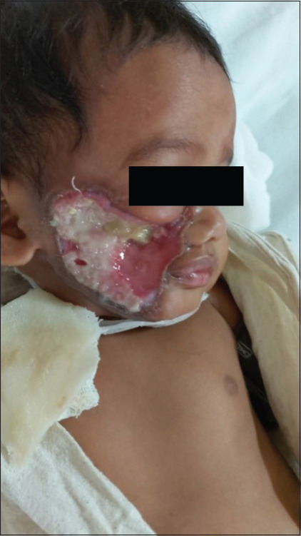
A 7-month-old child weighing 6 kg with ecthyma gangrenosum of the right cheek was scheduled for debridement and skin grafting. The patient was hospitalized with complaints of a rapidly growing vesicle on the cheek and developed sepsis with DIC (Disseminated intravascular coagulation) and ARF (Acute renal failure), which gradually improved. On examination, the patient had an excavated area on the right cheek with granulation tissue extending up to the margin of the angle of mouth [Figure 1]. As per the mother, the child could open his mouth and had no difficulty in feeding. We anticipated difficulty with mask ventilation due to a lack of tissues to ensure the seal. On the day of surgery, sterile gauzes were placed over the defect with a transparent adhesive dressing covering it to ensure a good mask seal. After sevoflurane induction and assessing the ability to ventilate, atracurium was administered. An oral airway was placed to facilitate ventilation with some difficulty as the mouth opening was narrow, which we attributed to a lack of muscle relaxation. During laryngoscopy with Macintosh no. 1 blade, the mouth opening was not sufficient to introduce the flange fully. Hence, ventilation was continued.
Figure 1.

Ecthyma gangrenosum in right cheek
With video laryngoscope (Miscomed) Miller blade 0 size, which had a narrow flange, glottis could just be visualized with external laryngeal manipulation. However, the styletted tube could not be passed through the glottis due to its larger diameter and because the entire oral orifice was occupied with the VL blade. So, an 8-French Pediatric Frova was passed through the glottis and a micro-cuff tube was railroaded over it. Tracheal placement was confirmed. Dexamethasone was given for repeated laryngoscopy and intubation attempts. Trachea was extubated following reversal, once the child was fully awake. Postoperative course was uneventful.
Airway management in infants is challenging. Increased oxygen consumption, higher alveolar ventilation versus FRC (functional residual capacity) ratio, increased chances of gastric insufflation, and anatomical features increase the chances of rapid desaturation while securing the airway.[2,3] Our attention toward the more prominent defect led us to miss the more significant problem of reduced mouth opening due to fibrosing lesion. A patient may have problems with multiple aspects of airway management, like mask ventilation, difficult laryngoscopy, and intubation. Active efforts should be taken to assess the airway in all aspects to avoid facing new problems. In this case, history from mother regarding any increase in mouth opening while crying and the duration of lesion should have been asked. This could have pointed toward the chronic nature of lesion causing fibrosis and reduced mouth opening. Also, mother’s help could have been taken to assess child’s mouth opening. A thorough approach to airway assessment is essential while managing pediatric airways.
Financial support and sponsorship
Nil.
Conflicts of interest
There are no conflicts of interest.
References
- 1.Hagberg CA. Benumof and Hagberg's Airway Management. 4th ed. Elsevier Inc; 2018. [Google Scholar]
- 2.Mortensen A, Lenz K, Abildstrøm H, Lauritsen TL. Anesthetizing the obese child. Paediatr Anaesth. 2011;21:623–9. doi: 10.1111/j.1460-9592.2011.03559.x. [DOI] [PubMed] [Google Scholar]
- 3.Sunder RA, Haile DT, Farrell PT, Sharma A. Pediatric airway management: Current practices and future directions. Paediatr Anaesth. 2012;22:1008–15. doi: 10.1111/pan.12013. [DOI] [PubMed] [Google Scholar]


