NIHPA Author Manuscripts logoLink to NIHPA Author Manuscripts
. Author manuscript; available in PMC: 2024 Jan 23.
Published in final edited form as: Drug Metab Rev. 2020 Jul 9;52(3):333–365. doi: 10.1080/03602532.2020.1772281

Novel Advances in Biotransformation and Bioactivation Research – 2019 year in review*

S Cyrus Khojasteh 1, James P Driscoll 2, Klarissa D Jackson 3, Grover P Miller 4, Kaushik Mitra 5, Ivonne MCM Rietjens 6, Donglu Zhang 1
PMCID: PMC10805366  NIHMSID: NIHMS1953669  PMID: 32645275

Abstract

Biotransformation is one of the main mechanisms used by the body to eliminate drugs. As drug molecules become more complicated, the involvement of drug metabolizing enzymes increases beyond those that are typically studied, such as the cytochrome P450 enzymes. In this review, we try to capture the many outstanding articles that were published in the past year in the field of biotransformation. We have divided the articles into two categories of (1) metabolites and drug metabolizing enzymes, and (2) bioactivation and safety.


This annual review is the fifth of its kind since 2016 (see references). This effort in itself also continues to evolve. We have followed the same format we used in previous years in terms of the selection of articles and the authoring of each section. I am pleased of the continued support of Rietjens, Miller, Zhang, Driscoll and Mitra to this review. We would like to welcome Klarissa D. Jackson as a new author for this year’s issue. We strive to maintain a balance of authors from academic and industry settings.

We would be pleased to hear your opinions of our commentary, and we extend an invitation to anyone who would like to contribute to a future edition of this review.

Cyrus Khojasteh, on behalf of the authors.

Figure 1.

Figure 1.

Glutathione conversion of AZD1979 in the active site of GST, proceeding via protonation of the cyclic aminyl nitrogen of the strained spiro-azetidinyl moiety and subsequent attack by deprotonated GSH (GS).

Figure 4.

Figure 4.

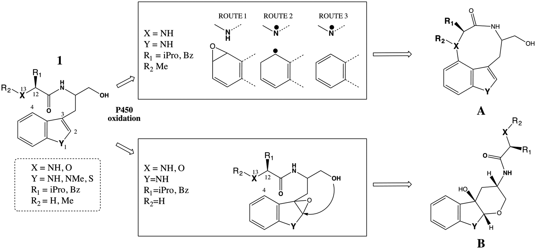
Bacterial P450-mediated oxidation for the formation of two cyclic products A and B. The specific requirement for the formation of these metabolites are captured in the middle boxes with the descriptions of X, Y, R1 and R2 substituents.

iPro=isopropyl and Bz= benzyl groups.

Figure 5.
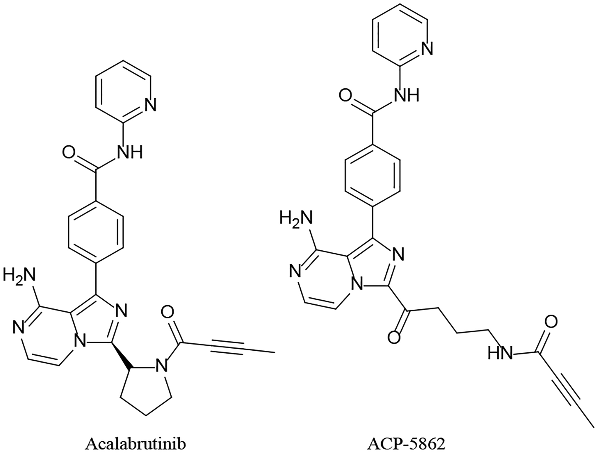
Figure 5.

Acalabrutinib and its active metabolite

Figure 6:
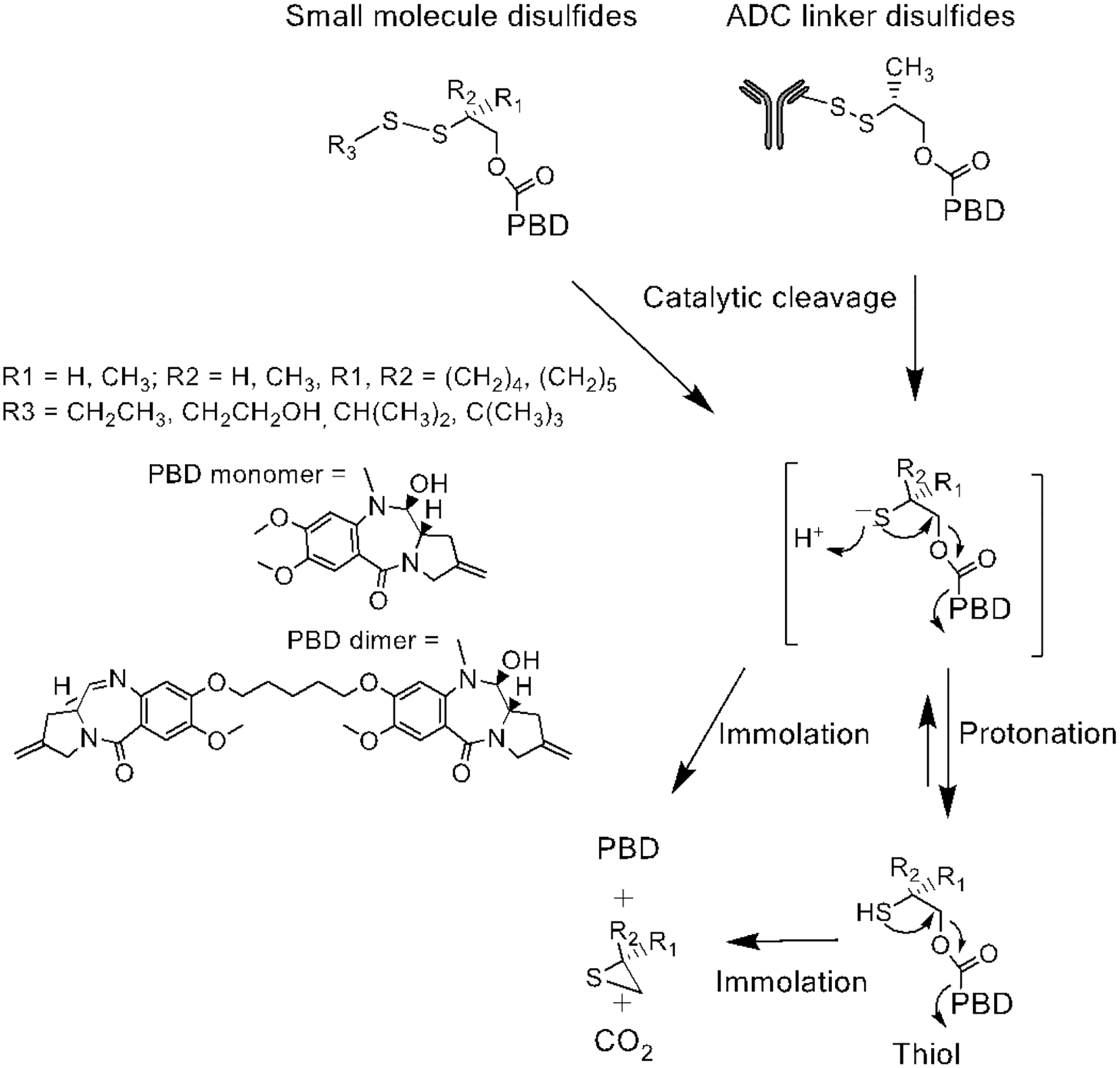
Figure 6:

Catalytic cleavage of disulfide bonds in small molecules and ADC linkers.

Figure 7.
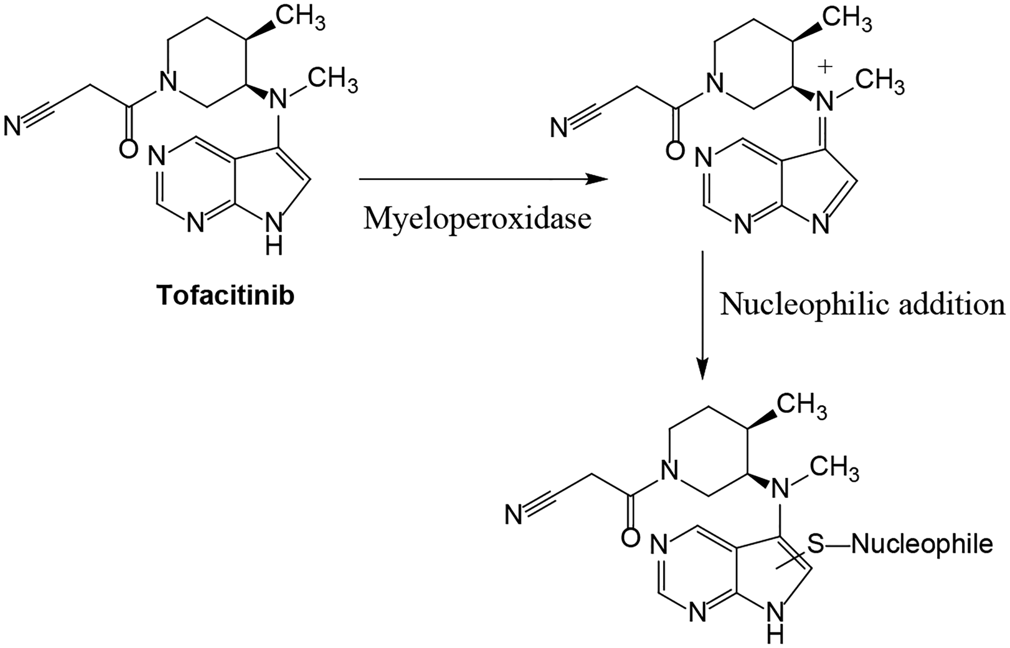
Figure 7.

Proposed bioactivation mechanism of tofacitinib by myeloperoxidase.

Figure 10.
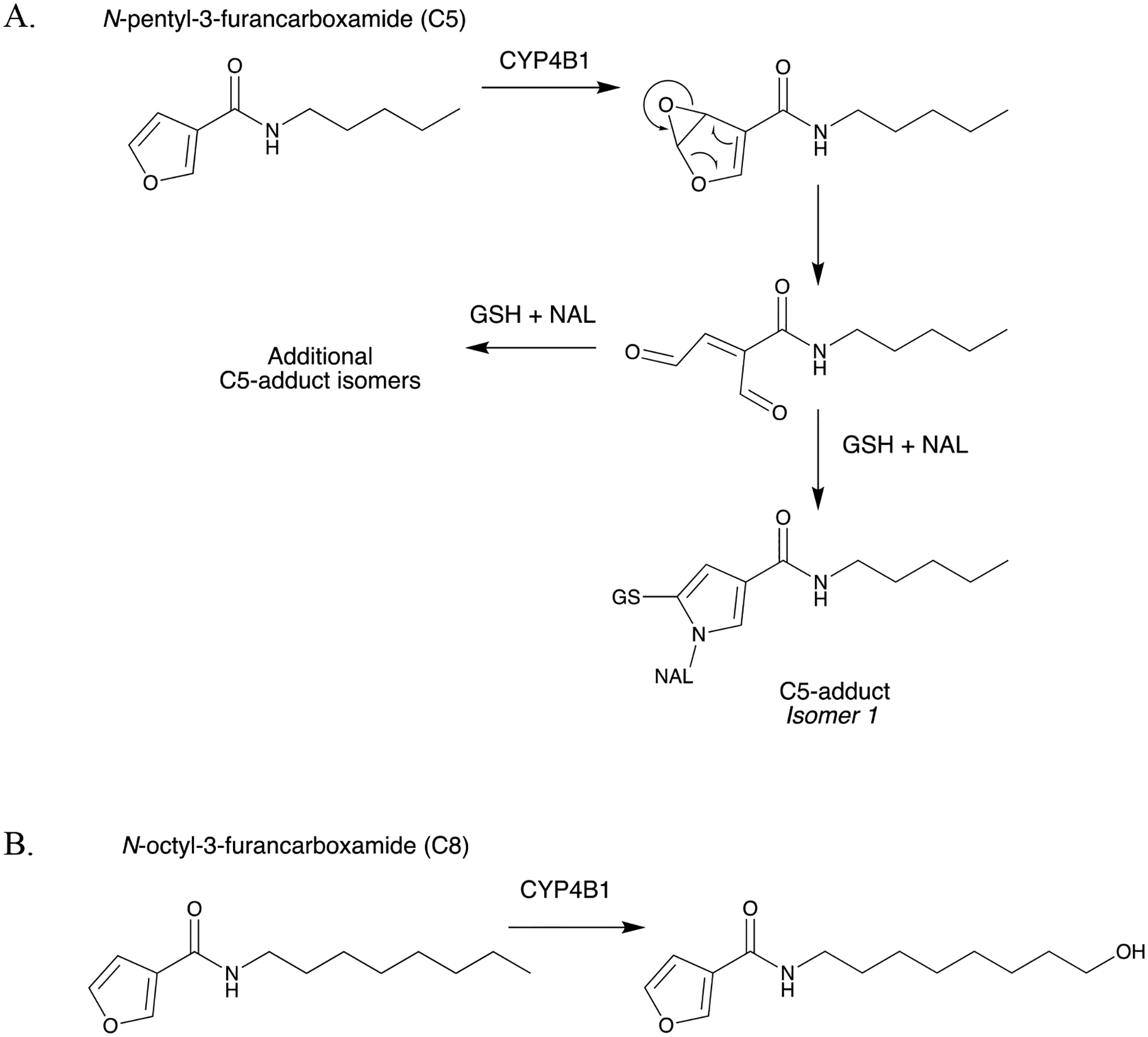
Figure 10.

CYP4B1-mediated bioactivation vs. ω-hydroxylation of N-alkyl-3-furancarboxamides. (A) Bioactivation of N-pentyl-3-furancarboxamide (C5) to form GSH/NAL-trapped C5-adducts, and (B) ω-hydroxylation of N-octyl-3-furancarboxamide (C8) (Kowlaski et al., 2019).

Figure 13.
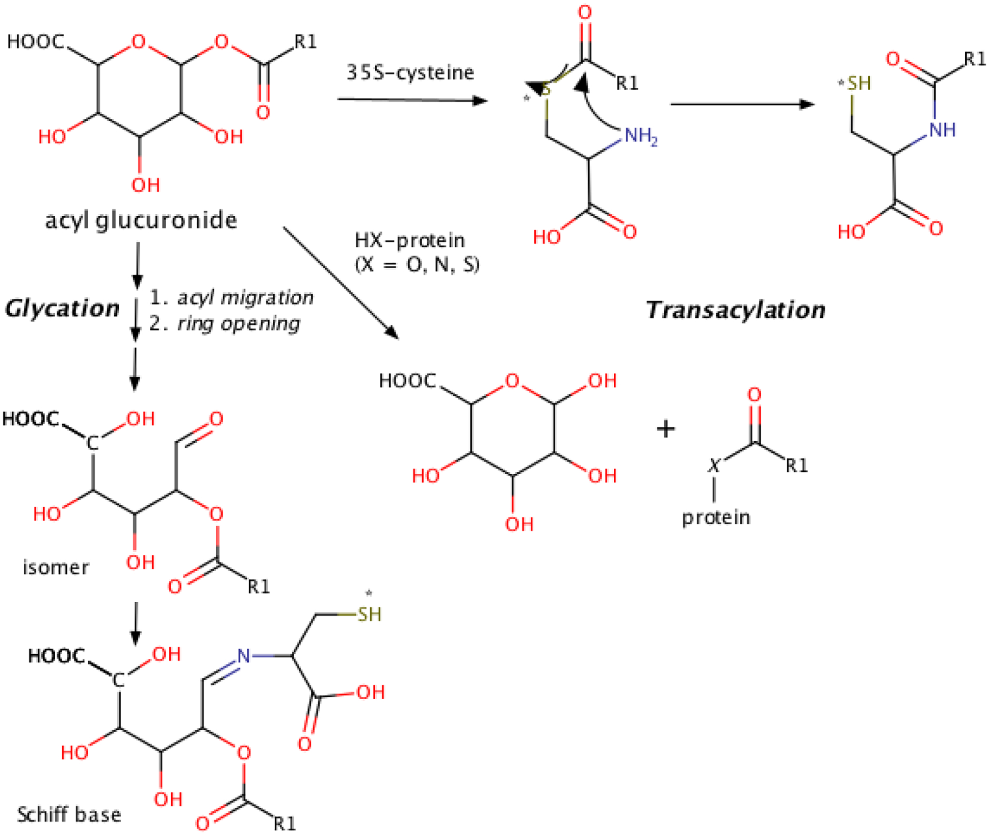
Figure 13.

Proposed pathways leading to trapping of the glycation adduct as a Schiff base and the transacylation adduct ultimately as an amide. 35S-label indicated by asterisk (*).

Table 1.

Articles covered in this review.

Title Authors Source
Metabolites and drug metabolizing enzymes
1 Metabolism of Strained Rings: Glutathione S-transferase-Catalyzed Formation of a Glutathione-Conjugated Spiro-azetidine without Prior Bioactivation Li XQ, Grönberg G, Bangur EH, Hayes MA, Catagnoli Jr. N, Weidolf L Drug Metabolism Disposition 47:1247–1256, 2019
2 Biotransformation of Myricetin: A Novel Metabolic Pathway to Produce Aminated Products in Mice Zhang S, Wang R, Zhao Y, Tareq FS, Sang FS Molecular Nutrition and Food Research 63(14):1900203, 2019
3 The Impact of Carboxylesterases in Drug Metabolism and Pharmacokinetics Di L Current Drug Metabolism 20:91–102, 2019
4 Biocatalytic Reversal of Advanced Glycation End Product Modification Kim NY, Goddard TN, Sohn S, Spiegel DA, Crawford JM ChemBioChem 20:2402–2410, 2019
5 Molecular basis for the P450-catalyzed C–N bond formation in indolactam biosynthesis He F, Mori T, Morita I, Nakamura H, Alblova M, Hoshino S, Awakawa T, Abe I Nature Chemical Biology 15:1206–1213, 2019
6 Bioavailability, Biotransformation, and Excretion of the Covalent Bruton Tyrosine Kinase Inhibitor Acalabrutinib in Rats, Dogs, and Humans Podoll T, Pearson PG, Evarts J, Ingallinera T, Bibikova E, Sun H, Gohdes M, Cardinal K, Sanghvi M, Slatter JG Drug Metabolism Disposition 47:145–154, 2019
7 Catalytic cleavage of disulfide bonds in small molecules and linkers of antibody- drug conjugates Zhang D, Fourie-O’Donohue A, Dragovich PS, Pillow TH, Sadowsky JD, Kozak KR, Cass RT, Liu L, Deng Y, Liu Y, Hop CECA, Khojasteh SC Drug Metabolism Disposition 47(10):1156–1163, 2019
Bioactivation and Safety
8 Metabolic Activation of Tofacitinib Mediated by Myeloperoxidase in Vitro Guo Y, Jia Y, Han L, Zhao Y, Li W, Zhang Z, Peng Y, Zheng J Chemical Research in Toxicology 32:2459–2465, 2019
9 Definition of Haptens Derived from Sulfamethoxazole: In Vitro and in Vivo Tailor A, Waddington J, Hamlett J, Maggs J, Kafu L, Farrell J, Dear G, Whitaker P, Naisbitt D, Park K, Meng X Chemical Research in Toxicology 32(10):2095–2106, 2019
10 Enzalutamide and Apalutamide: In Vitro Chemical Reactivity Studies and Activity in a Mouse Drug Allergy Model Ji C, Guha M, Zhu X, Whritenour J, Hemkens M, Tse S, Walker G, Evans E, Khan N, Finkelstein M, Callegari E, Obach R Chemical Research in Toxicology 32:2095–2106, 2019
11 Structure-activity Relationships of CYP4B1 Bioactivation of 4-ipomeanol Congeners: Direct Correlation Between Cytotoxicity and Trapped Reactive Intermediates. Kowalski JP, McDonald MG, Whittington D, Guttman M, Scian M, Girhard M, Hanenberg H, Wiek C, Rettie AE Chemical Research in Toxicology 32:2488–2498, 2019
12 A Metabolomic Perspective of Pazopanib-induced Acute Hepatotoxicity in Mice. Wang YK, Yang XN, Liang WQ, Xiao Y, Zhao Q, Xiao, Gonzalez FJ, Li F Xenobiotica 49(6):655–670, 2019
13 Rotenone Increases Isoniazid Toxicity but Does Not Cause Significant Liver Injury: Implications for the Hypothesis that Inhibition of the Mitochondrial Electron Transport Chain Is a Common Mechanism of Idiosyncratic Drug-Induced Liver Injury Cho T, Wang X, Uetrecht J Chemical Research in Toxicology 32:1423–1431, 2019
14 Mechanism of Idiosyncratic Drug-Induced Liver Injury Quantitative Evaluation of Reactivity and Toxicity of Acyl Glucuronides by [35S]-Cysteine Trapping Harada H, Toyoda Y, Abe Y, Endo T, Takeda H Chemical Research in Toxicology 32:1955–1964, 2019

Funding:

This work was supported by the US National Institutes of Health (NIH) grants No. R01LM012482 and R01LM012222, and the National Library of Medicine (LM).

Biographies

S Cyrus Khojasteh heads the Biotransformation Function at Genentech (South San Francisco). His research focuses on the mechanisms of biotransformation in drug discovery and development, from small molecules, antibody drug conjugates, and macrocyclic peptides. Cyrus received his Ph.D. in Medicinal Chemistry from the University of Washington under the direction of Professor Sidney D. Nelson.

James P. Driscoll is an accomplished Scientist and Project Leader in the Drug Metabolism and Pharmacokinetics department at MyoKardia, Inc, a clinical stage biopharmaceutical company focused on genetic heart disease. He has over 17 years of industry experience with Pfizer, Genentech, Theravance and MyoKardia. His expertise includes reactive metabolite identification, drug metabolism, and bioanalysis of small molecules. His responsibilities include driving early stage projects forward by identifying structure activity relationships, avoiding off-target toxicity, interrogating potential new targets in drug discovery, and leading a team focused on in vitro compound optimization. James received his B.S. in Human Biology from the State University of New York at Albany.

Klarissa D. Jackson is an assistant professor in the Division of Pharmacotherapy and Experimental Therapeutics at the University of North Carolina at Chapel Hill, UNC Eshelman School of Pharmacy. Jackson obtained her B.S. degree in Chemistry from Jackson State University and her Ph.D. in Pharmacology from Vanderbilt University in the laboratory of Drs. Jason Morrow and L. Jackson Roberts, II. She completed her postdoctoral training at the University of Washington School of Pharmacy in the Department of Medicinal Chemistry under the mentorship of Drs. Allan Rettie and Sidney Nelson. Jackson’s research interests focus on drug metabolism and toxicology to better understand the mechanisms and risk factors of adverse drug reactions.

Grover P. Miller is a full Professor in the Department of Biochemistry and Molecular Biology with a joint appointment in Biomedical Informatics at the University of Arkansas for Medical Sciences. He received BS degrees in Biochemistry and Chemistry with minors in English and French from the Louisiana State University. He obtained his PhD in Chemistry mentored by Stephen J. Benkovic at the Pennsylvania State University and subsequent postdoctoral training as an NIH NRSA fellow mentored by F. Peter Guengerich at Vanderbilt University. His research spans experimental and computational approaches to assess metabolic activation and detoxification of drugs, pollutants, and dietary compounds from the perspective of a chemist.

Kaushik Mitra heads the biotransformation group in Springhouse, PA at Janssen Pharmaceuticals. He is also the DMPK therapeutic area lead for the cardiovascular and metabolic disease portfolio. Previous to Janssen, Kaushik held successive positions at Merck as the head of biotransformation/ADME groups and the Molecular and Investigative Toxicology group. His research interests focus on mechanisms of drug-induced adverse effects such as liver injury and genetic toxicity. Kaushik received his undergraduate degree, with Honors in Chemistry, from St. Xavier’s College in Kolkata, India, Ph.D. in Organic Chemistry from the University of Missouri, Columbia under the mentorship of Prof. Kent S. Gates, and post-doctoral training in Biological Engineering with Prof. John M. Essigmann from the Massachusetts Institute of Technology.

Ivonne M.C.M. Rietjens is full professor and head of the division of Toxicology at Wageningen University, The Netherlands. She obtained her BSc and MSc degrees (cum laude) in Molecular Sciences with a major in Toxicology and a PhD in Toxicology under supervision of Prof. dr. Jan Koeman at Wageningen University. She completed post-doctoral trainings at the National Institute of Public Health and the Environment Health in The Netherlands and at the National Institutes of Health (Bethesda, USA). She is a member of the Royal Netherlands Academy of Arts and Sciences (KNAW), and chair or vice chair of many national and international advisory committees in the field of risk assessment. Her research focuses on the risk evaluation of food borne natural toxins with an emphasis on the role of reactive intermediates and DNA adduct formation, physiologically based kinetic (PBK) models for bioactivation and detoxification, genetic polymorphisms and consequences of life style factors for individual sensitivity and risk assessment and alternatives for animal testing.

Donglu Zhang a Principal scientist at Genentech. His research interests include applying drug metabolism studies in drug design and development of both small molecule drugs and antibody-drug conjugates (ADCs). He received the Sir James Black Award for discovery of and original research on Eliquis from British Pharmacological Society (2018) and the Ondetti and Cushman Award for invention of mass defect filtering method (MDF) from Bristol-Myers Squibb (2007). He received a Ph.D. in Organic Chemistry from University of Utah under direction of Professor C Dale Poulter.

Footnotes

*

This article is dedicated to the late Professor Judy L. Bolton from the University of Illinois at Chicago for her contribution to advancing the mechanistic understanding of estrogens and antiestrogens, and investigating natural alternatives to hormone replacement therapy.

References:

  1. Khojasteh SC, Bumpus NN, Driscoll JP, Miller GP, Mitra K, Rietjens IMCM, Zhang D. (2019) Biotransformation and bioactivation reactions – 2018 literature highlights. Drug Metab Rev. 51(2): 121–161. [DOI] [PubMed] [Google Scholar]
  2. Khojasteh SC, Miller GP, Mitra K, Rietjens IMCM. (2018) Biotransformation and bioactivation reactions – 2017 literature highlights. Drug Metab Rev. 50(3): 221–255. [DOI] [PubMed] [Google Scholar]
  3. Khojasteh SC, Rietjens IMCM, Dalvie D, Miller G. (2017) Biotransformation and bioactivation reactions - 2016 literature highlights. Drug Metab Rev. 49(3): 285–317. [DOI] [PubMed] [Google Scholar]
  4. Baillie TA, Dalvie D, Rietjens IMCM, Khojasteh SC. (2016) Biotransformation and bioactivation reactions - 2015 literature highlights. Drug Metab Rev. 48(2): 113–138. [DOI] [PubMed] [Google Scholar]
Drug Metab Rev. 2020 Jul 9;52(3):333–365.

Metabolism of Strained Rings: Glutathione S-transferase-Catalyzed Formation of a Glutathione-Conjugated Spiro-azetidine without Prior Bioactivation

XQ Li, G Grönberg, EH Bangur, MA Hayes, N Catagnoli Jr, L Weidolf

Synopsis:

This paper by Li et al. (2019) describes the metabolite profile in human hepatocytes of the melanin-concentrating hormone receptor 1 (MCHr1) antagonist AZD1979 [(3-(4-(2-oxa-6-azaspiro[3.3]heptan-6-ylmethyl)phenoxy)azetidin-1-yl)(5-(4-methoxyphenyl)-1,3,4-oxadiazol-2-yl)methanone] (Figure 1). A series of glutathione related metabolites was identified that were formed independent of cytochrome P450-mediated bioactivation, since co-incubation with the P450 inhibitor 1-aminobenzotrizaole did not affect their formation. The structure of the glutathionyl conjugate M12 was elucidated by LC/HRMS and NMR (Figure 1). The authors also propose a mechanism for the formation of M12 involving a protonated cyclic aminyl intermediate that is attacked by the glutathione thiolate anion (GS) in the active site of the GSTs (Figure 1). All GSTs tested were able to catalyse the reaction, with GSTA2–2 being the most efficient. In the absence of GSTs, no M12 formation was observed.

Commentary:

AZD1979 was designed in a drug discovery program for the treatment of obesity because of its appetite-modulating properties (Johansson et al. 2016). The compound contains a strained heterocycle in the form of a spiro-oxetanylazetidinyl-fused ring system. Such a spiro-fused ring system presents a structural motif often used in organic chemistry and drug discovery, in which two rings are fused via a single common atom. Introducing such a spiro-ring system aims to rigidify the ligand conformation, enabling optimal receptor interactions (Zheng et al. 2014). In an earlier study on the metabolic pathways of AZD1979, it was already demonstrated that the spiro-ring system facilitated a novel metabolic pathway proceeding via microsomal epoxide hydrolase-catalysed hydrolysis of the spiro-oxetanyl ring of AZD1979, generating the corresponding diol metabolite (Li et al. 2016). The present paper shows that conjugation with GSH does not occur at the spiro-oxetanyl ring of AZD1979 but results in azetidinyl ring-opening catalyzed by GSTs. The conversion appears to present a major metabolic pathway for AZD1979 in human hepatocytes. The reaction proceeds via a nucleophilic attack by the thiolate anion of GSH (GS), formed in the active site of GSTs, on the protonated strained heterocycle spiro-azetidinyl ring. In most cases, GSH conjugate formation is preceeded by cytochrome P50-mediated bioactivation, forming electrophilic reactive metabolites like epoxides (arene oxides), quinones, quinoneimines or quinone methides. The glutathione conjugation of AZD1979 was shown to occur without the need for cytochrome P450-mediated bioactivation and presents a novel metabolic conversion that may be relevant for other drug candidates with a similar strained spiro ring system.

The study by Li et al. (2019) also demonstrated that in the absence of GSTs, no M12 formation was observed, suggesting that the spontaneous chemical conjugation of AZD1979 was insignificant. The lack of such a spontaneous chemical reactivity of the AZD1979 azetidinyl moiety with GSH might be due to the requirement of GSH thiolate anion formation, which is facilitated in the active site of GSTs, in combination with the need for protonation of the azetidinyl moiety (Li et al. 2019). A similar situation occurred for the epoxide hydrolase mediated ring opening of the spiro-oxetanyl moiety that was shown to undergo ring opening only in the presence of epoxide hydrolase. Thus, the GSH conjugation now described for AZD1979 may not raise a toxicological concern.

References:

  1. Johansson A, Löfberg C, Antonsson M, von Unge S, Hayes MA, Judkins R, Ploj K, Benthem L, Lindén D, Brodin P, Wennerberg M, et al. 2016. Discovery of (3-(4-(2-Oxa-6-azaspiro 3.3 heptan-6-ylmethyl)phenoxy)azetidin-1-yl)(5-(4-methoxyphenyl)-1,3,4-oxadiazol-2-yl)methanone (AZD1979), a Melanin Concentrating Hormone Receptor 1 (MCHr1) Antagonist with Favorable Physicochemical Properties. J Med Chem. 59(6): 2497–2511. [DOI] [PubMed] [Google Scholar]
  2. Li XQ, Grönberg G, Bangur EH, Hayes MA, Castagnoli N Jr, Weidolf L. 2019. Metabolism of Strained Rings: Glutathione S-transferase-Catalyzed Formation of a Glutathione-Conjugated Spiro-azetidine without Prior Bioactivation. Drug Metab Dispos. 47(11): 1247–1256. [DOI] [PubMed] [Google Scholar]
  3. Li XQ, Hayes MA, Grönberg G, Berggren K, Castagnoli N Jr, Weidolf L. 2016. Discovery of a Novel Microsomal Epoxide Hydrolase-Catalyzed Hydration of a Spiro Oxetane. Drug Metab Dispos. 44(8):1341–1348. [DOI] [PubMed] [Google Scholar]
  4. Zheng YJ, Tice CM, Singh SB. 2014. The use of spirocyclic scaffolds in drug discovery. Bioorg Med Chem Lett. 24(16):3673–3682. [DOI] [PubMed] [Google Scholar]
Drug Metab Rev. 2020 Jul 9;52(3):333–365.

Biotransformation of Myricetin: A Novel Metabolic Pathway to Produce Aminated Products in Mice

S Zhang, R Wang, Y Zhao, RS Tareq, S Sang

Synopsis:

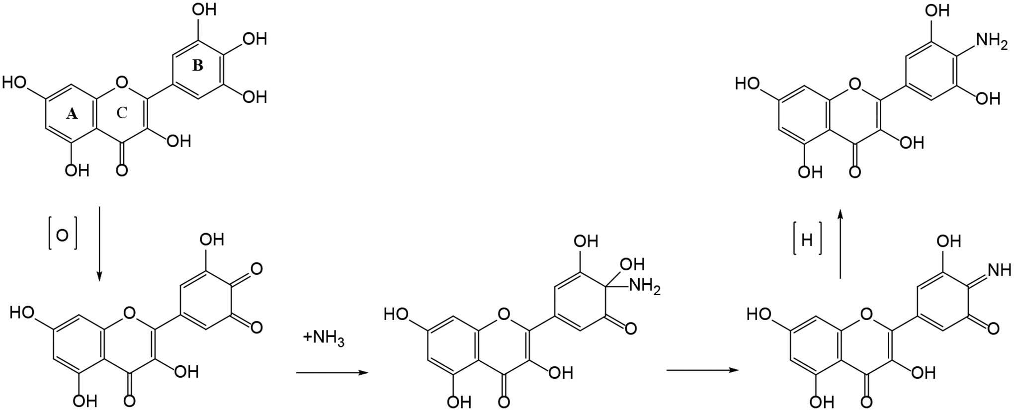
Zhang et al. (2019) report on a novel metabolic pathway for the dietary flavonoid myricetin in mice. In this pathway, the vic-trihydroxy moiety on the B-ring of myricetin reacts chemically with ammonia to form a quinoneimine intermediate that generates an aminated final product in which the hydroxyl moiety at C4’ is replaced by an amino group (Figure 2). The structure of this product, 4’-NH2-myricetin, was confirmed by LC-MS/MS and NMR. The metabolite was detected in fecal samples from mice dosed with myricetin. In addition, two other metabolites of myricetin, including mono-methylated myricetin and 2,4,5,-trihydroyphenylacetic acid, the latter formed by gut microbiota, were also detected in an aminated form. The authors concluded that this amination is a novel biotransformation pathway for myricetin.

Commentary:

Myricetin is a food-borne flavonoid that belongs to the subclass of flavonols. The major metabolites of myricetin are known to be 3,5-dihydroxyphenylacetic acid, 3,4,5-trihydroxyphenylacetic acid, mono-methylated myricetin, di-methylated myricetin and a mono- and diglucuronide. A novel metabolic pathway for myricetin reportedly involves formation of a metabolite resulting from a reaction of myricetin with ammonia. Ammonia is a toxic waste product ubiquitously present in the human body with concentrations in adult human blood amounting to 6–47 μmol/L. Previous studies already demonstrated that polyphenols that contain a vic-trihydroxyl moiety, such as pyrrogallol, epigallocatechin (EGC) and epigallocatechin-3-gallate (EGCG) can react with ammonia, generating aminated products in in vitro model systems (Hashida et al. 2009, Zhang S et al. 2019). The present study aimed to provide additional support for this amination as a general metabolic pathway for polyphenols, using myricetin as the additional model compound. It is of interest that the results of the present study not only confirmed the occurrence of this amination reaction for myricetin, but also detected 4’-NH2-myricetin in vivo in fecal samples collected from myricetin-treated mice. In addition, there was detection of aminated metabolites of the myricetin metabolites 3,4,5-trihydroxyphenylacetic acid and methylated myricetin, tentatively identified as 4-NH2-3,5-dihydroxyphenylacetic acid and 3’-methyl-4’-NH2-myricetin, respectively (Zhang et al. 2019). The study also reported detection of the aminated metabolites in plasma of the mice. An interesting topic for future research, pointed out by the study authors, would be to perform studies using germ-free mice to establish the role of the gut microbiota in this novel metabolic pathway for vic-trihydroxypolyphenols. The authors focus on describing the chemical reactions involved in the amination pathway and do not quantify the amount of dose levels that are converted into these aminated metabolites.

From the data presented in the article, however, an initial rough estimate can be made of the actual importance of this amination pathway for biotransformation of myricetin. Adding up the levels of the three aminated metabolites, 4’-NH2-myricetin (Figure 2), 4-NH2-3,5-dihydroxyphenylacetic acid and 3’-methyl-4’-NH2-myricetin, expressed in 4’-NH2-myricetin equivalents, indicates a fecal excretion of aminated metabolites amounting to approximately 0.3–0.6% of the administered dose. The plasma concentrations account for even lower percentages of the administered dose. The authors do not report these recovery data but this initial rough analysis of the results indicates that the metabolic pathway, albeit novel, may not present a substantial pathway for the respective polyphenols.

Figure 2.

Figure 2.

The mechanism proposed for the chemical amination of myricetin to 4’-NH2-myricetin.

References:

  1. Hashida K, Makino R, Ohara S. 2009. Amination of pyrogallol nucleus of condensed tannins and related polyphenols by ammonia water treatment. Holzforschung. 63(3): 319–326. [Google Scholar]
  2. Zhang S, Zhao Y, Ohland C, Jobin C, San S. 2019. Microbiota facilitates the formation of the aminated metabolite of green tea polyphenol (-)-epigallocatechin-3-gallate which trap deleterious reactive endogenous metabolites,. Free Radic Biol Med. 131:332–344. [DOI] [PMC free article] [PubMed] [Google Scholar]
  3. Zhang S, Wang R, Zhao Y, Tareq FS, Sang S. 2019. Biotransformation of Myricetin: A Novel Metabolic Pathway to Produce Aminated Products in Mice. Mol Nutr Food Res. 63(14):1900203. [DOI] [PubMed] [Google Scholar]
Drug Metab Rev. 2020 Jul 9;52(3):333–365.

The Impact of Carboxylesterases in Drug Metabolism and Pharmacokinetics

L Di

Synopsis:

The catalysis of hydrolysis reactions by carboxylesterase enzymes (CESs) is an important but perhaps under-appreciated pathway of drug biotransformation. Hydrolysis reactions are involved in the metabolism of about 20% of marketed drugs. Additionally, at least half of prodrug-to-drug conversions depend on a hydrolysis step. This paper by Di provides a review of current knowledge and makes a case for greater attention to CES-mediated metabolic pathways in drug discovery.

Commentary:

Not only are CESs involved in the elimination of multiple drugs (e.g. aspirin and oseltamivir), but they are also key players in the generation of the active entity from prodrugs (e.g. irinotecan). CES-mediated hydrolysis follows a general acid–base mechanism at its active site, which contains a catalytic triad of an acid (glutamate), a base (histidine) and a nucleophile (serine). Six different isoforms of human CES enzymes are known (CES 1–6). While these isoforms are widely distributed across tissues, some patterns in expression exist. For example, CES1 is at its highest concentration in the liver but is not present in the intestine, and CES2 levels are highest in the duodenum and small intestine and much lower in the liver. This distribution specificity requires a deep understanding of the isoform(s) involved in the metabolism of a particular drug to ascertain pharmacokinetic parameters or, in cases of prodrug activation at a certain organ, to determine pharmacology. Hepatic CES1 is located on the luminal side of the endoplasmic reticulum along with glucuronosyltransferase enzymes; a partnership between the two enzymes is often observed wherein hydrolysis by CES is followed by glucuronidation of the newly formed acid or alcohol.

The use of inhibitors in various in vitro models can be used to assess the contribution of CES pathways to overall drug metabolism (Shimizu et al. 2014). Permeability assays, such as those that use MDCK cells, can be used in conjunction with hydrolase inhibitors such as bis(4-nitrophenyl)phosphate (BNPP) to measure intestinal absorption. Gut stability of CES substrates can be evaluated in simulated gastric fluid, simulated intestinal fluid and intestinal S9, with or without CES inhibitors such as phenylmethylsulfonylfluoride (PMSF). CES inhibitors (e.g. BNPP and PMSF) can also be added to drug stability assays in blood or plasma when CES-mediated metabolism is suspected.

Cynomolgus monkeys appear to be the best preclinical species to predict human CES metabolism in vivo. In addition to having the most similar sequence homology to humans, monkeys lack CES enzymes in blood, which is also the case in humans. On the other hand, rat CES enzymes have a high catalytic capacity and efficiency compared to humans and are also present in blood. Dog CES is present in blood; however, there is no CES expression in the intestine.

Due to species differences in CES expression and metabolism, the prediction of human pharmacokinetics for a compound whose major route of elimination is CES-mediated poses a challenge. Overall, this paper presents a concise summary of CES chemistry and biochemistry and provides a high-level understanding of the investigative tools available for the study of CES-mediated biotransformation reactions.

References:

  1. Shimizu M, Fukami T, Nakajima M, Yokoi T. 2014. Screening of Specific Inhibitors for Human Carboxylesterases or Arylacetamide Deacetylase. Drug Metabol Dispos. 42:1103–1109. [DOI] [PubMed] [Google Scholar]
Drug Metab Rev. 2020 Jul 9;52(3):333–365.

Biocatalytic Reversal of Advanced Glycation End Product Modification

NY Kim, TN Goddard, S Sohn, DA Spiegel, JM Crawford

Synopsis:

Kim et al. (2019) report that MnmC, an enzyme involved in a bacterial tRNA-modification pathway, is capable of catalyzing the conversion of the advanced glycation endproducts (AGEs) N6-(carboxyethyl)lysine (CEL) and N6-(carboxymethyl)lysine (CML) back to the native lysine amino acid (Figure 3). Using site-directed mutagenesis and protein domain dissection combined with structural homology, the authors were also able to generate a C-terminal MnmC mutant (C-MnmC) with improved catalytic properties for the conversion of CEL into its free amino acid. This enzyme variant was active towards CEL-modified peptide models and an AGE-containing peptide that was an established ligand for the AGE receptor, RAGE. The authors conclude that their C-MnmC variants are promising lead catalysts for the development of AGE reversal tools and provide a better understanding of RAGE biology.

Commentary:

Advanced glycation end products (AGEs) are formed as condensation products between reducing sugars and nucleophilic amino acids during the Maillard reaction upon heating of food and are formed endogenously, through post-translational protein modifications. Their formation can be accelerated due to a dysfunction in glucose metabolism as observed in metabolic syndrome and type 2 diabetes. Accumulation of these AGEs has been linked to chronic inflammation and cellular damage and the pathogenesis of several metabolic and degenerative diseases, including neurodegenerative diseases, atherosclerosis and Alzheimer disease (Fishman et al. 2018, Kim et al. 2019, Li et al. 2012, and references therein). AGEs may increase degeneration processes by binding to different receptors, including the receptor for AGEs (RAGE). Some studies showed strong correlations between specific AGEs and the development of age-related diseases (Kim et al. 2019 and references therein). Recently, enzymes have been discovered able to “repair” glycated proteins and nucleic acids including amadoriases and DJ-1 (Kim et al. 2009, Richarme et al. 2017). These enzymes act on early intermediates in the glycation process rather than on mature AGEs. Kim et al. (2019) indicate that there are currently no known strategies available to repair mature AGE modifications in order to potentially ameliorate established AGE-associated pathologies.

The current study now reports an enzyme capable of reversing formation of the model compounds N6-(carboxyethyl)lysine (CEL) and N6-(carboxymethyl)lysine (CML) (Figure 3), representing two of the major physiological ligands of RAGE that are associated with progression of a range of diabetic complications and neurodegenerative diseases. The normal physiological role of MnmC is related to the modification of RNA in oxidase and methyltransferase type conversions, two steps that are involved in biosynthesis of the 5-methylaminomethyl-2-thiouridine nucleoside in tRNA (Kim et al. 2019). The study shows that especially the C-terminal FAD dependent oxidase domain of the enzyme converts CEL, albeit at relatively high concentrations (1 and 10 mM). The enzyme was isolated from an E.coli that was able to use CEL as a sole lysine source. Through homology analysis, site directed mutagenesis and protein domain dissection studies, the authors managed to generate variants of the C-terminal domain of MnmC (C-MnmC) with an enhanced capacity to convert CEL as well as CML, albeit at lower rates and a substantially higher (100 mM) concentration. This higher catalytic efficiency was mainly achieved by an about 5-fold increase in kcat, with the Km for CEL remaining in the mM range. The enzyme converted free CEL as well as CEL that was present in CEL-modified peptide substrates, including peptidomimetics and a linear CEL-modified peptide substrate able to bind to RAGE. The authors claim that their study is the first to characterize a biocatalyst capable of reversing a mature AGE in a peptide context. Given that MnmC itself acts on nucleic acids, the authors propose that glycated DNA residues, such as carboxyethyl/carboxymethyl-deoxyguanosine, might also be interesting substrates to test. Whether the C-MnmC variants will prove to be adequate leads for future development of new molecular AGE reversal strategies remains open for future research.

Figure 3.

Figure 3.

Conversion of CEL (N6-(carboxyethyl)lysine by the C-terminal oxidase domain of MnmC back to lysine.

References:

  1. Fishman SL, Sonmez H, Basman C, Singh V, Poretsky L. 2018. The role of advanced glycation end-products in the development of coronary artery disease in patients with and without diabetes mellitus: a review. Mol Med. 24(1):59. [DOI] [PMC free article] [PubMed] [Google Scholar]
  2. Kim NY, Goddard TN, Sohn S, Spiegel DA, Crawford JM. 2019. Biocatalytic Reversal of Advanced Glycation End Product Modification. Chembiochem. 20(18):2402–2410. [DOI] [PMC free article] [PubMed] [Google Scholar]
  3. Kim S, Miura S, Ferri S, Tsugawa W, Sode K. 2009. Cumulative effect of amino acid substitution for the development of fructosyl valine-specific fructosyl amine oxidase. Enzyme and Microbial Technology. 44(1):52–56. [Google Scholar]
  4. Li JL, 2012. Advanced glycation end products and neurodegenerative diseases: Mechanisms and perspective. J Neurol Sci. 317(1–2):1–5. [DOI] [PubMed] [Google Scholar]
  5. Richarme G, Liu C, Mihoub M, Abdallah J, Leger T, Joly N, Liebart JC, Jurkunas UV, Nadal M, Couloc P, et al. 2017. Guanine glycation repair by DJ-1/Park7 and its bacterial homologs. Science. 357:208–211. [DOI] [PubMed] [Google Scholar]
Drug Metab Rev. 2020 Jul 9;52(3):333–365.

Molecular Basis for the P450-catalyzed C–N Bond Formation in Indolactam Biosynthesis

F He, T Mori, I Morita, H Nakamura, M Alblova, S Hoshino, T Awakawa, I Abe

Synopsis:

Bacterial-derived cytochrome P450 enzymes (P450s) were studied for their ability to catalyze intermolecular C–N and C–O bond formation. The investigators performed many in-depth mechanistic studies to better understand the formation of an intermolecular bond in a series of indolactam molecules. The mechanistic understanding allowed for certain targeted modifications that resulted in cyclic product formation with specific structure–metabolism relationships.

The P450s studied were TleB (from Streptomyces blastmyceticus), LtxB (from Moorea producens) and HinD (from Streptoalloteichus hindustanus). Each isoform had a different degree of enzymatic activities for the conversion of series L-tryptophanol analogs (1) to form cyclic products indolactam V (A) or B (Figure 4). Three mechanisms were proposed for the formation of A (lrie et al, 1995).

  • Route 1 involves formation of an epoxide intermediate, followed by the nucleophilic attack of the secondary amine.

  • Route 2 is a diradical coupling between the indole C4 radical and the valyl amide N13 nitrogen radical.

  • Route 3 involves the generation of the N13 radical and subsequent addition to the indole ring, forming the C5 radical intermediate with re-aromatization.

Based on structure–activity relationships and structural analysis, the reaction mechanism was shown to follow Route 2, which requires diradical formation starting from the primary step of hydrogen atom abstraction from the N1 indole. This is followed by reorientation of the molecule in the active site to position the N13 such that it can be oxidized by the heme iron to a radical. Ultimately, the new formed diradical complex collapses to form A.

Several analogs formed by replacing the N13-methyl (Me) group with NH2, methoxy or hydroxy groups were examined. These combinations do not result in the formation of A. In these cases, the new metabolite detected was B, in which the primary alcohol forms a new unsaturated cyclic ring. This observation was confirmed by the use of labeled 18O water and oxygen molecules, indicating that the 3β-OH group is derived from molecular oxygen.

With N1 modified from NH to NMe or S (thiophene), the rate of formation of either A or B diminishes. This result suggests that the primary site of oxidation moves away from oxidation of N1 to oxidation of the side chain. It was also noted that substitution at the basic amine N13 influences the type of reactions performed by the P450. R1, however, had little influence on the type of products formed, which suggests that the enzyme accommodates the size of the substituents in that position.

Commentary:

One of the hardest reactions in chemistry is functionalizing C–H bonds. This reaction has mainly been accomplished by P450-mediated biotransformation, in which the heme, coordinated with a substrate, can activate molecular oxygen. These reactions proceed mostly via a one electron radical type pathway that allows for abstraction of a hydrogen atom. The investigators here deploy certain bacterial P450s that could abstract a hydrogen atom in two different ways, resulting in formation of new C–N or C–O bonds. Potentially, these types of reactions could facilitate the formation of specific bonds that otherwise would be difficult to functionalize.

References:

  1. He F, Mori T, Morita I, Nakamura H, Alblova M, Hoshino S, Awakawa T, Abe I. 2019. Molecular basis for the P450-catalyzed C–N bond formation in indolactam biosynthesis. Nat Chem Biol. 15:1206–1213. [DOI] [PubMed] [Google Scholar]
  2. lrie K, Iguchi M, Oda T, Suzuki Y, Okuno S, Ohigashi H, Koshimizu K, Hayashi H, Arai M, et al. 1995. Synthesis of 6-substituted indolactams by microbial conversion. Tetrahedron. 51:6255–6266. [Google Scholar]
Drug Metab Rev. 2020 Jul 9;52(3):333–365.

Bioavailability, Biotransformation, and Excretion of the Covalent Bruton Tyrosine Kinase Inhibitor Acalabrutinib in Rats, Dogs, and Humans

T Podoll, PG Pearson, J Evarts, T Ingallinera, E Bibikova, H Sun, M Gohdes, K Cardinal, M Sanghvi, JG Slatter

Synopsis:

The paper by Podoll et al. describes the human and preclinical tox species ADME profiles of a covalent inhibitor of bruton kinase (BTK), acalabrutinib (Calquence; Figure 5), indicated for mantle cell lymphoma. [14C]Acalabrutinib was metabolized to form more than 35 metabolites, with the primary routes being CYP3A-mediated oxidation of the pyrrolidine ring, thiol adduction to the butynamide warhead and hydrolysis of the amide moiety. One of the pyrrolidine ring-opened metabolites was found to circulate in all species and was pharmacologically active, consistent with the fact that it still retained the butynamide moiety (Figure 5). Recovery of compound-related radioactivity ranged from 88–97% across species (within 96 hr for preclinical species and 168 hr in humans). The commentary focuses on the butynamide chemistry as it relates to covalent modification of proteins.

Commentary:

Acalabrutinib (Calquence) belongs to the same covalent-modifier compound class as another clinical drug ibrutinib (Imbruvica); however, it is designed with a less reactive 2-butynamide moiety compared to the acrylamide group present in ibrutinib. (Jackson et al., 2017). The purpose of the electrophile is to selectively modify Cys481 in the ATP binding pocket of BTK. Compounds that exert their pharmacology via covalent modification of the target represents an interesting case study in metabolism since they contain a reactive moiety by design and they raise the possibility of indiscriminate covalent binding to proteins that might lead to adverse reactions such as drug-induced liver injury and skin toxicity. This paper describes a detailed study of the ADME properties of the compound in three species and identifies multiple metabolites. As expected, the formation of glutathione and cysteine adducts represents a major elimination pathway of the compound. The authors demonstrated target engagement via analysis of human peripheral blood mononuclear cells (PBMCs) harvested from the human ADME study. The PBMCs were found to contain high levels of radioactivity, consistent with the theory that acalabrutinib covalently binds to the target receptor (BTK) present in the B-cells. Based on calculations of radioactivity associated with a known number of cells, it was found that 10,000 molecules of acalabrutinib were associated per PBMC.

One of the concerns of covalent modifiers is the risk of adverse effects that might arise from indiscriminate protein labeling by the drug and subsequent hapten-mediated immune responses (Baillie, 2017). The authors estimated that acalabrutinib carries lesser risk for such toxicity by using ADME data from animals and humans to showcase the low propensity of protein covalent binding, in conjunction with earlier in vitro studies that show the relative stability of the 2-butynamide moiety (Barf et al., 2017). Reversible plasma protein binding studies with acalabrutinib and ACP-5862 showed recovery consistent with the lower reactivity of butynamide compared to acrylamide-based covalent modifiers. Also, the higher (>95%) recovery of radioactivity derived from acalabrutinib in the mass balance studies was used to argue that covalent binding to proteins does not represent a meaningful route of acalabrutinib elimination. Note, however, that cysteinyl adducts were observed as a metabolic route, and it is conceivable that they derived from degraded proteins, other than BTK, that were modified by the drug. Addition of a thiol-based nucleophile such as GSH, Cys-Gly or Cys with the alkyne functionality indicated the obligate engagement of GSTM1 and GSTM2.

Overall, this paper represents a systematic study of a covalent modifier with a ‘new’ electrophilic moiety, butynamide. The authors showed that acalabrutinib is less reactive to GSH, perhaps representing its lower reactivity compared to acrylamides; furthermore, mass recovery from in vitro reversible plasma protein binding studies and high dose recovery from human ADME studies support the in vitro conclusion. This design makes the case that butynamide should be more widely considered as a covalent modifier.

References:

  1. Baillie TA. 2016. Targeted covalent inhibitors for drug design. Angew Chem Int Ed Engl. 55(43):13408–13421. [DOI] [PubMed] [Google Scholar]
  2. Jackson PA, Widen JC, Harki DA, Brummond KM. 2017. Covalent modifiers: A chemical perspective on the reactivity of α,β-unsaturated carbonyls with tiols via hetero-michael addition reactions. J Med Chem. 60:839–885. [DOI] [PMC free article] [PubMed] [Google Scholar]
  3. Barf T, Covey T, Izumi R, van de Kar B, Gulrajani M, van Lith B, van Hoek M, de Zwart E, Mittag D, Demont D. 2017. Acalabrutinib (ACP-196): a covalent Bruton tyrosine kinase inhibitor with a differentiated selectivity and in vivo potency profile. J Pharmacol Exp Ther. 363:240–252. [DOI] [PubMed] [Google Scholar]
Drug Metab Rev. 2020 Jul 9;52(3):333–365.

Catalytic Cleavage of Disulfide Bonds in Small Molecules and Linkers of Antibody–Drug Conjugates

D Zhang, A Fourie-O’Donohue, PS Dragovich, TH Pillow, JD Sadowsky, KR Kozak, RT Cass, L Liu, Y Deng, Y Liu, CECA Hop, SC Khojasteh

Synopsis:

This paper by Zhang el al. (2019) reports on a catalytic cleavage mechanism for disulfides in small molecules and linkers of antibody-drug conjugates (ADCs) by thioredoxin (TRX) and glutaredoxin (GRX; Figure 6). The model molecules used in this study contained a self-immolating linker to prevent reformation of the disulfide bond after cleavage and facilitate assessment of cleavage efficiency and product identification. A similar immolation linker was used to connect a payload to an antibody. Incubations of the disulfide-containing small molecules or ADC compounds with recombinant TRX and GRX in the presence of cofactors produced the expected products that were consistent with catalytic cleavage of the disulfide bond. Incubations of these disulfide-containing compounds in whole blood with TRX and GRX produced similar products as those produced in incubations with recombinant enzymes.

Commentary:

In cells, catalytic disulfide cleavage is an essential mechanism in protein folding and synthesis. However, the catalytic mechanism that drives the cleavage of disulfide bonds in xenobiotics is not well understood. TRX and GRX are cytosolic enzymes of 10–12 kDa in size. TRX is located in cytoplasm, mitochondria, the nucleus or outside cells with a cellular concentration of 2–12 μM and a plasma concentration of up to 6 nM. TRX reductase and NADPH are required for TRX catalytic activity (Holmgren and Bjornstedt, 1995). GRX concentration in red blood cells can reach 1 μM and has an optimal pH 8 for catalytic activity. GRX also requires a reductase and NADPH or GSH as a cofactor. In the study by Zhang et al., incubations with recombinant TRX and GRX enzymes and whole blood demonstrated catalytic cleavage of disulfide bonds in xenobiotics. Interestingly, neither TRX nor GRX alone are expected to cleave inner disulfide bonds such as the interchain disulfides of an antibody. Detailed product identification by LC/MS showed that various incubations of the disulfide-containing small molecules or ADC compounds with recombinant TRX and GRX or with rat whole blood, which contains these enzymes, produced metabolites that were consistent with catalytic cleavage of the disulfide bond in these compounds.

Disulfide-containing drugs are not common, which may limit investigation of catalytic disulfide cleavage; however, TRX and GRX might also be involved in the metabolism of thiol-containing drugs such as romidepsin, a disulfide-containing HDAC inhibitor prodrug (Amengual et al., 2018), and albitiazolium (Caldarelli et al., 2012). The self-immolating disulfide linker (β-mercaptoethyl-carbamate, -SCH2CH2OCO), which can be directly attached to cysteine thiols of antibodies, is becoming an effective linker to release payload (Pillow et al., 2017). More applications of disulfide bonds in drug design are anticipated.

References:

  1. Zhang D, Fourie-O’Donohue A, Pillow T, Su D, Kozak KR, Xu K, Dragovich PS, Hop CECA, Khojasteh SC. 2019. Catalytic cleavage mechanism of xenobiotic disulfide. Drug Metab Dispos. 47(10):1156–1163. [DOI] [PubMed] [Google Scholar]
  2. Holmgren A, Bjornstedt M. 1995. Thioredoxin and thioredoxin reductase. Methods Enzymol. 252:199–208. [DOI] [PubMed] [Google Scholar]
  3. Amengual JE, Lichtenstein R, Lue J, Sawas A, Deng C, Lichtenstein E, Khan K, Atkins L, Rada A, Kim HA, et al. 2018. A phase 1 study of romidepsin and pralatrexate reveals marked activity in relapsed and refractory T-cell lymphoma. Blood. 131:397–407. [DOI] [PMC free article] [PubMed] [Google Scholar]
  4. Caldarelli SA, Hamel M, Duckert JF, Ouattara M, Calas M, Maynadier M, Wein S, Perigaud C, Pellet A, Vial HJ, Peyrottes S. 2012. Disulfide prodrugs of albitiazolium (T3/SAR97276): synthesis and biological activities. J Med Chem. 55:4619–4628. [DOI] [PubMed] [Google Scholar]
  5. Pillow TH, Sadowsky JD, Zhang D, Yu SF, Del Rosario G, Xu K, He J, Bhakta S, Ohri R, Kozak KR, Ha E, Junutula JR, Flygare JA. 2017a. Decoupling stability and release in disulfide bonds with antibody-small molecule conjugates. Chem Sci. 8:366–370. [DOI] [PMC free article] [PubMed] [Google Scholar]
Drug Metab Rev. 2020 Jul 9;52(3):333–365.

Metabolic Activation of Tofacitinib Mediated by Myeloperoxidase in Vitro

Y Guo, Y Jia, L Han, Y Zhao, W Li, Z Zhang, Y Peng, J Zheng

Synopsis:

Guo et al. (2019) used metabolite identification to show that tofacitinib, a JAK inhibitor, was oxidized to a chemically reactive nitrenium ion by myeloperoxidase (MPO) in neutrophils (Figure 7). Incubation of tofacitinib with leucocytes in the presence of N-acetyl-cysteine lead to reaction of the electrophilic nitrenium ion with N-acetyl-cysteine to form N-acetyl-cysteine conjugates. In addition, the nitrenium ion reacted with sulfhydryl groups of cysteine residues on cellular protein in leucocytes after being exposed to tofacitinib, as seen by LC/MS analysis of digested proteins. The generation of the nitrenium ion was also verified by HClO-mediated oxidation of tofacitinib.

Commentary:

Myeloperoxidase (MPO) is an important peroxidase expressed in neutrophils, with a chloride ion substrate that is oxidized to hypochlorous acid (HClO). The authors presented several lines of evidence to support that oxidation of tofacitinib is mediated by MPO. When tofacitinib was incubated with isolated leucocytes in the presence of N-acetyl-cysteine, two major N-acetyl-cysteine conjugates, M1 and M2, were detected by LC-MS/MS. After rat leukocytes were exposed to tofacitinib, the protein was completely digested by chymotrypsin and Pronase E, and two similar tofacitinib–cysteine conjugates were identified by LC-MS/MS. The oxidation of tofacitinib mediated by MPO was further investigated by incubation of tofacitinib with isolated MPO and H2O2 in the presence of N-acetyl-cysteine.

Metabolic oxidation of tofacitinib by MPO in neutrophils produced a reactive nitrenium ion that showed chemical reactivity toward sulfhydryl groups in cysteine residues of cellular protein. The findings facilitate the understanding of the mechanisms of leukopenia induced by tofacitinib. Tofacitinib has a similar structure to that of clozapine, with an aromatic secondary amine, which is the major structural motif responsible for clozapine-induced leukopenia. Other pharmaceutic agents that were reported to form a reactive nitrenium ion include vesnarinone, aminopyrine and amodiaquine. This study supports investigations to elucidate bioactivation mechanisms, through extrahepatic enzymes, that are not responsible for drug clearance but potentially lead to toxicity.

References:

  1. Guo Y, Jia Y, Han L, Zhao Y, Li W, Zhang Z, Peng Y, Zheng J. 2019. Metabolic activation of tofacitinib mediated by myeloperoxidase in vitro. Chem Res Toxicol. 32:2459–2465. [DOI] [PubMed] [Google Scholar]
Drug Metab Rev. 2020 Jul 9;52(3):333–365.

Definition of Haptens Derived from Sulfamethoxazole: in vitro and in vivo

A Tailor, J Waddington, J Hamlett, J Maggs, L Kafu, J Farrell, G Dear, P Whitaker, D Naisbitt, K Park, X Meng

Synopsis:

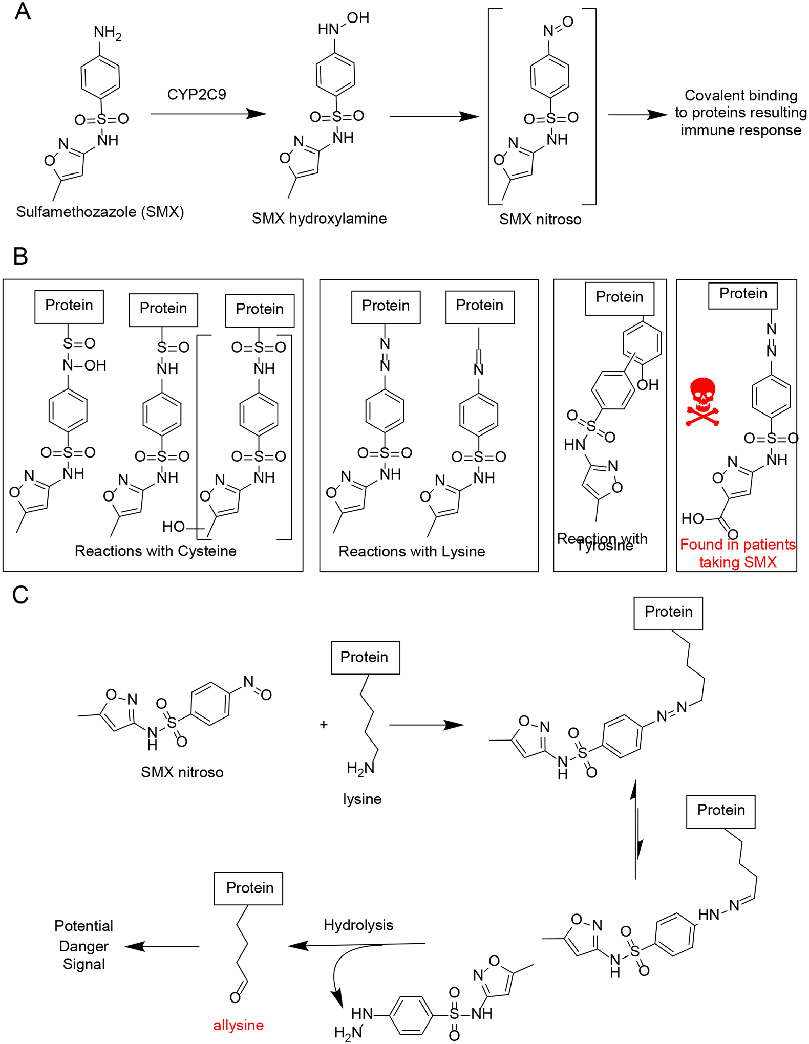
Sulfamethoxazole (SMX) is an antibiotic that has been associated with the occurrence of idiosyncratic adverse drug reactions (IADRs). IADRs are thought to be caused by the formation of the reactive intermediate, sulfamethoxazole nitroso (SMX-NO; Figure 8A), which is formed via the auto-oxidation of the hydroxylamine (Taylor et al., 2019). These studies investigate the potential of synthetic SMX-NO to covalently bind to amino acids using model proteins, with the hope of identifying the haptens involved in mediating the immune response. The results show that synthetic SMX-NO can bind covalently to cysteine, lysine and tyrosine residues in model proteins using mass spectrometry (Figure 8B). Three reactions were observed with cysteine side chains. The tyrosine adducts were formed by displacement of the nitroso group. Both lysine adducts were formed through nucleophilic addition reactions. For the first time, the authors were also able to identify protein modifications using protein digested albumin combined with mass spectrometry in patients taking SMX in which a carboxylic acid metabolite of SMX-NO was bound to lysine residues K195, K199, and K525 on albumin as seen in Figure 8B.

Commentary:

The findings from this work are of interest from a bioactivation perspective. Finding the potential lysine–sulfamethoxazole hapten on albumin is significant since it has never been observed before in patients taking the drug. The interaction of SMX-NO with lysine is a complicated pathway (Figure 8C). The reaction starts with nucleophilic addition of the amino moiety of the lysine to the nitroso group, which produces an N-hydroxyhydrazine intermediate and then loses water to form an arylazoalkane adduct. These adducts are not stable and can hydrolyze, leaving behind allysine. The aldehyde group of allysine is reactive and can undergo Schiff base reactions with other amines and the free amine on SMX. The formation of these Schiff base products could play an important role in the immune response associated with SMX. The authors speculate that the formation of reactive allysine on albumin could lead to intermolecular rearrangement, which could cause a conformational change in the protein and lead to the danger signal required for SMX hypersensitivity. It would be a worthwhile endeavor to investigate this phenomenon with other compounds with free amines that cause allergic reactions, as there is already evidence for nitroso-dapsone-associated activation of the immune system (Alzahrani et al., 2017).

Figure 8.

Figure 8.

Proposed bioactivation in vitro and in vivo of sulfamethoxazole to form drug-protein adducts.

References:

  1. Tailor A, Waddington J, Hamlett J, Maggs J, Kafu L, Farrell J, Dear G, Whitaker P, Naisbitt D, Park K, Meng X. 2019. Definition of haptens derived from sulfamethoxazole: in vitro and in vivo. Chem Res Toxicol. 32(10): 2095–2106. [DOI] [PubMed] [Google Scholar]
  2. Alzahrani A, Ogese M, Meng X, Waddington J, Tailor A, Farrell J, Maggs J, Betts C, Park B, Naisbitt D. 2017. Dapsone and nitroso dapsone activation of naïve T-cells from healthy donors. Chem Res Toxicol. 30(12):2174–2186. [DOI] [PubMed] [Google Scholar]
Drug Metab Rev. 2020 Jul 9;52(3):333–365.

Enzalutamide and Apalutamide: In Vitro Chemical Reactivity Studies and Activity in a Mouse Drug Allergy Model

C Ji, M Guha, X Zhu, J Whritenour, M Hemkins, S Tse, GS Walker, E Evans, NK Khan, MB Finkelstein, E Callegari, RS Obach

Synopsis:

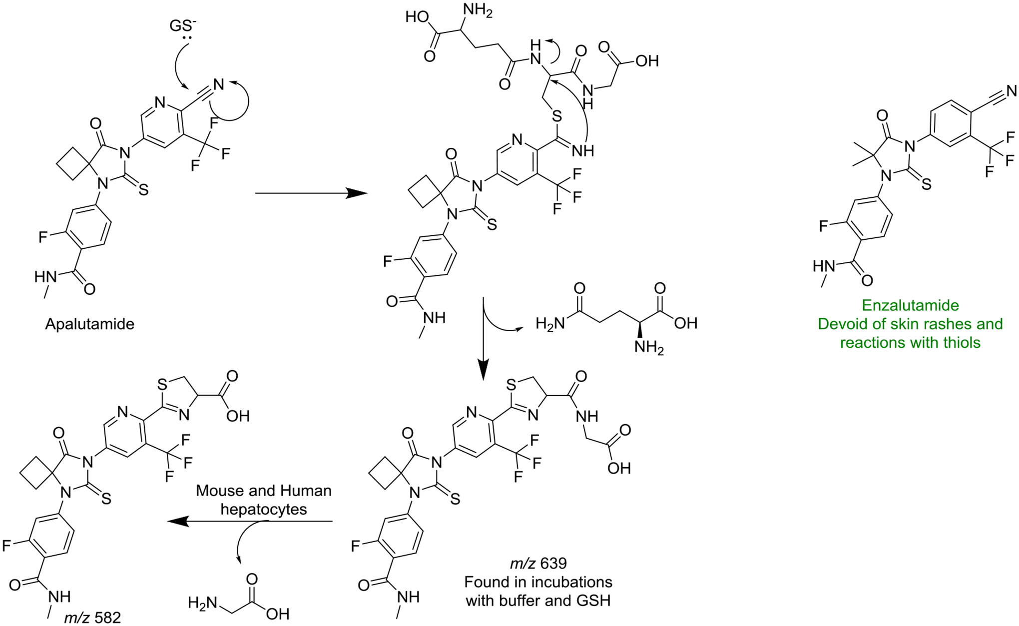
Castration resistant prostate cancer and nonmetastatic castration resistant prostate cancer are current unmet medical needs, and enzalutamide and apalutamide, respectively, are two androgen receptor inhibitors approved for their treatment. In clinical trials with apalutamide, 23.8% of patients reported a skin rash while on drug versus just 5.5% of the placebo-controlled group. Conversely, 2.3% of patients taking enzalutamide reported a skin rash compared to 2.4% in the control group. Given the high incidence of skin rash, an immune mediated drug hapten-protein hypothesis was proposed and tested (Ji et al., 2019). The 2-cyanopyridine moiety of apalutamide was identified to react with glutathione (Figure 9) in a manner that was proposed previously (Oballa et al., 2007). The proposed mechanism of the reaction starts with the nucleophilic attack of the thiol to the carbon in the cyano-moiety. The resulting imine forms a covalent bond with the α-carbon of cysteine and glutamine leaves forming a cyclic product (m/z 639; Figure 9). In human and mouse hepatocytes, further cleavage of the cys-gly adduct was found after loss of glycine (m/z 582). Enzalutamide did not undergo this reaction. The authors employed a mouse drug allergy model to test the immune mediated hypothesis. All the animals dosed with 100 mg/kg/day of apalutamide had a positive response in the model. The total numbers of T-cells and B-cells were higher in animals dosed with apalutamide. Animals dosed with enzalutamide, or the negative control, metformin, showed no increase compared to the vehicle control.

Commentary:

The mechanisms by which idiosyncratic drug reactions occur are not well understood and predictive models are difficult to develop, given that the underlying mechanisms are not always well-elucidated. Historically, rodent models for skin rashes were hard to develop due to their high immune tolerance to insult. The immune response leading to drug induced skin reaction involves many factors that include genetic risk factors and drug metabolizing enzymes (Sharma et al., 2019). Considering the complex mechanisms involved in developing skin rashes, the use of the model to differentiate drugs that cause skin rashes (ofloxacin and apalutamide) compared to compounds which don’t (metformin and enzalutamide) in the clinic is a significant finding. The results of the mouse studies help confirm the clinical skin rash findings for apalutamide. Advances in IADR predictive models help drug developers make safer and more effective drugs and the mouse drug allergy model could be used as an important tool.

References:

  1. Ji C, Guha M, Zhu X, Whritenour J, Hemkens M, Tse S, Walker G, Evans E, Khan N, Finkelstein M, Callegari E Obach RS 2019. Enzalutamide and Apalutamide: In Vitro Chemical Reactivity Studies and Activity in a Mouse Drug Allergy Model. Chem Res Toxicol. 32:2095–2106. [DOI] [PubMed] [Google Scholar]
  2. Oballa R, Truchon J, Bayly C, Chauret N, Day S, Crane S, Berthelette C. 2007. A generally applicable method for assessing the electrophilicity and reactivity of diverse nitrile-containing compounds. Bioorg Med Chem Lett. 17(4):998–1002. [DOI] [PubMed] [Google Scholar]
  3. Sharma A, Saito Y, Hung SI, Naisbitt D, Uetrecht J, Bussiere J. 2019. The skin as a metabolic and immune-competent organ: implications for drug-induced skin rash. J Immunotoxicol. 16(1):1–12. [DOI] [PubMed] [Google Scholar]
Drug Metab Rev. 2020 Jul 9;52(3):333–365.

Structure–Activity Relationships of CYP4B1 Bioactivation of 4-ipomeanol Congeners: Direct Correlation Between Cytotoxicity and Trapped Reactive Intermediates

JP Kowalski, MG McDonald, D Whittington, M Guttman, M Scian, M Girhard, H Hanenberg, C Wiek, AE Rettie

Synopsis:

The natural product 4-ipomeanol is produced in sweet potatoes infected with the fungus Fusarium solani, discovered by Prof. Ben Wilson at Vanderbilt University in the early 1970s (Boyd et al., 1974). The compound is one of many toxic furanoterpenoids Wilson and his associates found in nature. The metabolic activation pathway of 4-ipomeanol involves cytochrome P450-mediated oxidation of the furan ring to form an electrophilic enedial intermediate (α,β-unsaturated dialdehyde), which binds to tissue macromolecules (DNA and protein) and elicits toxic responses (Baer et al., 2005; Figure 10). Rabbit CYP4B1 is the most active enzyme involved in catalyzing 4-ipomeanol bioactivation (Czerwinski et al., 1991). In the present study, Kowalski et al. (2019) investigated structural features of the metabolic liability of 4-ipomeanol by synthesizing a series of N-alkyl-3-furancarboxamide analogs with varying alkyl chain lengths (C1-C8) to characterize the structure–metabolism and –toxicity relationships of CYP4B1-mediated bioactivation (Figure 10). The investigators used HepG2 cells expressing rabbit CYP4B1 to screen N-alkyl-3-furancarboxamide analogs for cytotoxicity, and they evaluated the potential of each analog to form reactive metabolites trapped as glutathione (GSH)/N-α-acetyl-L-lysine (NAL)-pyrrole adducts. N-Pentyl-3-furancarboxamide (C5) was one of the most potent analogs in cytotoxicity assays, and C5 formed the highest levels of GSH/NAL-trapped pyrrole adducts. The relative levels of GSH/NAL-pyrrole adducts formed from C1-C8 were inversely correlated with LD50 values in CYP4B1-expressing HepG2 cells. When comparing the kinetics of C2, C5, and C8 ω-hydroxylation, the catalytic efficiency of CYP4B1 increased with aliphatic chain length, with C8 ω-hydroxylation having the highest Vmax/Km. Docking studies with CYP4B1 indicated that the furan ring of C2 and C5 was well positioned above the heme for epoxidation, whereas the terminal alkyl tail of C8 was positioned above the heme for ω-hydroxylation. Collectively, the cytotoxicity, metabolism, kinetic and modeling data in this study support the conclusion that the orientation of N-alkyl-3-furancarboxamides in the CYP4B1 active site influences the propensity toward bioactivation of the furan versus ω-hydroxylation of the alkyl chain.

Commentary:

4-Ipomeanol displays species- and tissue-specific organ toxicity, causing pulmonary toxicity in cattle and rodents and hepatotoxicity in humans via cytochrome P450-mediated bioactivation (Boelsterli, 2003). 4-Ipomeanol has been previously investigated as a cytotoxic prodrug for the treatment of lung cancer; however, it showed dose-limiting hepatotoxicity in humans (Rowinsky et al., 1993) due to bioactivation by hepatic P450s. A novel approach has been proposed to specifically express functional CYP4B1 as a suicide gene in target tissues, such as in T-cell therapy, for the treatment of cancer (Roellecke et al., 2016). The study by Kowalski et al. (2019) provides unique insights into mechanistic toxicology and prodrug design. The N-alkyl-3-furancarboxamides investigated as CYP4B1 pro-toxicants retain the furan “warhead” but avoid the metabolic liability of rapid glucuronidation at the alcohol moiety of 4-ipomeanol (Statham et al., 1982; Parkinson et al., 2016; Teitelbaum et al., 2019). This work contributes significantly to the field as it clearly demonstrates a quantitative relationship between cytotoxicity and trapped reactive metabolites, which has not been reported previously. The combination of quantitative metabolism and kinetic data along with docking studies and cytotoxicity data strongly support the authors’ conclusions regarding the structure–activity relationships of N-alkyl-3-furancarboxamides and CYP4B1 catalytic orientation. Time-dependent inhibition studies indicated that the putative enedial reactive intermediate of N-pentyl-3-furancarboxamide (C5) escapes the CYP4B1 active site because addition of the trapping agents GSH/NAL protected CYP4B1 from time-dependent inactivation. Increased alkyl chain length from C2 to C8 was predicted to increase substrate binding to CYP4B1, as evidenced by higher rates of C8 ω-hydroxylation. A parabolic relationship was shown between GSH/NAL-trapped adducts and cytotoxicity as well as adduct formation and alkyl tail length. While C4-C6 were found to be the most potent (LD50 = 5 μM) of the compounds tested in this study, the authors suggest additional strategies to further optimize the metabolic properties of CYP4B1 pro-toxicants because ω-hydroxylation at the alkyl tail could also lead to the formation of inactive products.

References:

  1. Baer BR, Rettie AE, Henne KR. 2005. Bioactivation of 4-ipomeanol by CYP4B1: Adduct characterization and evidence for an enedial intermediate. Chem Res Toxicol. 18:855–864. [DOI] [PubMed] [Google Scholar]
  2. Boelsterli UA. 2003. Bioactivation of xenobiotics to reactive metabolites, in Mechanistic Toxicology: The Molecular Basis of How Chemicals Disrupt Biological Targets. pp 62–63, Taylor and Francis, New York. [Google Scholar]
  3. Boyd MR, Burka LT, Harris TM, Wilson BJ. 1974. Lung-toxic furanoterpenoids produced by sweet potatoes (Ipomoea batatas) following microbial infection. Biochim Biophys Acta. 337:184–195. [DOI] [PubMed] [Google Scholar]
  4. Czerwinski M, McLemore TL, Philpot RM, Nhamburo PT, Korzekwa K, Gelboin HV, Gonzalez FJ. 1991. Metabolic activation of 4-ipomeanol by complementary DNA-expressed human cytochromes-P450: Evidence for species-specific metabolism. Cancer Res. 51:4636–4638. [PubMed] [Google Scholar]
  5. Kowalski JP, McDonald MG, Whittington D, Guttman M, Scian M, Girhard M, Hanenberg H, Wiek, and Rettie AE. 2019. Structure-activity relationships of CYP4B1 bioactivation of 4-ipomeanol congeners: Direct correlation between cytotoxicity and trapped reactive intermediates. Chem Res Toxicol. 32:2488–2498. [DOI] [PubMed] [Google Scholar]
  6. Rowinsky EK, Noe DA, Ettinger DS, Christian MC, Lubejko BG, Fishman EK, Sartorius SE, Boyd MR, Donehower RC. 1993.Phase I and pharmacological study of the pulmonary cytotoxin 4-ipomeanol on a single dose schedule in lung cancer patients: Hepatotoxicity is dose limiting in humans. Cancer Res. 53:1794–1801. [PubMed] [Google Scholar]
Drug Metab Rev. 2020 Jul 9;52(3):333–365.

A Metabolomic Perspective of Pazopanib-induced Acute Hepatotoxicity in Mice

YK Wang, XN Yang, WQ Liang, Y Xiao, Q Zhao, Xiao, FJ Gonzalez, F Li

Synopsis:

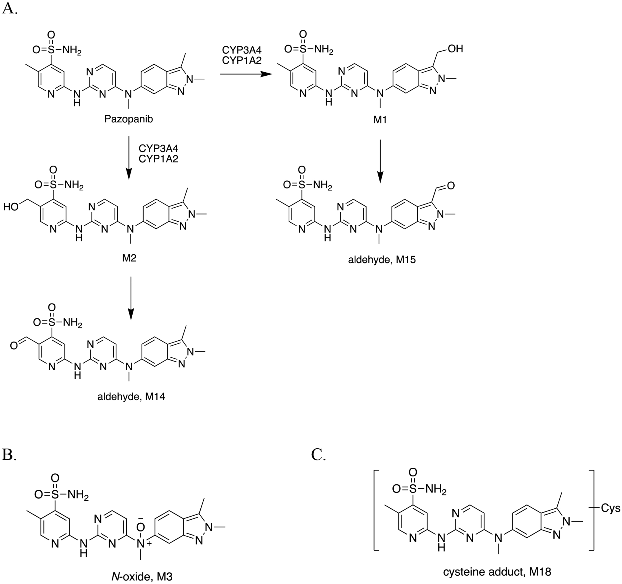
Pazopanib is a tyrosine kinase inhibitor approved for the treatment of advanced or metastatic renal cell carcinoma; however, this drug carries a boxed warning for idiosyncratic hepatotoxicity (Teo et al., 2013). The mechanisms of the liver toxicity are not well defined. Wang et al. (2019) conducted a comprehensive analysis of the pazopanib metabolites in vitro and in vivo and evaluated changes in endogenous metabolite profiles in mice treated with pazopanib at human-relevant scaled doses. LC-MS-based metabolomic analyses of plasma, urine and feces were combined with evaluation of liver injury markers (alanine aminotransferase (ALT) and aspartate aminotransferase (AST)), histological assessment, and gene expression data. Several novel metabolites of pazopanib were identified in excreta, including N-oxides (M3 and M12), aldehydes (M14 and M15), and cysteine- (M18), N-acetylcysteine- (M19), and cysteinylglycine-adducts (M20). The detection of thiol conjugates (M18–M20) suggests that pazopanib undergoes bioactivation in vivo to form reactive metabolites, which are eliminated through the mercapturic acid pathway. Interestingly, the levels of M3 N-oxide and M18 cysteine adducts in urine were positively correlated with serum ALT and AST. Through metabolomic analysis, L-carnitine was found to be elevated in mice treated with a high dose of pazopanib, and levels of proline and lysophosphatidylcholine (LPC, 18:1) were decreased. Mice treated with pazopanib showed evidence of oxidative stress (i.e. decreased hepatic superoxide dismutase and glutathione and increased malondialdehyde). Pazopanib-induced elevations in ALT and AST were reversed by pretreatment of mice with the pan-P450 inhibitor 1-aminobenzotriazole (ABT), and the levels of metabolites and cysteine adducts were reduced by pretreatment with ABT. These findings suggest that P450-mediated metabolism may be involved in pazopanib-induced liver toxicity.

Commentary:

This study contributes to the growing literature regarding the role of drug metabolism in tyrosine kinase inhibitor-associated hepatotoxicity and provides insight into the potential pathways involved in the toxicity (Duckett and Cameron, 2010; Teo et al., 2015; Jackson et al., 2018). The article by Wang et al. (2019) demonstrates that metabolomics along with other mechanistic approaches are useful tools to gain a more comprehensive understanding of the biotransformation pathways and molecular mechanisms involved in drug-induced organ toxicity. A metabolic pathway was proposed for the formation of pazopanib aldehyde metabolites (Figure 11A). CYP3A4 and CYP1A2 mediated hydroxylation of the 3-ethyl-2-methyl-2H-indazole moiety of pazopanib followed by further oxidation to form the M15 aldehyde, whereas hydroxylation of the 3-methylpyridine moiety followed by further oxidation yielded the M14 aldehyde (Figure 11A). These findings are consistent with a recent study (Paludetto et al., 2020), which detected pazopanib aldehyde metabolites in patient plasma samples.

Figure 11.

Figure 11.

Proposed metabolic pathways and novel metabolites of pazopanib. (A) Formation of aldehyde metabolites M14 and M15. (B) Structures of N-oxide metabolite, M3, and (C) cysteine adduct, M18. Pazopanib metabolite structures were elucidated by high-resolution mass spectrometry and tandem MS fragmentation patterns (Wang et al., 2019).

The bioactivation pathway of pazopanib leading to formation of thiol-reactive metabolites remains unclear. The detection of cysteine-, N-acetylcysteine- and cysteinylglycine-adducts of pazopanib suggests initial glutathione conjugation to an electrophilic intermediate; however, the site of thiol conjugation to pazopanib reactive metabolites was not determined in this study (Figure 11C). As described by Kalgutkar (2017), aldehydes can react with sulfhydryl nucleophiles to form cyclized thiazolidine adducts. The cysteinylglycine- and cysteine adducts of pazopanib reported by Wang et al. (2019) may arise from reaction of glutathione with the aldehyde metabolites of pazopanib. Loss of water and cleavage of the γ-glutamate and glycine moieties is a proposed pathway to yield cyclized cysteinylglycine- (M20) and cysteine adducts (M18), respectively. Additional studies are needed to fully elucidate the mechanisms.

Reference:

  1. Duckett DR, Cameron MD. 2010. Metabolism considerations for kinase inhibitors in cancer treatment. Expert Opin Drug Metab Toxicol. 6(10):1175–1193. [DOI] [PMC free article] [PubMed] [Google Scholar]
  2. Jackson KD, Durandis R, Vergne MJ. 2018. Role of cytochrome P450 enzymes in the metabolic activation of tyrosine kinase inhibitors. Int J Mol Sci. 19(8). [DOI] [PMC free article] [PubMed] [Google Scholar]
  3. Paludetto MN, Stigliani JL, Robert A, Bernardes-Genisson V, Chatelut E, Puisset F, Arellano C. 2020. Involvement of pazopanib and sunitinib aldehyde reactive metabolites in toxicity and drug-drug interactions in vitro and in patient samples. Chem Res Toxicol. 33(1):181–190. [DOI] [PubMed] [Google Scholar]
  4. Teo YL, Ho HK, Chan A. 2013. Risk of tyrosine kinase inhibitors-induced hepatotoxicity in cancer patients: A meta-analysis. Cancer Treat Rev. 39:199–206. [DOI] [PubMed] [Google Scholar]
  5. Teo YL, Ho HK, Chan A. 2015. Formation of reactive metabolites and management of tyrosine kinase inhibitor-induced hepatotoxicity: a literature review. Expert Opin Drug. Metab Toxicol 11(2):231–242. [DOI] [PubMed] [Google Scholar]
  6. Wang YK, Yang XN, Liang WQ, Xiao Y, Zhao Q, Xiao, Gonzalez FJ, Li F. 2019. A metabolomic perspective of pazopanib-induced acute hepatotoxicity in mice. Xenobiotica. 49(6):655–670. [DOI] [PMC free article] [PubMed] [Google Scholar]
Drug Metab Rev. 2020 Jul 9;52(3):333–365.

Rotenone Increases Isoniazid Toxicity but Does Not Cause Significant Liver Injury: Implications for the Hypothesis that Inhibition of the Mitochondrial Electron Transport Chain Is a Common Mechanism of Idiosyncratic Drug-Induced Liver Injury

T Cho, X Wang, J Uetrecht

Synopsis:

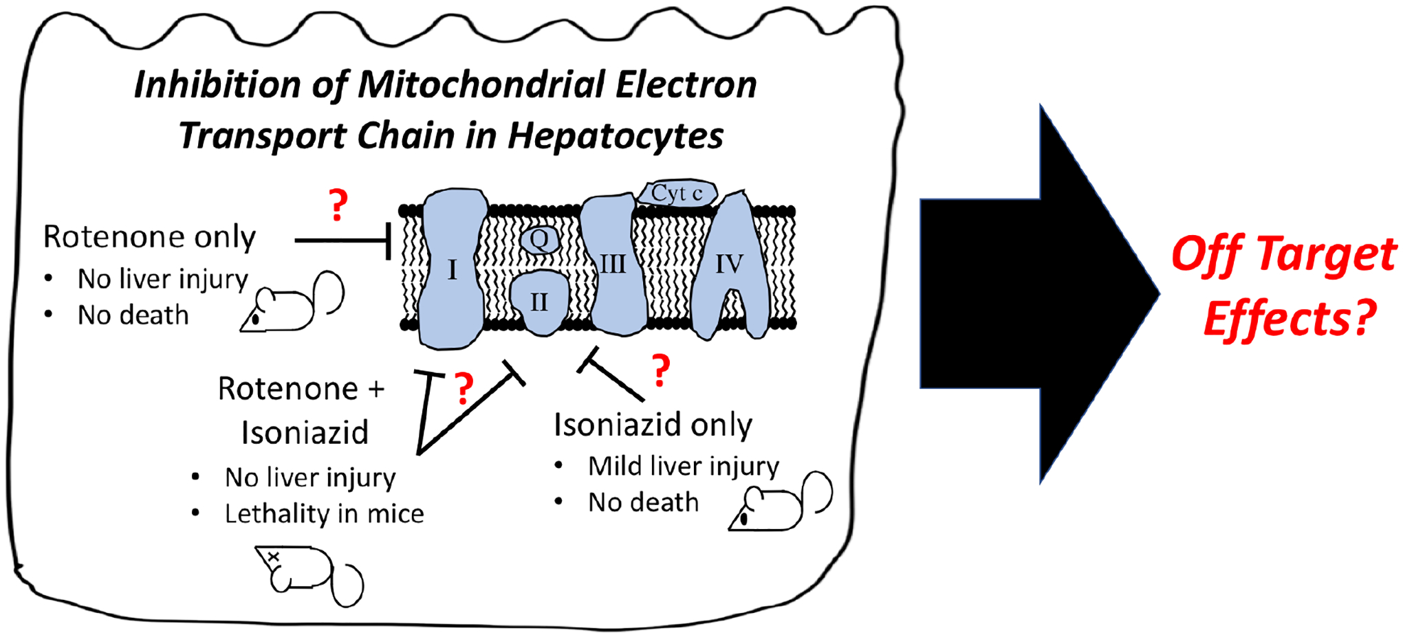
A major cause of drug failure is idiosyncratic drug-induced liver injury (IDILI), and thus, the elimination of drug leads prone to inducing liver toxicity is a critical step during the development stage. The flagging of those molecules often involves in vitro screens for mitochondrial toxicity due to its association with IDILI; however, in vivo demonstrations of the mechanistic role of mitochondrial dysfunction in IDILI are lacking. In this study, wild type mice and an impaired immune tolerance mouse model were given varying concentrations of rotenone alone or together with isoniazid in food or water. The combination of rotenone and isoniazid has previously been reported to inhibit the mitochondrial electron transport chain in in vitro murine hepatocyte studies and, therefore, was suspected to induce IDILI. Treatment with rotenone or isoniazid alone did not cause death in animals, but their co-administration led to lethality in 100% of the mice. Toxicity was not related to liver injury as assessed by hepatic histology and serum glutamate dehydrogenase activity. Based on those findings, inhibition of the mitochondrial electron transport chain was not a significant contributor to the IDILI mechanism.

Commentary:

Accurate decision-making in drug attrition requires knowledge of IDILI mechanisms to design effective screens for problematic drug leads, such as those that might cause mitochondrial toxicity. Although strong evidence supports a role for the immune system in IDILI, in vitro studies have shown that mitochondrial dysfunction might also be implicated in IDILI. Despite the in vitro data, in vivo evidence showing the relationship between IDILI and mitochondrial toxicity is lacking. In response to this shortcoming, Cho et al. (2019) report a critical expansion of evidence for the potential role of mitochondrial dysfunction in IDILI, sparking a lively discussion in the literature (Cho and Uetrecht, 2020; Fromenty, 2020). The authors of the in vivo study employed a clever strategy to target mitochondrial dysfunction while amplifying the effect for improved observation of liver injury (Figure 9). First, the synergy between rotenone and isoniazid relies on a common toxicological mechanism; both molecules inhibit the mitochondrial electron transport chain but through different complexes of that system. Second, the combined administration of these two drugs results in a higher response than expected for the individual molecules, which is helpful with complex biological systems. Third, the treatment approach included PD-1(−/−) mice that were administered anti-CTLA4 as a novel animal model for IDILI (Mak and Uetrecht, 2015). This impaired immune tolerance mouse model was expected to be a more sensitive responder to the rotenone and isoniazid combination than wildtype C57BL/6 mice. Fourth, the combination of molecules is representative of the exposures occurring in everyday life, although the results are more challenging to interpret. The study design also included histological analyses, the gold standard for liver injury, as well as measures of serum glutamate dehydrogenase activity rather than that for the more traditional alanine aminotransferase (ALT). The latter assay was a critical inclusion given that isoniazid depletes the pyridoxal phosphate cofactor used by ALT. Taken together, the combined treatment did induce higher lethality but without indications of liver toxicity, suggesting that mitochondrial dysfunction induced by those chemicals was not a critical driver in toxicity.

Figure 9.

Figure 9.

Reactivity of apalutamine with glutathione.

Nevertheless, the interpretation of the results may be more complicated than that. First, there was no specific assessment of mitochondrial dysfunction. It is unclear if rotenone and isoniazid treatments altered complex I and II activities, respectively, in any appreciable way (Figure 12) regardless of outcomes for the mice. Importantly, exposure responses by C57BL/6 mice may be strain specific and, thus, complicate the extrapolation of the findings. For example, isoniazid induces microvesicular steatosis injury in the liver for BALB/cJ, DBA/2J and LG/J mice but not C57BL/6J mice (Labbe et al., 2008; Bailey et al., 2009; Lucena et al., 2010; Stewart et al., 2010), indicating underlying and varied genetic predisposition to liver injury in animal models. Second, the focus on liver injury ignored other possible off target effects that may induced lethality in mice while not causing liver injury or possibly mitochondrial dysfunction (Figure 12). Rotenone effects are not limited to the liver; rather, damage extends to the heart, liver, kidneys and spleen (Jiang et al., 2017). Similarly, isoniazid exposure causes hypotension, renal failure and metabolic acidosis (Watkins et al., 1990; Gokhale et al., 2009). Although not studied, those off target effects may have played a role in lethal outcomes for mice exposed to the combination of molecules. Despite those issues, the current study provides an additional data set to interrogate the role of mitochondrial dysfunction in IDILI that contrasts with prior reports of mitochondrial dysfunction associated with liver injury for valproic acid (Zimmerman and Ishak, 1982), perhexiline (Deschamps et al., 1994), troglitazone (Ong et al., 2007), amiodarone (Fromenty et al., 1990; Felser et al., 2013) and buprenorphine (Berson et al., 2001). Granted, correlation does not prove causation, but the converse is also true; absence of evidence as in the current test case does not necessarily refute the possibility of mitochondrial dysfunction playing a role in IDILI. Further research and conversations on in vivo assessments of mitochondrial dysfunction in liver injury are clearly necessary to truly advance an understanding of IDILI mechanisms and subsequently screening for those possibilities during drug development.

Figure 12.

Figure 12.

Interrogation of whether mitochondrial dysfunction induces liver injury in mice models of idiosyncratic liver injury (Cho et al., 2019). C57BL/6 mice treated with rotenone and isoniazid led to minimal liver injury, yet the combined exposure to both drugs induced lethality. Rotenone and isoniazid inhibit complex I and II of the mitochondrial electron transport chain, respectively, and are thought to induce mitochondrial dysfunction (not assayed in this study; red question marks). Moreover, the study did not consider off target effects of the molecules as possible contributors to observed lethality.

References:

  1. Bailey CM, Kasiviswanathan R, Copeland WC, and Anderson KS (2009) R964C mutation of DNA polymerase gamma imparts increased stavudine toxicity by decreasing nucleoside analog discrimination and impairing polymerase activity. Antimicrob Agents Chemother 53:2610–2612. [DOI] [PMC free article] [PubMed] [Google Scholar]
  2. Berson A, Fau D, Fornacciari R, Degove-Goddard P, Sutton A, Descatoire V, Haouzi D, Lettéron P, Moreau A, Feldmann G, and Pessayre D (2001) Mechanisms for experimental buprenorphine hepatotoxicity: major role of mitochondrial dysfunction versus metabolic activation. J Hepatol 34:261–269. [DOI] [PubMed] [Google Scholar]
  3. Cho T, and Uetrecht J (2020) Response to the Letter to the Editor Concerning the Article “Rotenone Increases Isoniazid Toxicity but Does Not Cause Liver Injury: Implications for the Hypothesis That Inhibition of the Mitochondrial Electron Transport Chain Is a Common Mechanism of Idiosyncratic Drug-Induced Liver Injury” by Bernard Fromenty. Chem Res Toxicol 33:5–6. [DOI] [PubMed] [Google Scholar]
  4. Cho T, Wang X, and Uetrecht J (2019) Rotenone Increases Isoniazid Toxicity but Does Not Cause Significant Liver Injury: Implications for the Hypothesis that Inhibition of the Mitochondrial Electron Transport Chain Is a Common Mechanism of Idiosyncratic Drug-Induced Liver Injury. Chem Res Toxicol 32:1423–1431. [DOI] [PubMed] [Google Scholar]
  5. Deschamps D, DeBeco V, Fisch C, Fromenty B, Guillouzo A, and Pessayre D (1994) Inhibition by perhexiline of oxidative phosphorylation and the beta-oxidation of fatty acids: possible role in pseudoalcoholic liver lesions. Hepatology 19:948–961. [PubMed] [Google Scholar]
  6. Felser A, Blum K, Lindinger PW, Bouitbir J, and Krähenbühl S (2013) Mechanisms of hepatocellular toxicity associated with dronedarone--a comparison to amiodarone. Toxicol Sci 131:480–490. [DOI] [PubMed] [Google Scholar]
  7. Fromenty B (2020) Letter to the Editor Regarding the Article Rotenone Increases Isoniazid Toxicity but Does Not Cause Significant Liver Injury: Implications for the Hypothesis that Inhibition of the Mitochondrial Electron Transport Chain Is a Common Mechanism of Idiosyncratic Drug-Induced Liver Injury by Cho and Co-Workers, 2019. Chem Res Toxicol 33:2–4. [DOI] [PubMed] [Google Scholar]
  8. Fromenty B, Fisch C, Berson A, Letteron P, Larrey D, and Pessayre D (1990) Dual effect of amiodarone on mitochondrial respiration. Initial protonophoric uncoupling effect followed by inhibition of the respiratory chain at the levels of complex I and complex II. J Pharmacol Exp Ther 255:1377–1384. [PubMed] [Google Scholar]
  9. Gokhale YA, Vaidya MS, Mehta AD, and Rathod NN (2009) Isoniazid toxicity presenting as status epilepticus and severe metabolic acidosis. J Assoc Physicians India 57:70–71. [PubMed] [Google Scholar]
  10. Jiang X-W, Qiao L, Feng X-X, Liu L, Wei Q-W, Wang X-W, and Yu W-H (2017) Rotenone induces nephrotoxicity in rats: oxidative damage and apoptosis. Toxicol Mech Methods 27:528–536. [DOI] [PubMed] [Google Scholar]
  11. Labbe G, Pessayre D, and Fromenty B (2008) Drug-induced liver injury through mitochondrial dysfunction: mechanisms and detection during preclinical safety studies. Fundam Clin Pharmacol 22:335–353. [DOI] [PubMed] [Google Scholar]
  12. Lucena MI, García-Martín E, Andrade RJ, Martínez C, Stephens C, Ruiz JD, Ulzurrun E, Fernandez MC, Romero-Gomez M, Castiella A, Planas R, Durán JA, De Dios AM, Guarner C, Soriano G, Borraz Y, and Agundez JAG (2010) Mitochondrial superoxide dismutase and glutathione peroxidase in idiosyncratic drug-induced liver injury. Hepatology 52:303–312. [DOI] [PubMed] [Google Scholar]
  13. Mak A, and Uetrecht J (2015) The Combination of Anti-CTLA-4 and PD1−/− Mice Unmasks the Potential of Isoniazid and Nevirapine To Cause Liver Injury. Chem Res Toxicol 28:2287–2291. [DOI] [PubMed] [Google Scholar]
  14. Ong MMK, Latchoumycandane C, and Boelsterli UA (2007) Troglitazone-induced hepatic necrosis in an animal model of silent genetic mitochondrial abnormalities. Toxicol Sci 97:205–213. [DOI] [PubMed] [Google Scholar]
  15. Stewart JD, Horvath R, Baruffini E, Ferrero I, Bulst S, Watkins PB, Fontana RJ, Day CP, and Chinnery PF (2010) Polymerase γ gene POLG determines the risk of sodium valproate-induced liver toxicity. Hepatology 52:1791–1796. [DOI] [PMC free article] [PubMed] [Google Scholar]
  16. Watkins RC, Hambrick EL, Benjamin G, and Chavda SN (1990) Isoniazid toxicity presenting as seizures and metabolic acidosis. J Natl Med Assoc 82:57, 62,, 64. [PMC free article] [PubMed] [Google Scholar]
  17. Zimmerman HJ, and Ishak KG (1982) Valproate-induced hepatic injury: analyses of 23 fatal cases. Hepatology 2:591–597. [DOI] [PubMed] [Google Scholar]
Drug Metab Rev. 2020 Jul 9;52(3):333–365.

Mechanism of Idiosyncratic Drug-Induced Liver Injury Quantitative Evaluation of Reactivity and Toxicity of Acyl Glucuronides by [35S]-Cysteine Trapping

H Harada, Y Toyoda, Y Abe, T Endo, H Takeda

Synopsis:

A major cause of drug failure is idiosyncratic drug-induced liver injury (IDILI), and thus, the elimination of drug leads prone to inducing liver toxicity is a critical step during the development stage. The flagging of those molecules often involves in vitro screens for mitochondrial toxicity due to its association with IDILI; however, in vivo demonstrations of the mechanistic role of mitochondrial dysfunction in IDILI are lacking. In this study, wild type mice and an impaired immune tolerance mouse model were given varying concentrations of rotenone alone or together with isoniazid in food or water. The combination of rotenone and isoniazid has previously been reported to inhibit the mitochondrial electron transport chain in in vitro murine hepatocyte studies and, therefore, was suspected to induce IDILI. Treatment with rotenone or isoniazid alone did not cause death in animals, but their co-administration led to lethality in 100% of the mice. Toxicity was not related to liver injury as assessed by hepatic histology and serum glutamate dehydrogenase activity. Based on those findings, inhibition of the mitochondrial electron transport chain was not a significant contributor to the IDILI mechanism.

Commentary:

Accurate decision-making in drug attrition requires knowledge of IDILI mechanisms to design effective screens for problematic drug leads, such as those that might cause mitochondrial toxicity. Although strong evidence supports a role for the immune system in IDILI, in vitro studies have shown that mitochondrial dysfunction might also be implicated in IDILI. Despite the in vitro data, in vivo evidence showing the relationship between IDILI and mitochondrial toxicity is lacking. In response to this shortcoming, Cho et al. (2019) report a critical expansion of evidence for the potential role of mitochondrial dysfunction in IDILI, sparking a lively discussion in the literature (Cho and Uetrecht, 2020; Fromenty, 2020). The authors of the in vivo study employed a clever strategy to target mitochondrial dysfunction while amplifying the effect for improved observation of liver injury (Figure 9). First, the synergy between rotenone and isoniazid relies on a common toxicological mechanism; both molecules inhibit the mitochondrial electron transport chain but through different complexes of that system. Second, the combined administration of these two drugs results in a higher response than expected for the individual molecules, which is helpful with complex biological systems. Third, the treatment approach included PD-1(−/−) mice that were administered anti-CTLA4 as a novel animal model for IDILI (Mak and Uetrecht, 2015). This impaired immune tolerance mouse model was expected to be a more sensitive responder to the rotenone and isoniazid combination than wildtype C57BL/6 mice. Fourth, the combination of molecules is representative of the exposures occurring in everyday life, although the results are more challenging to interpret. The study design also included histological analyses, the gold standard for liver injury, as well as measures of serum glutamate dehydrogenase activity rather than that for the more traditional alanine aminotransferase (ALT). The latter assay was a critical inclusion given that isoniazid depletes the pyridoxal phosphate cofactor used by ALT. Taken together, the combined treatment did induce higher lethality but without indications of liver toxicity, suggesting that mitochondrial dysfunction induced by those chemicals was not a critical driver in toxicity.

Nevertheless, the interpretation of the results may be more complicated than that. First, there was no specific assessment of mitochondrial dysfunction. It is unclear if rotenone and isoniazid treatments altered complex I and II activities, respectively, in any appreciable way (Figure 12) regardless of outcomes for the mice. Importantly, exposure responses by C57BL/6 mice may be strain specific and, thus, complicate the extrapolation of the findings. For example, isoniazid induces microvesicular steatosis injury in the liver for BALB/cJ, DBA/2J and LG/J mice but not C57BL/6J mice (Labbe et al., 2008; Bailey et al., 2009; Lucena et al., 2010; Stewart et al., 2010), indicating underlying and varied genetic predisposition to liver injury in animal models. Second, the focus on liver injury ignored other possible off target effects that may induced lethality in mice while not causing liver injury or possibly mitochondrial dysfunction (Figure 12). Rotenone effects are not limited to the liver; rather, damage extends to the heart, liver, kidneys and spleen (Jiang et al., 2017). Similarly, isoniazid exposure causes hypotension, renal failure and metabolic acidosis (Watkins et al., 1990; Gokhale et al., 2009). Although not studied, those off target effects may have played a role in lethal outcomes for mice exposed to the combination of molecules. Despite those issues, the current study provides an additional data set to interrogate the role of mitochondrial dysfunction in IDILI that contrasts with prior reports of mitochondrial dysfunction associated with liver injury for valproic acid (Zimmerman and Ishak, 1982), perhexiline (Deschamps et al., 1994), troglitazone (Ong et al., 2007), amiodarone (Fromenty et al., 1990; Felser et al., 2013) and buprenorphine (Berson et al., 2001). Granted, correlation does not prove causation, but the converse is also true; absence of evidence as in the current test case does not necessarily refute the possibility of mitochondrial dysfunction playing a role in IDILI. Further research and conversations on in vivo assessments of mitochondrial dysfunction in liver injury are clearly necessary to truly advance an understanding of IDILI mechanisms and subsequently screening for those possibilities during drug development.

References:

  1. Bailey CM, Kasiviswanathan R, Copeland WC, and Anderson KS (2009) R964C mutation of DNA polymerase gamma imparts increased stavudine toxicity by decreasing nucleoside analog discrimination and impairing polymerase activity. Antimicrob Agents Chemother 53:2610–2612. [DOI] [PMC free article] [PubMed] [Google Scholar]
  2. Berson A, Fau D, Fornacciari R, Degove-Goddard P, Sutton A, Descatoire V, Haouzi D, Lettéron P, Moreau A, Feldmann G, and Pessayre D (2001) Mechanisms for experimental buprenorphine hepatotoxicity: major role of mitochondrial dysfunction versus metabolic activation. J Hepatol 34:261–269. [DOI] [PubMed] [Google Scholar]
  3. Cho T, and Uetrecht J (2020) Response to the Letter to the Editor Concerning the Article “Rotenone Increases Isoniazid Toxicity but Does Not Cause Liver Injury: Implications for the Hypothesis That Inhibition of the Mitochondrial Electron Transport Chain Is a Common Mechanism of Idiosyncratic Drug-Induced Liver Injury” by Bernard Fromenty. Chem Res Toxicol 33:5–6. [DOI] [PubMed] [Google Scholar]
  4. Cho T, Wang X, and Uetrecht J (2019) Rotenone Increases Isoniazid Toxicity but Does Not Cause Significant Liver Injury: Implications for the Hypothesis that Inhibition of the Mitochondrial Electron Transport Chain Is a Common Mechanism of Idiosyncratic Drug-Induced Liver Injury. Chem Res Toxicol 32:1423–1431. [DOI] [PubMed] [Google Scholar]
  5. Deschamps D, DeBeco V, Fisch C, Fromenty B, Guillouzo A, and Pessayre D (1994) Inhibition by perhexiline of oxidative phosphorylation and the beta-oxidation of fatty acids: possible role in pseudoalcoholic liver lesions. Hepatology 19:948–961. [PubMed] [Google Scholar]
  6. Felser A, Blum K, Lindinger PW, Bouitbir J, and Krähenbühl S (2013) Mechanisms of hepatocellular toxicity associated with dronedarone--a comparison to amiodarone. Toxicol Sci 131:480–490. [DOI] [PubMed] [Google Scholar]
  7. Fromenty B (2020) Letter to the Editor Regarding the Article Rotenone Increases Isoniazid Toxicity but Does Not Cause Significant Liver Injury: Implications for the Hypothesis that Inhibition of the Mitochondrial Electron Transport Chain Is a Common Mechanism of Idiosyncratic Drug-Induced Liver Injury by Cho and Co-Workers, 2019. Chem Res Toxicol 33:2–4. [DOI] [PubMed] [Google Scholar]
  8. Fromenty B, Fisch C, Berson A, Letteron P, Larrey D, and Pessayre D (1990) Dual effect of amiodarone on mitochondrial respiration. Initial protonophoric uncoupling effect followed by inhibition of the respiratory chain at the levels of complex I and complex II. J Pharmacol Exp Ther 255:1377–1384. [PubMed] [Google Scholar]
  9. Gokhale YA, Vaidya MS, Mehta AD, and Rathod NN (2009) Isoniazid toxicity presenting as status epilepticus and severe metabolic acidosis. J Assoc Physicians India 57:70–71. [PubMed] [Google Scholar]
  10. Jiang X-W, Qiao L, Feng X-X, Liu L, Wei Q-W, Wang X-W, and Yu W-H (2017) Rotenone induces nephrotoxicity in rats: oxidative damage and apoptosis. Toxicol Mech Methods 27:528–536. [DOI] [PubMed] [Google Scholar]
  11. Labbe G, Pessayre D, and Fromenty B (2008) Drug-induced liver injury through mitochondrial dysfunction: mechanisms and detection during preclinical safety studies. Fundam Clin Pharmacol 22:335–353. [DOI] [PubMed] [Google Scholar]
  12. Lucena MI, García-Martín E, Andrade RJ, Martínez C, Stephens C, Ruiz JD, Ulzurrun E, Fernandez MC, Romero-Gomez M, Castiella A, Planas R, Durán JA, De Dios AM, Guarner C, Soriano G, Borraz Y, and Agundez JAG (2010) Mitochondrial superoxide dismutase and glutathione peroxidase in idiosyncratic drug-induced liver injury. Hepatology 52:303–312. [DOI] [PubMed] [Google Scholar]
  13. Mak A, and Uetrecht J (2015) The Combination of Anti-CTLA-4 and PD1−/− Mice Unmasks the Potential of Isoniazid and Nevirapine To Cause Liver Injury. Chem Res Toxicol 28:2287–2291. [DOI] [PubMed] [Google Scholar]
  14. Ong MMK, Latchoumycandane C, and Boelsterli UA (2007) Troglitazone-induced hepatic necrosis in an animal model of silent genetic mitochondrial abnormalities. Toxicol Sci 97:205–213. [DOI] [PubMed] [Google Scholar]
  15. Stewart JD, Horvath R, Baruffini E, Ferrero I, Bulst S, Watkins PB, Fontana RJ, Day CP, and Chinnery PF (2010) Polymerase γ gene POLG determines the risk of sodium valproate-induced liver toxicity. Hepatology 52:1791–1796. [DOI] [PMC free article] [PubMed] [Google Scholar]
  16. Watkins RC, Hambrick EL, Benjamin G, and Chavda SN (1990) Isoniazid toxicity presenting as seizures and metabolic acidosis. J Natl Med Assoc 82:57, 62,, 64. [PMC free article] [PubMed] [Google Scholar]
  17. Zimmerman HJ, and Ishak KG (1982) Valproate-induced hepatic injury: analyses of 23 fatal cases. Hepatology 2:591–597. [DOI] [PubMed] [Google Scholar]

RESOURCES