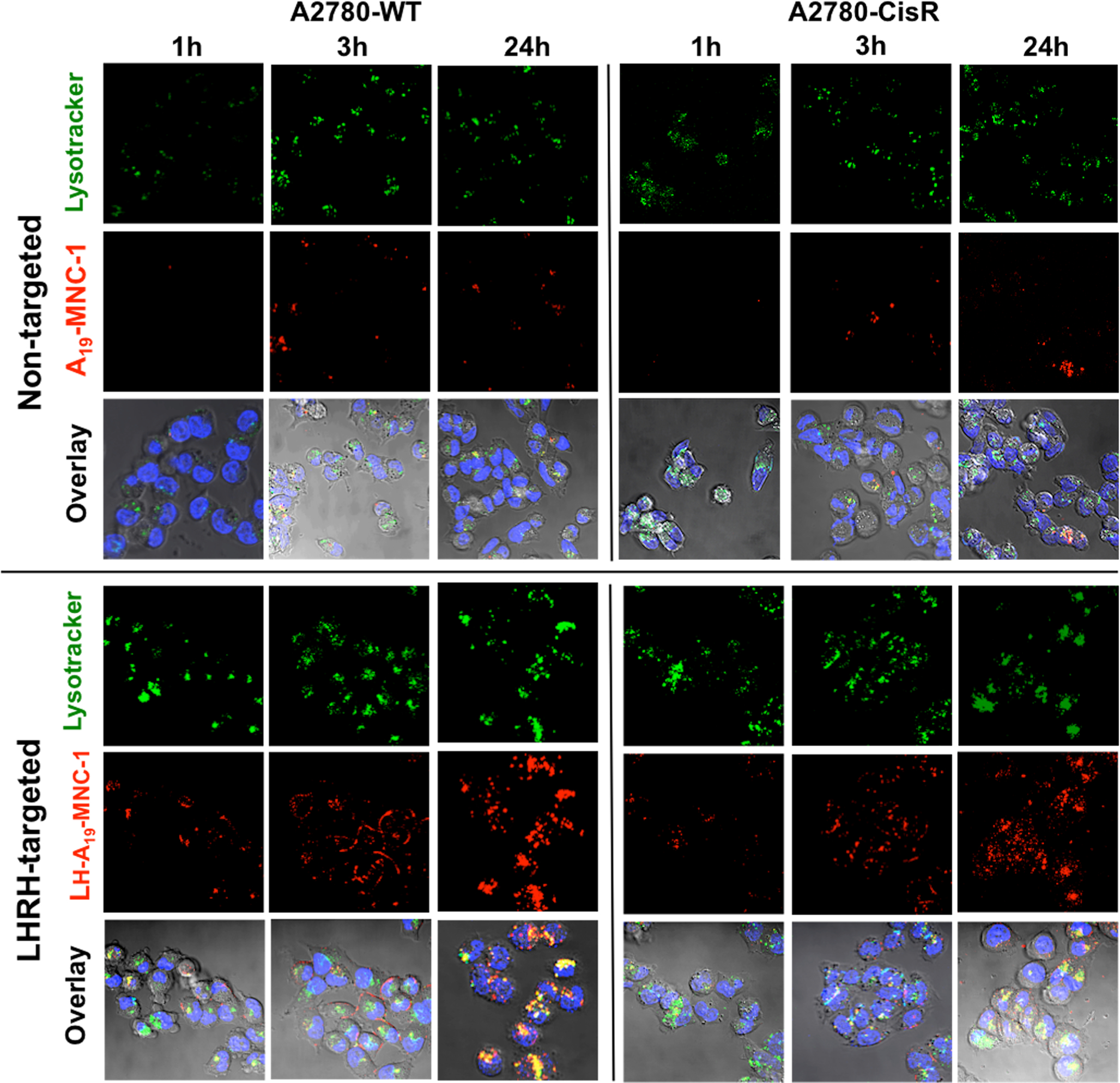
The authors regret that in the initially published version of this article, an error occurred in the Figure 7 that illustrates wild-type A2780-WT and cisplatin-resistant A2780-CisR human ovarian cancer cells at three different time points during their incubation with nontargeted A19-MNC-1 and LHRH-conjugated A19-MNC-1. In the original publication, the images for non-targeted A2780-CisR cells at the 1 h and 24 h time points were accidentally mixed up with the (correct) images for “non-targeted” A2780 for the same time points, resulting in duplicate image sets for these time points for both cell lines. Therefore, here we present the corrected version of Figure 7. For each treatment and time point, there is a set of three images: (1) lysotracker green stain, (2) Alexa-Fluor 647-labeled MNCs, and (3) overlay (with Hoechst nuclear stain). The conclusion that “untargeted MNCs showed very low cellular uptake even after 24 h in both wild type and cisplatin-resistant cells” has not changed.
Figure 7.

Confocal microscopy of the wild-type A2780-WT and cisplatin-resistant A2780-CisR human ovarian cancer cells at different time points during their incubation with nontargeted A19-MNC-1 and LHRH-conjugated A19-MNC-1. Live cells were exposed to the said Alexa-Fluor-647-labeled (red) MNCs and stained with the lysotracker green (green) and Hoechst nuclear stain (blue) for 30 min. The colocalization of the labeled MNCs in lysosomes is seen in the overlay (yellow punctate regions) (magnification 63×).


