

Abstract
Burn injuries are of serious concern worldwide not only because of the physical impact but also because of severe mental and emotional distress and reduced quality of life. The usual management comprises topical and systemic medications, and supportive care; however, conventional therapy may be expensive and insufficient for many cases. Thus, we describe herein a unique case in which a low-level laser was used concomitantly with the conventional approach for the management of head and neck burn wounds and could improve the patient's clinical condition within a short period.
Keywords: Burns, explosions, low-level light therapy
INTRODUCTION
Burns result from tissue exposure to chemical, physical, or biological agents[1] and might be accompanied by slow healing, infection, pain, hypertrophic scarring, and even death.[2] The healing of these wounds depends on a series of interdependent and simultaneous cellular events involving cytokines and the extracellular matrix that can be divided into inflammation, proliferation, and remodeling.[1]
In Brazil, an upper-middle-income country with 202 million people, burn injuries are a serious concern. In the period between 2008 and 2014, there was a mean annual rate of 18,551 admissions to the Brazilian universal health-care system (Sistema Único de Saúde-SUS) and 2,466 deaths related to burns.[3] Despite efforts in implementing preventive strategies and advances in treatment options, burns are still associated with significant morbidity and mortality.[4] Burned patients may also present severe mental and emotional distress,[2] besides a detrimental impact on their quality of life.[5]
Having in mind the highly complex biological response to a burn injury[5] and the high costs of treatment,[1] some alternatives such as phototherapies have been recently investigated. Although the management of burn wounds by photobiomodulation (lasers and LED) in both animal model studies[1,6] and case reports[2,4,7] have proven to yield suitable results, not enough evidence-based information is available for clinical application. In light of these facts, the present study reports a clinical case in which a low-level laser was used concomitantly with the conventional approach for the management of head and neck burn wounds.
CASE REPORT
A 33-year-old male patient was brought to the Emergence Service of Hospital Associação Beneficente de Campo Grande-Santa Casa (MS, Brazil) after being involved in an explosion. At the admission, he presented second-degree burn injuries, affecting a large area on the head, neck, thorax, and upper limbs, resulting directly from the exposition to fire, and pharyngeal burns due to steam inhalation. Other injuries were ruled out by the trauma team. Because of the risk of inhalation injuries, the patient received early (i.e. prehospital) orotracheal intubation with mechanical ventilation by the life-support team.
After being admitted to the intensive care unit (ICU), he received amitriptyline, haloperidol, and alprazolam for pain control. Other drugs were also administered systemically in order to control inflammation (dexamethasone), prevent infections (piperacillin with tazobactam and moxifloxacin), and improve healing (Vitamin C). Regencel™ (Cristália Prod. Quím. Farm. Ltda., SP, Brazil) and artificial tears (dextran and hypromellose) were used topically to manage injuries to the patient's eyes.
Once he was completely stabilized clinically, a nasoenteral feeding tube was installed and the conventional approach was adopted according to the hospital's protocol for burned patients, i.e. serial tissue debridement following the placement of either silver sulfadiazine or collagenase ointment over the burn wounds.
On the 1 day of hospitalization, the oral medicine team was asked to evaluate the patient and noted an extensive facial area presenting painful burn wounds, besides edema and bleeding ulcerative ulcers on the lips, which were certainly associated with the orotracheal intubation [Figure 1a]. Initially, the method of fixation of the orotracheal tube was proposed to be changed for reducing mechanical trauma to the patient's perioral region. Furthermore, low-level laser therapy (LLLT) sessions were then proposed for modulation of inflammatory processes, tissue repair, and pain relief. For that, an InGaAIP low-level laser (LASER duo/MMOptics, SP, Brazil) was used perpendicularly in a defocused mode at 1 cm from the lesions. Every lesion area of 1 cm2 was irradiated for 30 s, with 100 mW of power, and energy of approximately 3 J. After every LLLT session, debridement procedures were performed, collagenase ointment was applied onto the burn wounds, and Aquacel Ag® (ConvaTec, NJ, USA) was used to cover them [Figure 1b]. These procedures were repeated every 24 h, totalizing 5 sessions.
Figure 1.
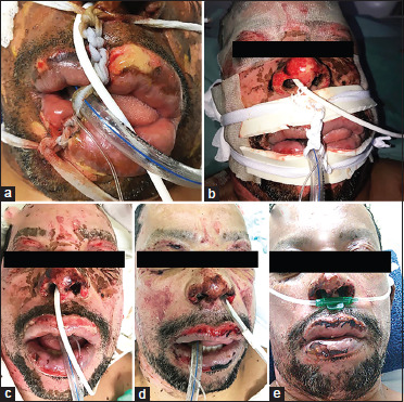
Clinical evaluation. (a) Initial presentation – bleeding ulcerative ulcers on the lips. (b) Initial presentation with the method of fixation of the orotracheal tube changed for reducing mechanical trauma to the patient's perioral region. (c) 24 h after second low-level laser therapy session showing improvement of facial edema and little bleeding. (d) 72 h after third low-level laser therapy session, showing improvement in healing process. (e) 5th low-level laser therapy session showing burn wounds in an advanced stage of healing
On the 2 day, facial edema was markedly improved, and very little bleeding was present. The burn wounds also showed a gentle improvement in healing [Figure 1c]. On the 3 day, neither facial edema nor bleeding was observed. The burn wounds were slightly painful and presented a marked improvement in healing [Figure 1d]. On the 5 day, the patient was extubated and placed on noninvasive ventilation (nasal oxygen catheter). Furthermore, the nasoenteral feeding tube was removed. Clinically, the burn wounds were in an advanced stage of healing, no longer inducing spontaneous pain [Figure 1e]. The patient recovered his orofacial basic functions satisfactorily, being discharged from the ICU within 2 days. He was then transferred to the Hospital's Burn Unit for the treatment of the other burn wounds (thorax and upper limbs).
DISCUSSION
In the current case report, adding LLLT to the conventional approach for burn wound care resulted in an important clinical improvement of the patient's orofacial conditions within approximately 5 days. To the best of the authors' knowledge, this is the first case on the benefits of photobiomodulation to burns on the head and neck; however, there have been studies with similar approaches for other body areas. For example, a series of cases presented the use of LED at 658 nm in patients with bilateral second- or third-degree burns on the limbs[4] and a case report combined high-power laser debridement with chemical debridement and LLLT at 808 nm for third-degree necrotic burn ulcer on the sacral area.[7] There are no previous comparative studies.
The rationale for applying laser light in burns was based on the light absorption by the tissues,[2] resulting in a positive effect on the healing process, mostly related to modulation of inflammation and cell proliferation,[1] and pain relief.[4] Photobiomodulation is also noninvasive, safe,[7] low-cost, and has reliable scientific evidence on all phases of tissue healing.[1] Furthermore, it has no side effects and can be used concomitantly with drug therapy.[1]
To sum up and considering all the difficulties in treating burns and the great discrepancy among the studies available, the current case report suggests that the combination of LLLT with the conventional approach for head-and-neck burn care is useful for improving the patients' clinical conditions.
Declaration of patient consent
The authors certify that they have obtained all appropriate patient consent forms. In the form, the legal guardian has given his consent for images and other clinical information to be reported in the journal. The guardian understands that names and initials will not be published and due efforts will be made to conceal identity, but anonymity cannot be guaranteed.
Financial support and sponsorship
Nil.
Conflicts of interest
There are no conflicts of interest.
Ethics statement
Patient anonymity was assured.
REFERENCES
- 1.Brassolatti P, de Andrade AL, Bossini PS, Otterço AN, Parizotto NA. Evaluation of the low-level laser therapy application parameters for skin burn treatment in experimental model: A systematic review. Lasers Med Sci. 2018;33:1159–69. doi: 10.1007/s10103-018-2526-5. [DOI] [PubMed] [Google Scholar]
- 2.Ocon CA, Dos Santos SA, Caires JR, de Oliveira MF, Serra AJ, Leal-Junior EC, et al. Effects and parameters of the photobiomodulation in experimental models of third-degree burn: Systematic review. Lasers Med Sci. 2019;34:637–48. doi: 10.1007/s10103-018-2633-3. [DOI] [PubMed] [Google Scholar]
- 3.Citron I, Amundson J, Saluja S, Guilloux A, Jenny H, Scheffer M, et al. Assessing burn care in Brazil: An epidemiologic, cross-sectional, nationwide study. Surgery. 2018;163:1165–72. doi: 10.1016/j.surg.2017.11.023. [DOI] [PubMed] [Google Scholar]
- 4.De Oliveira RA, Boson LL, Portela SM, Filho AL, de Oliveira Santiago D. Low-intensity LED therapy (658 nm) on burn healing: A series of cases. Lasers Med Sci. 2018;33:729–35. doi: 10.1007/s10103-017-2399-z. [DOI] [PubMed] [Google Scholar]
- 5.Jeschke MG, van Baar ME, Choudhry MA, Chung KK, Gibran NS, Logsetty S. Burn injury. Nat Rev Dis Primers. 2020;6:11. doi: 10.1038/s41572-020-0145-5. [DOI] [PMC free article] [PubMed] [Google Scholar]
- 6.Yadav A, Verma S, Keshri GK, Gupta A. Role of 904 nm superpulsed laser-mediated photobiomodulation on nitroxidative stress and redox homeostasis in burn wound healing. Photodermatol Photoimmunol Photomed. 2020;36:208–18. doi: 10.1111/phpp.12538. [DOI] [PubMed] [Google Scholar]
- 7.Kazemikhoo N, Hashemi Pour S, Nilforoushzadeh MA, Mokmeli S, Dahmardehei M. The efficacy of carbone dioxide laser debridement along with low-level laser therapy in treatment of a grade 3 necrotic burn ulcer in a paraplegic patient (a case report) J Lasers Med Sci. 2019;10:338–41. doi: 10.15171/jlms.2019.54. [DOI] [PMC free article] [PubMed] [Google Scholar]