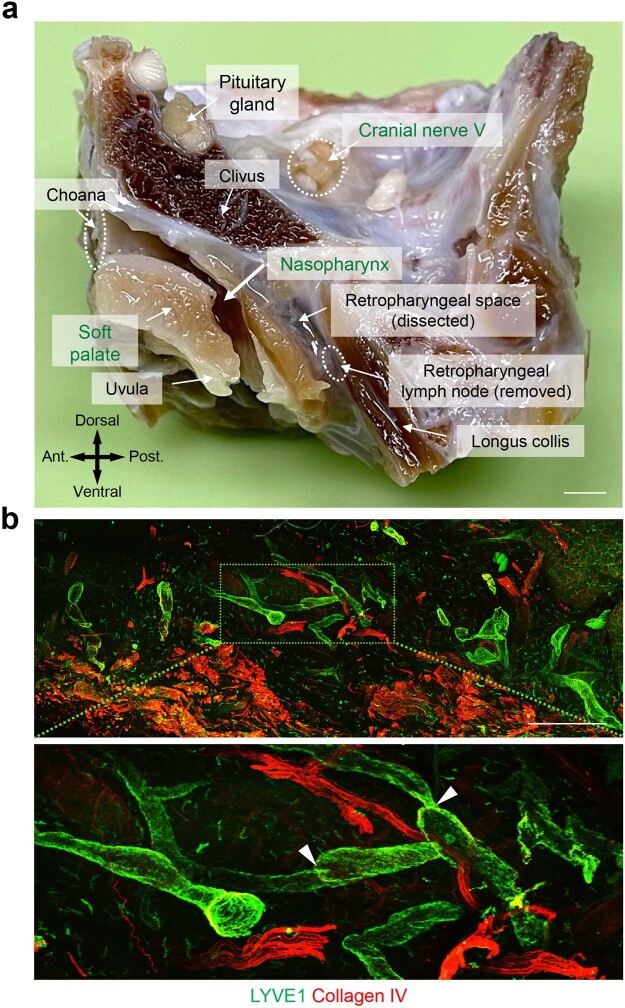
Extended Data Fig. 2. Location of the nasopharyngeal lymphatic plexus in the mucosa of the nasopharynx of the primate, Macaca fascicularis.
a, Mid-sagittal section of the head and neck of Macaca fascicularis with relevant landmarks labelled. Anatomical positions are indicated in the lower left corner. Scale bar, 3 mm. Ant., anterior; Post., posterior anatomical position. b, Immunofluorescence images of thick sections of nasopharyngeal mucosa stained for LYVE1+ (green) of lymphatics and collagen IV+ (red) on vessels. Green dashed-line box in upper panel is enlarged in the lower panel. White arrowheads mark lymphatic valves. Scale bar, 1 mm. Similar findings were obtained from n = 5 Macaca fascicularis in two independent experiments.

