Abstract
Capnocytophaga spp. have been implicated as putative periodontal pathogens associated with various periodontal diseases. Although the genus is known to contain five human oral isolates, accurate identification to species level of these organisms recovered from subgingival plaque has been hampered by the lack of a reliable method. Hence, most studies to date have reported these isolates as Capnocytophaga spp. Previous attempts at identification were based on biochemical tests; however, the results were inconclusive. Considering the differing virulence features of the respective isolates, it is crucial to identify these isolates to species level. The universal and conservative nature of the 16S rRNA gene has provided an accurate method for bacterial identification. The aim of this study was to identify Capnocytophaga spp. via restriction enzyme analysis of this gene (16S rRNA PCR-restriction fragment length polymorphism). The results (backed up by 16S rRNA gene sequencing) showed that this method reliably identifies all named Capnocytophaga spp. to species level.
Traditionally bacterial identification was established using a broad range of phenotypic characteristics. Organisms were assigned to groups based on morphological features (macro- and microscopic, including staining characteristics) and physiological attributes such as nutritional requirements, fermentation products, growth conditions (oxygen, temperature, and inhibitory products), and the ability to form spores. This formed the basis of classification for bacterial genera (9). These phenotypic features are shared by many species and are therefore nonspecific. They vary depending on the cultural conditions and/or age of colonial growth. The number of tests, their duration, the labor involved, and their costs often limit the range of tests chosen for routine identification. The choice of tests undertaken and their standardization varies between laboratories, which in turn may be a potential source of misclassification (12, 34). In addition, some strain-specific tests that claim to differentiate between species within a genus fail to do so conclusively (17, 30).
The accessibility and ease of use of bacterial genetic information heralded a new era in bacterial systematics. The major breakthrough in determining the evolution and phylogeny of prokaryotes came with the introduction of rRNA sequencing techniques (37). The 16S rRNA gene is universally distributed and highly conserved (38). Due to its conserved nature and ease of manipulation, it has been extensively used to establish accurate identification of clinical bacterial isolates. Amplification of the 16S rRNA gene using PCR (25) allows generation of high copy numbers of this gene, which may subsequently be used for bacterial identification. Cleavage of PCR-generated 16S rRNA gene amplicons by a restriction enzyme(s) (RE) results in differentiation by restriction fragment length polymorphisms (RFLP). The 16S rRNA PCR-RFLP may be separated by electrophoresis and visualized on an agarose gel. This procedure has been used extensively as a method for bacterial species identification (8, 23, 31, 36).
Identification of Capnocytophaga spp. from clinical isolates is crucial in epidemiological typing and clinical diagnosis. The genus comprises a group of capnophilic, facultatively anaerobic, gram-negative, slender fusiform rods, which perform fermentative metabolism and exhibit gliding motility when grown on solid culture media (20). It contains five human oral species, Capnocytophaga gingivalis, C. ochracea, C. sputigena, C. granulosa, and C. haemolytica (5, 20), and two species (C. canimorsus and C. cynodegmi) that form part of the canine and feline oral flora (3). The clinical significance of the latter two species is that C. canimorsus can cause human systemic infections (21) while C. cynodegmi may lead to localized infections (3); both types of infection arise via dog or cat scratches or bites. Investigations into possible associations of species within this genus with various pathological conditions have been hampered by the lack of a reliable scheme for species identification. Previous studies have attempted to distinguish between these species by biochemical tests (19, 29), protein profiles (16, 35), multilocus enzyme electrophoresis and serotyping of immunoglobulin A1 protease (11), DNA probes (7), 16S rRNA PCR-RFLP (36), and 16S rRNA sequence analysis (6, 35). Most of these methods (other than 16S rRNA PCR-RFLP) are labor-intensive, costly, and time-consuming and therefore not suitable for most microbiology laboratories, especially when several hundred clinical isolates are to be identified.
The aim of this study, therefore, was to develop a strategy for the molecular identification of Capnocytophaga spp. using 16S rRNA PCR-RFLP. The study was performed in two parts. (i) The first part entailed screening different endonuclease RE in order to select one that yielded a different restriction pattern for each of the Capnocytophaga type strains. The identities of the type strains obtained by 16S rRNA PCR-RFLP were confirmed by 16S rRNA sequencing. (ii) The second part involved identification of 187 clinical isolates of Capnocytophaga spp. by 16S rRNA PCR-RFLP, with subsequent verification by 16S rRNA gene sequencing.
MATERIALS AND METHODS
The seven Capnocytophaga type strains used were obtained from the American Type Culture Collection (ATCC), Manassas, Va. They were C. gingivalis ATCC 33624, C. ochracea ATCC 27872, C. sputigena ATCC 33612, C. granulosa ATCC 51502, C. haemolytica ATCC 51501, C. canimorsus ATCC 35979, and C. cynodegmi ATCC 49044.
Clinical isolates of Capnocytophaga spp. (n = 187) from subgingival plaque were obtained as described by Ciantar et al. (4). Briefly, subgingival plaque was collected from patients diagnosed with chronic adult periodontitis (periodontal pockets of ≥5 mm and radiographic evidence of bone loss) and were placed in a vial containing Trypticase soy broth. The sample was immediately transported to the microbiology laboratory, serially diluted, inoculated onto fastidious anaerobe agar (FAA; Lab M, Bury, United Kingdom), and incubated anaerobically (MACS Anaerobic Workstation; Don Whitley Scientific, Shipley, West Yorkshire, United Kingdom) for 5 days. Capnocytophaga spp., selected on the basis of their colony morphology and negative Gram staining, were subcultured to purity on FAA incubated under 5% (vol/vol) CO2 at 37°C for 3 days.
Bacterial DNA was extracted by a 2-min boiling procedure. Briefly, a few bacterial colonies were aseptically suspended in sterile nuclease-free water (Anachem, Luton, Bedfordshire, United Kingdom) in sterile, labeled 0.5-ml Eppendorf tubes (Sarstedt, Leicester, United Kingdom). The tubes were then sealed, vigorously agitated, and boiled for 2 min. Immediately after boiling, the tubes were placed on ice. A 1:10 dilution of this bacterial suspension in sterile nuclease-free water was subsequently prepared and kept on ice. It was later used as the nucleic acid template for the PCR amplification (18).
The PCR master mix contained 5 mM Taq NH4 buffer; 200 μM (each) dATP, dCTP, dGTP, and dTTP (all deoxynucleoside triphosphates from Promega, Madison, Wis.); 2.5 mM MgCl2, 25 pmol of each PCR primer μl−1; and 1 U of Taq polymerase (Bioline, London, United Kingdom). The PCR primers used were 27f (5′ AGAGTTTGATCMTGGCTCAG 3′; Genosys, Cambridge, United Kingdom), 1492r (5′ TACGGYTACCTTGTTACGACTT 3′; Genosys), and 357f (5′ CTCCTACGGGAGGCAGCAG 3′) (in the sequences listed here, M stands for C or A, and Y stands for C or T) (18). Fifty-microliter volumes (each) of the bacterial DNA template and the master mix were placed in PCR tubes (ABgene, Epsom, Surrey, United Kingdom). Two reactions per strain were performed, one for 16S rRNA PCR-RFLP analysis and the other for 16S rRNA gene sequencing.
PCR amplification was performed in a Biometra Uno II thermal cycler (Anachem) under the following conditions: 95°C for 5 min, followed by 29 cycles at 94°C for 1 min, 54°C for 1 min, and 72°C for 1 min, with a final extension period of 72°C for 5 min. Negative and positive controls using sterile, nuclease-free water and an Escherichia coli template, respectively, were also prepared. PCR amplicons (ca. 1,500 bp) were assessed by loading 10 μl of the PCR product into separate wells of a 0.8% (wt/vol) agarose gel (Agarose I; Anachem) containing ethidium bromide (0.5 μg/ml). A molecular weight marker (2,000 to 50 bp; Anachem) was loaded into the end well. The gel was immersed in Tris-acetate EDTA buffer (BDH, Poole, Dorset, United Kingdom) and subjected to a voltage difference of 70 V that led to separation of the fragments. The gel was visualized after excitation under UV transillumination by placing it in a MultiImage light cabinet (Alpha Innotech Corp., Cannock, Staffordshire, United Kingdom), and the resulting image was captured by a computer software program (AlphaEase; Alpha Innotech).
The PCR-RFLP reaction mixture for each of the Capnocytophaga ATCC strains (25 μl) contained 21.5 μl of the PCR amplicon, 1 U of RE (either CfoI, HaeIII, or RsaI; Promega), and 2.5 μl of the corresponding RE buffer. RE digestion was performed in sterile, labeled Eppendorf tubes. The RE mixture was briefly mixed, pulse spun, and incubated overnight at 37°C. The resulting 16S rRNA PCR-RFLP digests (25 μl) were transferred to separate wells of a 2% (wt/vol) superfine resolution agarose gel (Anachem) containing ethidium bromide (0.5 μg ml−1). The same molecular weight marker (10 μl) was loaded into each of the end wells of the gel. The fragments were allowed to separate electrophoretically and were visualized as described above.
PCR products were purified by using the QIAGEN (Crawley, West Sussex, United Kingdom) purification kit and were sequenced by using an ABI Prism BigDye Terminator cycle sequencing kit (Applied Biosystems, Warrington, United Kingdom) and primer 357f (see above). Sequenced products were further purified by standard ethanol precipitation. Sequence separation took place in an automated DNA sequencer (ABI Prism 310 Genetic Analyzer, Applied Biosystems). The resulting electrophoretograms were analyzed with the Chromas computer software program (version 1.43; Techelysium Pty Ltd., Tewantin, Australia). Only sequences of 300 bases or longer were analyzed by comparison with the 16S rRNA gene databases located at the Ribosomal Database Project (RDP-II; Michigan State University, East Lansing) (22); they were also analyzed with the basic local alignment search tool (BLAST; version 2.2.1) (1, 2, 28). Stringent criteria (tall, distinct peaks, minimal number of “Ns” (non-called bases), and very low background noise) were set for the electrophoretogram prior to submission of each sequence to both databases.
The 187 Capnocytophaga clinical isolates and the 7 type strains were processed as described above. Since analysis of the 16S rRNA PCR-RFLP patterns of the Capnocytophaga ATCC type strains with CfoI (Promega) revealed seven different patterns (see Results), the RE digests of the clinical isolates were performed using only this enzyme.
The 16S rRNA gene sequences (n = 37) of all the Capnocytophaga spp. available on the RDP-II and BLAST databases were downloaded and saved. In silico 16S rRNA PCR-RFLP analysis of these sequences was performed based on the restriction site for CfoI (GCG▾C). The sequences available on the RDP-II and BLAST databases are listed in Table 1.
TABLE 1.
List of all Capnocytophaga 16S rRNA gene sequences located in the RDP-II and BLAST databases as of May 2002
| Capnocytophaga taxona | Presence of the 16S rRNA gene sequenceb at:
|
Accession no. | |
|---|---|---|---|
| RDP | BLAST | ||
| C. gingivalis ATCC 33624T* | √ | √ | X67608 |
| C. gingivalis 16S rRNA gene | √ | √ | L14639 |
| C. gingivalis LMG 12118 | √ | √ | U41346 |
| Capnocytophaga sp. strain S3 | √ | √ | AY005073 |
| Capnocytophaga sp. strain S1 | √ | √ | U42008 |
| Capnocytophaga sp. strain LMG 12116 | √ | √ | U41352 |
| C. ochracea ATCC 27872T* | √ | √ | U41350 |
| C. ochracea ATCC strain. 25 | √ | √ | X67610 |
| C. ochracea ATCC 33596T | √ | √ | L14635 |
| C. ochracea LMG 12117 | √ | √ | U41351 |
| C. ochracea LMG 12115 (FDC 7) | √ | √ | U41353 |
| C. ochracea FDC 7b | √ | √ | U41354 |
| Capnocytophaga sp. strain S1 | √ | √ | U42008 |
| Capnocytophaga sp. strain S1b | √ | √ | U42009 |
| C. sputigena ATCC 33612T* | √ | √ | L14636 |
| C. sputigena 16S rRNA gene | √ | √ | X67609 |
| C. sputigena strain 897 CIP100 | √ | √ | AF133536 |
| C. granulosa ATCC 51502T* | √ | √ | X97248 |
| C. granulosa LMG 16022T | √ | √ | U41347 |
| C. granulosa LMG 12119 | √ | √ | U41348 |
| C. haemolytica ATCC 51501T* | √ | √ | X97247 |
| C. haemolytica LMG 16021T | √ | √ | U41349 |
| C. canimorsus ATCC 35979T* | √ | √ | L14637 |
| C. canimorsus 16S rRNA gene | √ | √ | X97246 |
| C. cynodegmi ATCC 49044T* | √ | √ | L14638 |
| C. cynodegmi 16S rRNA gene | √ | √ | X97245 |
| Capnocytophaga sp. oral clone X089 | √ | AY05080 | |
| Capnocytophaga sp. oral clone EL043 | √ | AY008312 | |
| Capnocytophaga sp. oral clone DS022 | √ | AF366270 | |
| Capnocytophaga sp. oral clone AA032 | √ | AY005079 | |
| Capnocytophaga sp. oral clone AH015 | √ | AY005074 | |
| Capnocytophaga sp. oral clone X066 | √ | AY005078 | |
| Capnocytophaga sp. oral clone A47ROY | √ | AY005077 | |
| Capnocytophaga sp. oral clone BB167 | √ | AY005076 | |
| Capnocytophaga sp. oral clone DZ074 | √ | AF385494 | |
| Capnocytophaga sp. oral clone BM058 | √ | AY005075 | |
| Capnocytophaga sp. oral clone BU084 | √ | AF385569 | |
Asterisks indicate Capnocytophaga ATCC type strains used in this study to produce the 16S rRNA PCR-RFLP shown in Fig. 1.
A check mark indicates that the sequence is present in the respective database. Note that the oral clones could be located only in the BLAST database.
The 16S rRNA gene sequences for Capnocytophaga spp. (except for Capnocytophaga sp. oral clone BM058) located on the RDP-II and BLAST databases were used to construct a phylogenetic tree for these species. The oral clone BM058 was omitted from the analysis because the sequence available on the database was a very short partial sequence. The sequences were aligned by using ClustalX (32). The resulting sequence alignment was edited with BioEdit, version 5.0.9 (14), and was subsequently used to produce a phylogenetic tree by the neighbor-joining method using ClustalX. Tree rooting was established by including the 16S rRNA gene for Porphyromonas gingivalis.
RESULTS
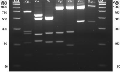
Of the three RE (CfoI, HaeIII, and RsaI) used for RFLP analysis of the seven ATCC type strains of Capnocytophaga species, CfoI was the only one that yielded seven different 16S rRNA PCR-RFLP, with each RFLP characterizing a particular species (Fig. 1). Thus, CfoI was the RE chosen for RFLP analysis of the clinical isolates. Gram staining of the ATCC strains confirmed their cellular morphology as gram-negative, slender fusiform rods.
FIG. 1.
16S rRNA PCR-RFLP analysis of ATCC type strains of Capnocytophaga species with restriction endonuclease CfoI. MM, molecular weight marker; Cg, C. gingivalis; Co, C. ochracea; Cs, C. sputigena; Cgr, C. granulosa; Ch, C. haemolytica; Ccn, C. canimorsus; Ccy, C. cynodegmi.
The identities of the Capnocytophaga type strains were confirmed by submitting the respective sequenced 16S rRNA genes to the RDP-II and BLAST databases.
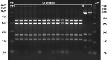
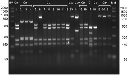
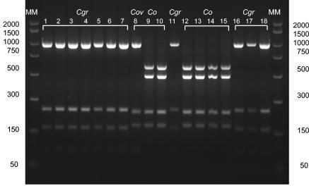
Preliminary identification of all clinical isolates of Capnocytophaga to genus level was performed by examination of their colony morphology, growth atmosphere, and Gram stain reaction (gram-negative, fusiform rods). Identification of all clinical isolates was initially accomplished by comparing their 16S rRNA PCR-RFLP with those of the Capnocytophaga ATCC type strains used in this study (Fig. 1). The 16S rRNA PCR-RFLP of a representative group of Capnocytophaga clinical isolates are depicted in Fig. 2, 3, and 4. The large majority of the clinical isolates' 16S rRNA PCR-RFLP conformed exactly to those of the ATCC type strains (see, e.g., those of C. ochracea [typical] and C. granulosa in Fig. 2; C. gingivalis in Fig. 3, lanes 2, 3, and 4; and C. sputigena in Fig. 3, lane 18). An advantage of this method was that strain contamination was easily detected (Fig. 3, lane 17).
FIG. 2.
16S rRNA PCR-RFLP analysis of clinical isolates of Capnocytophaga species with restriction endonuclease CfoI. Co, C. ochracea; Cgr, C. granulosa; MM, molecular weight marker.
FIG. 3.
16S rRNA PCR-RFLP analysis of clinical isolates of Capnocytophaga species with restriction endonuclease CfoI. Co, C. ochracea; Cg, C. gingivalis; Cgr, C. granulosa; Cgv, C. gingivalis variant; Cs, C. sputigena; C, contaminant; MM, molecular weight marker.
FIG. 4.
16S rRNA PCR-RFLP analysis of clinical isolates of Capnocytophaga species with restriction endonuclease CfoI. Cgr, C. granulosa; Cov, C. ochracea variant; Co, C. ochracea; MM, molecular weight marker.
Some strains showed subtle differences in their RFLP and were hence labeled as “variants,” e.g., C. ochracea variant (Fig. 3, lane 6; Fig. 4, lane 8). Isolates conforming to this pattern were identified as oral clones of Capnocytophaga spp. via 16S rRNA gene sequencing. An RFLP pattern similar to that of C. canimorsus was observed (Fig. 3, lane 14); however, identification by 16S rRNA gene sequencing revealed this to be a variant of C. gingivalis.
Definitive identification of Capnocytophaga clinical isolates was achieved by comparing the sequenced 16S rRNA genes with those available in the RDP-II and BLAST databases. In the case of the RDP-II data, although a similarity value cutoff of ≥0.8 had been set, some data points with a similarity value of ≤0.8 had to be accepted, because reprocessing of these strains, e.g., C. ochracea variant strains, failed to yield better similarity values in spite of the fact that the electrophoretogram fulfilled the predetermined criteria. The Capnocytophaga 16S rRNA gene sequences contained in each of the two databases are listed in Table 1.
Table 2 presents the validation by 16S rRNA gene sequencing of the identification of clinical isolates by 16S rRNA PCR-RFLP. The results showed that the sequencing data confirmed the initial identification of clinical isolates obtained by 16S rRNA PCR-RFLP only when the RFLP exactly matched that of the ATCC strains used in this study. The identification results obtained with BLAST demonstrated better corroboration of the RFLP results than did the RDP-II data. The BLAST sequence results were more accurate and, unlike the RDP-II data, included all the Capnocytophaga oral clones deposited by Paster et al. (26). This explains why some RDP-II sequence results gave low similarity values, e.g., those for C. ochracea variants, which were incorrectly identified as C. sputigena in spite of a high-quality electrophoretogram. The lower number of sequences in the RDP-II database accounted for the low level of correlation in identification between RFLP analysis and RDP for the C. ochracea variant strains (41.8%). The corresponding comparison between RFLP and BLAST gave 100% correlation.
TABLE 2.
Identification correlation of Capnocytophaga strains identified by 16S rRNA PCR-RFLP versus 16S rRNA sequencinga
| Species identified by RFLP | No. of strains | No. (%) of strains with same identification result by RFLP vs sequencing and comparison to:
|
|
|---|---|---|---|
| RDP | BLAST | ||
| C. gingivalis (typical) | 8 | 8 (100.0) | 8 (100.0) |
| C. gingivalis (variant) | 3 | 3 (100.0) | 3 (100.0) |
| C. ochracea (typical) | 46 | 44 (95.0) | 46 (100.0) |
| C. ochracea (variant) | 43 | 18 (41.8) | 43 (100.0) |
| C. sputigena | 7 | 7 (100.0) | 7 (100.0) |
| C. granulosa | 67 | 62 (93.9) | 67 (100.0) |
| C. haemolytica | 8 | 8 (100.0) | 8 (100.0) |
| C. canimorsus | 2 | 2 (100.0) | 2 (100.0) |
| C. cynodegmi | 3 | 2 (66.0) | 3 (100.0) |
Restriction endonuclease CfoI was used for 16S rRNA PCR-RFLP. After the 16S rRNA genes of clinical isolates were sequenced, the isolates were identified by comparison of their 16S rRNA sequences to those in the RDP-II and BLAST databases.
One C. ochracea (typical) strain (processed twice; hence n = 2) was incorrectly identified as C. sputigena by RDP in spite of the fact that the similarity values were 0.858 and 0.827. The same strain was identified as Capnocytophaga sp. oral clone X089 by BLAST.
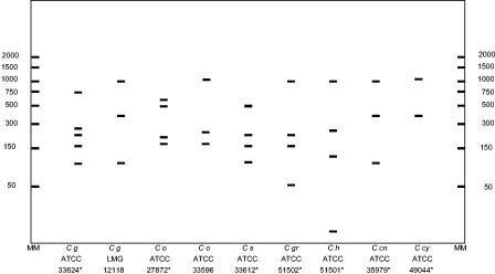
The strains initially identified as C. canimorsus by RFLP were identified as either C. gingivalis 33624T, Capnocytophaga sp. strain S3, or C. gingivalis LMG 12118 when submitted to both databases; hence, the corresponding RFLP analysis pattern was labeled “C. gingivalis variant.” As can be seen from Fig. 5 (in silico analysis), the RFLP pattern of these strains simulated the RFLP for either C. gingivalis LMG 12118 or C. canimorsus ATCC 35979 more closely than it did that of C. gingivalis ATCC 33624. Sequence alignment by ClustalW (version 1.8, 1999) (33) of C. gingivalis LMG 12118 with C. canimorsus yielded 91.2% similarity, demonstrating a species difference rather than strain variation. The C. gingivalis variant (C. gingivalis LMG 12118) could be differentiated from C. canimorsus by performing a second, separate digest of the 16S rRNA gene using the RE HaeIII. Alignment of C. gingivalis ATCC 33624 versus C. gingivalis LMG 12118 revealed 97.8% similarity (implying strain variation).
FIG. 5.
In silico analysis of 16S rRNA PCR-RFLP of representative Capnocytophaga species obtained from the RDP-II and BLAST databases. C g, C. gingivalis; C o, C. ochracea; C s, C. sputigena; C gr, C. granulosa; C h, C. haemolytica; C cn, C. canimorsus; C cy, C. cynodegmi. Asterisks indicate type strains used in this study. MM, molecular weight marker.
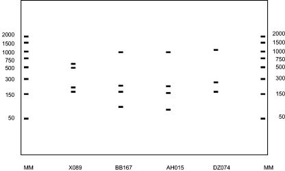
The results of the in silico analysis of 16S rRNA genes of representative Capnocytophaga strains available in the RDP-II and BLAST databases are shown in Fig. 5. Within C. gingivalis and C. ochracea, strains conformed to two patterns, while C. sputigena, C. granulosa, C. haemolytica, C. canimorsus, and C. cynodegmi strains conformed to a single RFLP fingerprint each. Strains with RFLP fingerprints similar to those of the following representative oral strains are given in parentheses: C. gingivalis LMG 12118 (Capnocytophaga sp. strain S3), C. ochracea ATCC 27872T (C. ochracea FDC 7b, LMG 12115, and LMG 12117, and Capnocytophaga sp. strain S1), C. sputigena 33612T (C. sputigena strain 897 CIP100, C. sputigena 16S rRNA gene), C. granulosa ATCC 51502 (C. granulosa LMG 16022 and LMG 12119), and C. haemolytica ATCC 51501 (C. haemolytica LMG 16021). Oral clones of Capnocytophaga spp. were also analyzed in silico (Fig. 6). Sequence alignment of C. gingivalis, C. ochracea, and Capnocytophaga sp. oral clones showed some degree of intraspecies diversity. Clones EL043, DS022, AA032, X066, A47ROY, and BU084 had fingerprints similar to that of Capnocytophaga sp. oral clone X089.
FIG. 6.
In silico analysis of 16S rRNA PCR-RFLP of representative oral Capnocytophaga clones obtained from the BLAST database (26). MM, molecular weight marker.
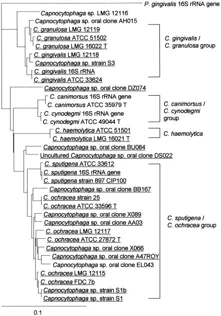
The results of the phylogenetic analysis are presented in Fig. 7. The dendrogram showed that the strains were very closely related, which was to be expected, because these species are all members of the same genus. However, subdivisions within the genus can be observed, resulting in the formation of three large clades: the C. gingivalis-C. granulosa group, the C. canimorsus-C. cynodegmi group, and the C. ochracea-C. sputigena group. C. haemolytica formed a small, separate group on its own.
FIG. 7.
Phylogenetic tree of Capnocytophaga species based on sequence similarity of 16S rRNA genes from the RDP-II and BLAST databases. Underlined taxa were isolated during this study. P. gingivalis was used as an outgroup.
C. ochracea and C. sputigena formed the largest group, and most of the oral clones fell into this group. Capnocytophaga oral clone BU084 and the uncultured Capnocytophaga oral clone DS022, although closely related to the C. ochracea-C. sputigena group, formed a separate, distinct branch in the dendrogram. This suggested that these could form a separate species. The results also showed that, except for seven strains, all representatives of these 16S rRNA gene sequences were isolated during the course of this study.
DISCUSSION
Traditionally, identification of Capnocytophaga spp. has been performed using conventional biochemical methods (10, 15, 27, 29). However, such methods failed to distinguish between the more closely phylogenetically related species within this genus, e.g., C. ochracea and C. sputigena (17, 30). Such findings were corroborated by preliminary experiments in this investigation (data not shown), the results of which led to the cessation of biochemical testing as a method of strain identification.
Several molecular methods (e.g., full gene sequencing, genomic restriction analysis, conventional ribotyping, 16S-23S intergenic spacer gene sequencing) are currently available for intrageneric differentiation of bacterial species. Indeed, 16S rRNA sequencing is regarded as the “gold standard” for bacterial identification. Most of these procedures, however, are lengthy, time-consuming, and expensive and therefore not accessible to most microbiology laboratories. Conventional ribotyping methods usually involve the hybridization of restriction fragments of chromosomal DNA to respective probes by Southern blotting. Modification of the conventional ribotyping obviates the use of Southern blotting, which is a labor-intensive procedure. The main advantages of identification by 16S rRNA PCR-RFLP using this modified ribotyping method are as follows: (i) it can be performed by the majority of laboratories, (ii) it allows a high throughput of strains, (iii) it is less expensive, and (iv) results can be obtained within 24 h (maximum).
The known human oral strains (to date) of Capnocytophaga are C. gingivalis, C. ochracea, C. sputigena, C. granulosa, and C. haemolytica. In this study the C. canimorsus and C. cynodegmi type strains were included, because although they are inhabitants of the canine and feline oral flora (3), they are capable of causing severe and sometimes fatal infections in humans (13). Thus, a method for their rapid identification would be clinically important.
A large number of strains (187) were identified by two methods: (i) 16S rRNA PCR-RFLP analysis and (ii) 16S rRNA gene sequencing and submission to the RDP-II and BLAST databases.
Bacterial DNA was easily extracted by a 2-min boiling procedure. The DNA served adequately as a template for PCR, precluding the need for lengthy DNA extraction protocols and thus further reducing procedure time.
The Capnocytophaga ATCC strains were sequenced in order to authenticate the identities of the type strains and to evaluate the quality of the sequences deposited in the 16S rRNA gene databases, which were subsequently to be used to identify the clinical isolates. These ATCC strains were concurrently subjected to 16S rRNA PCR-RFLP analysis, and the results obtained from the two processes were compared in order to verify the suitability of RFLP as a method for identification of Capnocytophaga species. The primers used for the PCR were 27f and 1492r. These were chosen because they are ideal primers for PCR amplification of 16S rRNA genes (18, 24).
The number of clinical isolates (187) identified by using this strategy was deemed to constitute a representative sample of the collection of Capnocytophaga isolates (n = 848) obtained as a result of this investigation. The two parts of the study, 16S rRNA PCR-RFLP and 16S rRNA gene sequencing, were performed blindly so as to avoid any bias in interpretation of the results. The sequence obtained for each strain was submitted to both the RDP-II and BLAST databases. This eliminated the possibility that any differences in results from the database could have been due to differences in DNA sequencing results rather than to actual differences between strains. Some strains were processed twice, for two reasons: (i) as a quality control during 16S rRNA gene sequencing and (ii) due to low RDP-II similarity values in spite of high-quality electrophoretograms.
The 16S rRNA gene sequences for Capnocytophaga spp. available in the RDP-II and BLAST databases are summarized in Table 1. The number of 16S rRNA gene sequences in the BLAST database totals 37, compared to the 26 sequences in the RDP-II database. None of the oral clones of Capnocytophaga spp. located in the BLAST database were available in the RDP-II database. This explains why some strains identified as C. ochracea (typical) by RFLP and as oral clones of Capnocytophaga spp. by BLAST (one of which also gave a C. ochracea typical RFLP following in silico analysis [Fig. 6]) were identified as C. sputigena by RDP. These strains were reprocessed on different occasions by using different sequencing primers (27f and 1492r were used as the different sequencing primers); however, the same results were obtained. This could not have been the result of amplicon contamination, because the negative control (water) was in fact negative. Rather, these strains were identified as C. sputigena by the RDP database because they are closely related phylogenetically to C. ochracea.
This study was based on an initial 16S rRNA PCR-RFLP analysis by Wilson et al. (36), in which they described eight RFLP patterns for Capnocytophaga spp. Although some of those RFLP patterns (including variants) were similar to patterns found in this study, comparison of RFLP fingerprints between the studies was precluded, because the authors had used a 2-kb DNA ladder in which the lowest fragment size was 154 bases. This could easily have obscured fragments of smaller sizes found in most strains (e.g., C. gingivalis ATCC 33624, C. gingivalis LMG 12118, C. sputigena, C. granulosa, C. haemolytica, and C. canimorsus) and therefore might have given a distorted RFLP fingerprint. In addition, no sequencing data were available.
The division of the genus Capnocytophaga into seven species was confirmed by Vandamme et al. (35) using protein profile electrophoresis. They, too, reported on the division of C. ochracea into two groups. In that study, as in this one, considerable genotypic heterogeneity was noted within the genus Capnocytophaga in spite of minimal phenotypic differences. While this might be due to previously unidentified species within this genus, 16S rRNA similarities greater than 97% are insufficient to guarantee a new species. It is not known whether these genetically different species are associated with specific pathological conditions.
The differences between the 16S rRNA PCR-RFLP of “typical” and “variant” strains could be explained following close examination of the results of in silico sequencing analysis. The latter showed that the variant RFLP fingerprints resulted either from a different base or from the presence of an insertion-deletion (indel) sequence at the restriction site. These nucleotide differences prevented restriction by the enzyme, thus resulting in a different RFLP pattern.
The oral clones of Capnocytophaga spp. deposited by Paster et al. (26) in the BLAST database seemed to be most closely related to C. ochracea when sequence alignment was performed using ClustalW. Those clones were obtained from subgingival plaque retrieved from patients with different periodontal conditions, which did not include adult periodontitis, as distinct from this study, which seems to be the first to report the isolation of these species from subgingival plaque.
Comparison of the 16S rRNA sequencing results obtained from RDP-II and BLAST emphasized the importance of using a database that is more up-to-date and contains a more diverse range of sequences. The main drawback is that although 16S rRNA gene sequencing is considered the gold standard for bacterial identification, sequences submitted for bacterial identification are compared with those available on the database, which, in some cases, might be partial sequences. Furthermore, the quality of the deposited sequences is not vetted in any way, and the query sequence might not be available on the database, thus giving a poor correlation because of its absence rather than because of inferior sequence quality.
The phylogenetic relatedness of C. ochracea and C. sputigena explained the similar biochemical features shared by these two species. This similarity made biochemical differentiation between them impossible. This finding, obtained during the early course of this study (unpublished data), has been corroborated by other investigators (17, 30). The majority of the clones deposited in the BLAST database by Paster et al. (26) have been cultured and identified during this study. These included the uncultured Capnocytophaga oral clone DS022. The phylogenetic relationships of the species explained the discrepancies in bacterial identification when the same sequence was submitted to the RDP-II and BLAST databases. Since the RDP-II database contained a smaller range of Capnocytophaga 16S rRNA gene sequences than the BLAST database (Table 1) and did not contain any of the cloned sequences, all the sequences identified as oral Capnocytophaga clones by the BLAST database were identified as a closely related species within that cluster of organisms by the RDP-II database. Thus, for example, sequences identified as C. ochracea strain 25 by the RDP-II database were identified as Capnocytophaga sp. oral clone BB167, X066, or A47ROY by the BLAST database. The absence of oral Capnocytophaga clones from the RDP-II database helped to explain the poor identification correlation (41.8%) between 16S rRNA PCR-RFLP and RDP (Table 2).
A C. gingivalis variant (C. gingivalis LMG 12118) and C. canimorsus gave similar 16S rRNA PCR-RFLP when CfoI was used for restriction digestion. These strains could be easily differentiated with HaeIII as the RE. In addition, they showed only 91.2% similarity when aligned by using ClustalW, indicating that they were completely different species.
In conclusion, the results of this investigation have shown that 16S rRNA PCR-RFLP analysis using CfoI as the restriction enzyme is a reliable, rapid, and accurate method for the identification of clinical isolates of Capnocytophaga spp., especially when large numbers of clinical isolates need to be identified. More importantly, such methods are cost-effective and available to most routine microbiology laboratories, thus allowing for more accurate identification of clinical isolates.
Acknowledgments
We acknowledge the support for this project provided by the Shirley Glasstone Memorial Prize (awarded though the British Dental Association) and Eli Lilly Diabetes Research, UK.
REFERENCES
- 1.Altschul, S. F., W. Gish, W. Miller, E. W. Myers, and D. J. Lipman. 1990. Basic local alignment search tool. J. Mol. Biol. 215:403-410. [DOI] [PubMed] [Google Scholar]
- 2.Altschul, S. F., T. L. Madden, A. A. Schäffer, J. Zhang, Z. Zhang, W. Miller, and D. J. Lipman. 1997. Gapped BLAST and PSI-Blast: a new generation of protein database search programs. Nucleic Acids Res. 25:3389-3402. [DOI] [PMC free article] [PubMed] [Google Scholar]
- 3.Brenner, D. J., D. G. Hollis, G. R. Fanning, and R. E. Weaver. 1989. Capnocytophaga canimorsus sp. nov. (formerly CDC group DF-2), a cause of septicemia following dog bite, and C. cynodegmi sp. nov., a cause of localized wound infection following dog bite. J. Clin. Microbiol. 27:231-235. [DOI] [PMC free article] [PubMed] [Google Scholar]
- 4.Ciantar, M., D. A. Spratt, H. N. Newman, and M. Wilson. 2001. Assessment of five culture media for the growth and isolation of Capnocytophaga species. Clin. Microbiol. Infect. 7:158-160. [DOI] [PubMed] [Google Scholar]
- 5.Ciantar, M., D. A. Spratt, H. N. Newman, and M. Wilson. 2001. Capnocytophaga granulosa and Capnocytophaga haemolytica: novel species in subgingival plaque. J. Clin. Periodontol. 28:701-705. [DOI] [PubMed] [Google Scholar]
- 6.Conrads, G., and R. Lutticken. 1992. Nucleotide sequences of 16S rRNA encoding genes from Capnocytophaga ochracea ATCC 33596, Capnocytophaga sputigena ATCC 33612 and Capnocytophaga gingivalis ATCC 33624. Nucleic Acids Res. 20:5847. [DOI] [PMC free article] [PubMed] [Google Scholar]
- 7.Conrads, G., R. Mutters, I. Seyfarth, and K. Pelz. 1997. DNA-probes for the differentiation of Capnocytophaga spp. Mol. Cell. Probes 11:323-328. [DOI] [PubMed] [Google Scholar]
- 8.Conville, P. S., S. H. Fischer, C. P. Cartwright, and F. G. Witebsky. 2000. Identification of Nocardia species by restriction endonuclease analysis of an amplified portion of the 16S rRNA gene. J. Clin. Microbiol. 38:158-164. [DOI] [PMC free article] [PubMed] [Google Scholar]
- 9.Cowan, S. T., and K. J. Steel. 1993. Manual for the identification of medical bacteria, 3rd ed. Cambridge University Press, New York, N.Y.
- 10.Forlenza, S. W., M. G. Newman, A. L. Horikoshi, and U. Blachman. 1981. Antimicrobial susceptibility of Capnocytophaga. Antimicrob. Agents Chemother. 19:144-146. [DOI] [PMC free article] [PubMed] [Google Scholar]
- 11.Frandsen, E. V. G., and W. G. Wade. 1996. Differentiation of human Capnocytophaga species by multilocus enzyme electrophoretic analysis and serotyping of immunoglobulin A1 proteases. Microbiology 142:441-448. [DOI] [PubMed] [Google Scholar]
- 12.Goodfellow, M., and C. H. Dickinson. 1985. Delineation and description of microbial populations using numerical methods, p. 165-225. In M. Goodfellow, D. Jones, and F. G. Priest (ed.), Computer-assisted bacterial systematics. Academic Press, London, United Kingdom.
- 13.Griego, R. D., T. Rosen, I. F. Orengo, and J. E. Wolf. 1995. Dog, cat and human bites: a review. J. Am. Acad. Dermatol. 33:1019-1029. [DOI] [PubMed] [Google Scholar]
- 14.Hall, T. A. 1999. BioEdit: a user-friendly biological sequence alignment editor and analysis program for Windows 95/98/NT. Nucleic Acids Symp. 41:95-98. [Google Scholar]
- 15.Jolivet-Gougeon, A., A. Buffett, C. Dupuy, J. L. Sixou, M. Bonnaure-Mallet, S. David, and M. Cormier. 2000. In vitro susceptibilities of Capnocytophaga isolates to β-lactam antibiotics and β-lactamase inhibitors. Antimicrob. Agents Chemother. 44:3186-3188. [DOI] [PMC free article] [PubMed] [Google Scholar]
- 16.Khwaja, K. J., P. Parish, M. J. Aldred, and W. R. Wade. 1990. Protein profiles of Capnocytophaga species. J. Appl. Bacteriol. 68:385-390. [DOI] [PubMed] [Google Scholar]
- 17.Kristiansen, J. E., A. Bremmelgard, H. E. Busk, O. Heltberg, W. Frederiksen, and T. Justersen. 1984. Rapid identification of Capnocytophaga isolated from septicaemic patients. Eur. J. Clin. Microbiol. 3:236-240. [DOI] [PubMed] [Google Scholar]
- 18.Lane, D. G. 1991. Nucleic acids techniques, p. 115-175. In E. Stackebrandt and M. Goodfellow (ed.), Bacterial systematics. John Wiley, Chichester, United Kingdom.
- 19.Laughon, B. E., S. A. Syed, and W. J. Loesche. 1982. API ZYM system for identification of Bacteroides spp., Capnocytophaga spp., and spirochetes of oral origin. J. Clin. Microbiol. 15:97-102. [DOI] [PMC free article] [PubMed] [Google Scholar]
- 20.Leadbetter, E. R., S. C. Holt, and S. S. Socransky. 1979. Capnocytophaga: new genus of Gram-negative gliding bacteria. I. General characteristics, taxonomic considerations and significance. Arch. Microbiol. 122:9-16. [DOI] [PubMed] [Google Scholar]
- 21.Mahrer, S., and E. Raik. 1992. Capnocytophaga canimorsus septicaemia associated with cat scratch. Pathology 24:194-196. [DOI] [PubMed] [Google Scholar]
- 22.Maidek, B. L., J. R. Cole, T. G. Lilburn, C. T. Parker, Jr., P. R. Saxman, R. J. Farris, G. M. Garrity, G. J. Olsen, T. M. Schmidt, and J. M. Tiedje. 2001. The RDP-II (Ribosomal Database Project). Nucleic Acids Res. 1:173-174. [DOI] [PMC free article] [PubMed] [Google Scholar]
- 23.Marshall, S. M., P. I. Melito, D. L. Woodward, W. M. Johnson, F. G. Rodgers, and M. R. Mulvey. 1999. Rapid identification of Campylobacter, Arcobacter, and Helicobacter isolates by PCR-restriction fragment length polymorphism analysis of the 16S rRNA gene. J. Clin. Microbiol. 37:4158-4160. [DOI] [PMC free article] [PubMed] [Google Scholar]
- 24.Medlin, L., H. J. Elwood, S. Stickel, and M. L. Sogin. 1998. The characterisation of enzymatically amplified eukaryotic 16S-like rRNA coding regions. Gene 71:491-499. [DOI] [PubMed] [Google Scholar]
- 25.Mullis, K. B., and F. Faloona. 1987. Specific synthesis of DNA in vitro via a polymerase catalysed chain reaction. Methods Enzymol. 155:335-350. [DOI] [PubMed] [Google Scholar]
- 26.Paster, B. J., S. K. Boches, J. L. Galvin, R. E. Ericson, C. N. Lau, V. A. Levanos, A. Sahasrabudhe, and F. E. Dewhirst. 2001. Bacterial diversity in human subgingival plaque. J. Bacteriol. 183:3770-3783. [DOI] [PMC free article] [PubMed] [Google Scholar]
- 27.Rummens, J. L., B. Gordts, and H. W. van Landuyt. 1986. In vitro susceptibility of Capnocytophaga species to 20 antimicrobial agents. Antimicrob. Agents Chemother. 30:739-742. [DOI] [PMC free article] [PubMed] [Google Scholar]
- 28.Schäffer, A. A., L. Aravind, T. L. Madden, S. Shavirin, J. L. Spouge, Y. I. Wolf, E. V. Koonin, and S. F. Altschul. 2001. Improving the accuracy of PSI-BLAST protein database searches with composition-based statistics and other refinements. Nucleic Acids Res. 15:2994-3005. [DOI] [PMC free article] [PubMed] [Google Scholar]
- 29.Socransky, S. S., S. C. Holt, E. R. Leadbetter, A. C. R. Tanner, E. Savitt, and B. F. Hammond. 1979. Capnocytophaga: new genus of Gram-negative gliding bacteria. III. Physiological characterisation. Arch. Microbiol. 122:29-33. [DOI] [PubMed] [Google Scholar]
- 30.Speck, H., R. M. Kroppenstedt, and W. Mannheim. 1987. Genomic relationships and species differentiation in the genus Capnocytophaga. Zentbl. Bakteriol. Hyg. A 266:390-402. [DOI] [PubMed] [Google Scholar]
- 31.Steinhauserova, I., J. Ceskova, K. Fojtikova, and I. Obrovska. 2001. Identification of thermophilic Campylobacter spp. by phenotypic and molecular methods. J. Appl. Microbiol. 90:470-475. [DOI] [PubMed] [Google Scholar]
- 32.Thompson, J. D., T. J. Gibson, F. Plewniak, F. Jeanmougin, and D. G. Higgins. 1997. The Clustal_X Windows interface: flexible strategies for multiple sequence alignment aided by quality analysis tools. Nucleic Acids Res. 25:4876-4882. [DOI] [PMC free article] [PubMed] [Google Scholar]
- 33.Thompson, J. D., D. G. Higgins, and T. J. Gibson. 1994. CLUSTAL W: improving the sensitivity of progressive multiple sequence alignment through sequence weighting, position-specific gap penalties and weight matrix choice. Nucleic Acids Res. 22:4673-4680. [DOI] [PMC free article] [PubMed] [Google Scholar]
- 34.Totten, P. A., C. M. Patton, F. C. Tenover, T. J. Barrett, W. E. Stamm, A. G. Steigerwalt, J. Y. Lin, K. K. Holmes, and D. J. Brenner. 1987. Prevalence and characterization of hippurate-negative Campylobacter jejuni in King County, Washington. J. Clin. Microbiol. 25:1747-1752. [DOI] [PMC free article] [PubMed] [Google Scholar]
- 35.Vandamme, P., M. Vancanneyt, A. Van Belkum, P. Segers, W. G. V. Quint, K. Kersters, B. J. Paster, and F. E. Dewhirst. 1996. Polyphasic analysis of strains of the genus Capnocytophaga and Centers for Disease Control group DF-3. Int. J. Syst. Bacteriol. 46:782-791. [DOI] [PubMed] [Google Scholar]
- 36.Wilson, M. J., W. G. Wade, and A. J. Weightman. 1995. Restriction fragment length polymorphism analysis of PCR amplified 16S ribosomal DNA of human Capnocytophaga. J. Appl. Bacteriol. 78:394-401. [DOI] [PubMed] [Google Scholar]
- 37.Woese, C. R., G. E. Fox, L. Zablen, T. Uchida, L. Bonen, K. Pechman, and B. J. Lewis. 1975. Conservation of primary structure in 16S ribosomal RNA. Nature 254:83-86. [DOI] [PubMed] [Google Scholar]
- 38.Woese, C. R. 1987. Bacterial evolution. Microbiol. Rev. 51:221-271. [DOI] [PMC free article] [PubMed] [Google Scholar]


