Abstract
In an attempt to improve the specificity of an enzyme-linked immunosorbent assay (ELISA) for the diagnosis of paracoccidioidomycosis (PCM), sera from patients with PCM were tested using various approaches, such as sodium metaperiodate antigen (gp43) treatment, a serum absorption process with Candida albicans or Histoplasma capsulatum antigens, and dilution of serum in galactose, the main common epitope among pathogenic fungi. The maximum specificity found in this ELISA was 84%. All of these procedures proved inefficient for eliminating all cross-reacting antibodies and obtaining an ELISA specific for PCM diagnosis.
Paracoccidioidomycosis (PCM) is a mycotic disease caused by the dimorphic fungus Paracoccidioides brasiliensis and causes a deep mycosis, resulting in a severe chronic granulomatous infection of the skin, mucous membranes, lymph nodes, and internal organs. Almost all South and Central America countries have large regions where PCM is endemic. The definitive diagnosis of PCM includes direct observation of the characteristic multiple-budding cells in biological fluids and tissue sections or isolation of the fungus from clinical materials. For cases in which P. brasiliensis is not observed through direct examination, several serological tests have been used to detect antibodies against the fungus in order to establish the diagnosis (2). gp43, a 43,000-Da glycoprotein from P. brasiliensis, is a major diagnostic antigen in serological assays (1, 3, 15, 18, 23, 24). It has been demonstrated that gp43 is as good as crude exoantigen preparations (concentrated, dialyzed culture supernatants) (3, 4) for detecting precipitating antibody in immunodiffusion tests, and purified gp43 has also been adapted to several other serological tests, such as immunoprecipitation of 125I-labeled gp43 (IPP), conventional enzyme-linked immunosorbent assay (ELISA), capture enzyme immunoassay (EIA), and passive hemagglutination (5, 20, 23). Cross reactivity in serological assays from patients with histoplasmosis depends on the test used and seems to involve a carbohydrate epitope. While IPP and capture EIA using gp43 appear specific for PCM diagnosis, experiments were performed to determine the nature of the cross reactivity in sera from those patients with histoplasmosis that reacted with gp43 in the IPP tests. Treatment of gp43 with 0.2 M d-galactose inhibited such cross reactivity, and no cross reactivity was observed with the deglycosylated form of gp43, with EH38, or with the 38,000-Da protein which had been treated with periodate (18). Owing to the N-linked high-mannose nature of the gp43 glycan, deglycosylation was efficiently carried out with endo-β-N-acetylglucosaminidase H. Since sera from patients with histoplasmosis generally reacted with the high-molecular-weight glycocomplex present in the crude antigen of P. brasiliensis, this reactivity and that with gp43 likely involved recognition of terminal β-galactofuranosyl units (24).
Travassos et al. (24) related that in general, specific reactions with gp43 are obtained when the antigen is in solution, such as in IPP, immunodiffusion, and immunoelectrophoresis, and that fixing gp43 onto solid phase substrates, e.g., plastic, increases the number of nonspecific reactions. The authors also related that it is possible that the spatial distribution of the gp43 glycan containing terminal β-galactofuranosyl units when fixed on a plastic substrate contributed to the increased accessibility of these units to heterologous cross-reacting antibodies.
Mendes-Gianinni et al. (15) found that none of the sera from patients with heterologous mycotic diseases reacted with the gp43 in ELISA after sera had been absorbed once with a culture filtrate of Histoplasma capsulatum. Similarly, Camargo et al. (5) related the same phenomenon when they used heterologous sera preabsorbed with Candida albicans yeast cells. However, contradicting results are found in the literature (17, 18).
ELISA has been employed in the serology of various mycotic diseases (7, 10, 11, 12, 13, 21, 22). It is an extremely sensitive method and is reagent saving compared with other assays. This method can be automated and may be less time-consuming and more economical for routine laboratories if specific reactions could be obtained. However, it has important limitations that arise from cross reactivity. Regarding the serology of PCM, this fact has been well known since the first serological studies using complement fixation tests, in the 1960s (8, 9). In relation to the use of ELISA for PCM diagnosis, various studies have shown that the main problem presented by the use of histoplasmosis sera is cross reaction (17, 18).
In this study, gp43 was assayed by ELISA, and we tried to standardize specific ELISA for the diagnosis of PCM by using various approaches. Then, gp43 was treated with sodium metaperiodate in an attempt to abolish the carbohydrate epitopes responsible for cross reactions; on the other hand, PCM and histoplasmosis sera were previously absorbed with heterologous fungal antigens (H. capsulatum or C. albicans) in order to eliminate cross-reactive antibodies; also, sera were diluted with galactose (0.2 M, final concentration) in order to eliminate anti-galactose antibodies, the main P. brasiliensis epitope in common with the H. capsulatum epitope (17), in an attempt to obtain specific reactions for PCM. Finally, two assay procedures were combined, absorption of sera with C. albicans and H. capsulatum followed by dilution of sera in galactose.
Serum specimens from 25 patients with PCM were selected on the basis of a positive direct examination of biological fluids; 14 specimens from patients with proven acute histoplasmosis and 20 serum specimens from healthy individuals were tested as controls in each scenario. All patient sera came from Hospital São Paulo, São Paulo, Brazil. Gp43 was purified according to the method of Puccia et al. (19), and optimization of solid-phase gp43 antigen concentration was determined by a peroxidase saturation technique (16). Once the ideal gp43 concentration to coat ELISA plates was determined, this molecule was treated with sodium metaperiodate at 10, 20, and 40 mM in order to determine the best concentration to eliminate carbohydrate epitopes without causing damage to the adsorbed molecule.
Sera were absorbed first with whole mycelium of H. capsulatum, Alnara isolate, and second with C. albicans yeast cells (ATCC 90028). Briefly, (i) 0.5 ml of serum was mixed with 0.5 ml of compacted C. albicans yeast cells and incubated under rotatory slow movement for 2 h at 37°C and overnight at 4°C. The mixture was then centrifuged at 2,000 rpm (10 min, 4°C), and the supernatant was recovered. (ii) The same protocol was used with compacted H. capsulatum mycelia. (iii) Sera were absorbed with both antigens. (iv) Sera were diluted in galactose (0.2 M, final concentration) in order to eliminate anti-galactose antibody epitopes. (v) Sera were previously absorbed with H. capsulatum and C. albicans followed by dilution in galactose (0.2 M, final concentration). Flat 96-well polystyrene plates were coated with gp43 in 0.1 M carbonate buffer [pH 9.6] and incubated for 1 h at 37°C and overnight at 4°C, followed by an immunoenzymatic reaction (ELISA) according to the method of Camargo et al. (5).
The ideal concentration of gp43 for coating the ELISA plates and obtaining the best sensitivity was found to be 2.5 μg/ml (250 ng/well). On the other hand, the treatment of gp43 with 40 mM sodium metaperiodate was found to be ideal in order to obtain a better separation between homologous and heterologous sera.
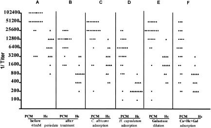
Figure 1 shows the overall results of ELISA using PCM and histoplasmosis sera, before and after absorption processes, reacting against gp43 treated with 40 mM sodium metaperiodate. First, Fig. 1A and B show the reactivity of sera from both groups of patients reacting with treated gp43 before the absorption process. Sera from patients with PCM reacted with the gp43 at dilutions of 1:3,200 to 1:102,400, and sera from patients with histoplasmosis reacted at dilutions of 1:800 to 1:25,600. In this case, ELISA was specific for 84% of PCM sera. As shown in Fig. 1C, when both groups of sera were absorbed with C. albicans, sera from patients with PCM reacted at dilutions of 1:200 to 1:51,200, and sera from patients with histoplasmosis reacted at dilutions of 1:400 to 1:12,800, indicating a 68% specificity of ELISA for PCM. As shown in Fig. 1D, when both groups of sera were absorbed with H. capsulatum antigen, sera from patients with PCM reacted at dilutions of 1:100 to 1:25,600 and sera from patients with histoplasmosis reacted at dilutions of 1:100 to 1:800, indicating a specificity of 80%. Similarly, as shown in Fig. 1E, when sera were diluted only in 0.2 M galactose, PCM sera reacted at dilutions of 1:100 to 1:51,200, and sera from histoplasmosis patients reacted at dilutions of 1:400 to 1:12800, showing a specificity of 60%. Figure 1F shows the reactivity of both groups of sera which were absorbed first with C. albicans, and then absorbed with H. capsulatum, and finally diluted in 0.2 M galactose. In this case, the specificity of ELISA was about 68%. Note that control sera from healthy individuals reacted in each scenario until the 1:200 dilution. All ELISAs were repeated at least three times with good reproducibility.
FIG. 1.
Results of ELISAs using PCM and histoplasmosis sera, before and after absorption processes. A and B, sera from both groups of patients reacting with gp43, before and after treatment with 40 mM sodium metaperiodate; C, patient sera absorbed with C. albicans; D, patient sera absorbed with H. capsulatum; E, patient sera diluted with galactose; F, patient sera absorbed with C. albicans and H. capsulatum antigens and diluted in galactose.
Nowadays, ELISA is a very popular test for the detection and quantitation of antibodies in medical research and in the clinical laboratory. In general, ELISA has a high sensitivity, but that sensitivity is not necessarily coupled with high specificity. For many years, ELISA has been the subject of many publications about antibody detection in many fungal diseases, mainly PCM (2, 6, 7, 10, 11, 12, 13, 21, 22). In this context, it has been used with a range of different kinds of antigenic preparations, such as crude mixtures (exoantigens and cytoplasmic antigens), partially purified preparations, and purified molecule, such as gp43. However, in a series of studies on PCM serology, and even in our previous paper (5), the authors described how a previous serum absorption process can eliminate antibodies responsible for cross reactivity (5, 14). During many years of experience in serology of PCM, however, we have observed that it is very difficult to obtain specific ELISA results and that many results are not reproducible. Sometimes, in some lots of PCM sera, we can obtain good differentiation between homologous and heterologous sera; at other times, these results are not obtained. This instability of results leads us to think that other factors, e.g., individual characteristics of each serum, the P. brasiliensis isolate that caused the disease, the individual immune response against a specific isolate that may carry different antigenic epitopes, are influencing the reactions.
In general, sodium metaperiodate can partially eliminate carbohydrate epitopes responsible for cross reactions and then may increase the specificity of the reaction; also, the absorption process with heterologous fungus cells may lead to a better specificity by eliminating common antibodies responsible for the cross reactivity.
In an attempt to obtain specific ELISA reactions for the diagnosis of PCM, we tried to use purified gp43 antigen, the immunodominant molecule which is specific when used in immunodiffusion tests, treated with sodium metaperiodate and sera individually preabsorbed with C. albicans or H. capsulatum or simultaneously absorbed with both antigens or sera diluted with galactose, the main cross-reacted carbohydrate. On the other hand, completely specific results were not obtained with tests of the C. albicans and H. capsulatum serum absorption processes followed by dilution of sera in galactose or by EIA reactivity tests with gp43 metaperiodate treatment.
Although gp43 is firmly established as a specific molecule for the serological diagnosis of PCM, this specificity depends on the test used, such as immunodiffusion. However, only a thorough understanding of its structure allows us to develop other approaches in order to obtain specific results by means of ELISA.
In conclusion, we related here the difficulty in obtaining an ELISA for specific and definitive diagnosis of PCM, even after treatment of gp43 with metaperiodate and the absorption process. These kinds of results lead us to reflect on how difficult it is to work in this area and to try to find new procedures to achieve our objectives.
Acknowledgments
This work was supported by FAPESP and CNPq.
REFERENCES
- 1.Blotta, M. H. S. L., and Z. P. Camargo. 1993. Immunological response to cell-free antigens of Paracoccidioides brasiliensis: relationships with clinical forms of paracoccidioidomycosis. J. Clin. Microbiol. 31:671-676. [DOI] [PMC free article] [PubMed] [Google Scholar]
- 2.Camargo, Z. P., and M. F. Franco. 2000. Current knowledge on pathogenesis and immunodiagnosis of paracoccidioidomycosis. Rev. Iberoam. Micol. 17:41-48. [PubMed] [Google Scholar]
- 3.Camargo, Z. P., C. Unterkircher, S. P. Campoy, and L. R. Travassos. 1988. Production of Paracoccidioides brasiliensis exoantigens for immunodiffusion tests. J. Clin. Microbiol. 26:2147-2151. [DOI] [PMC free article] [PubMed] [Google Scholar]
- 4.Camargo, Z. P., R. Berzaghi, C. C. Amaral, and S. H. Marques-da-Silva. 2004. Simplified method for producing Paracoccidioides brasiliensis exoantigens for use in immunodiffusion tests. Med. Mycol. 41:539-542. [DOI] [PubMed] [Google Scholar]
- 5.Camargo, Z. P., J. L. Guesdon, E. Drouhet, and L. Improvisi. 1984. Enzyme-linked immunosorbent assay (ELISA) in the paracoccidioidomycosis. Comparison with counterimmunoelectrophoresis and erythro-immunoassay. Mycopathologia 81:31-37. [DOI] [PubMed] [Google Scholar]
- 6.Camargo, Z. P., J. L. Gesztesi, E. C. O. Saraiva, C. P. Taborda, A. P. Vicentini, and J. D. Lopes. 1994. Monoclonal antibody capture enzyme immunoassay for detection of Paracoccidioides brasiliensis antibodies in paracoccidioidomycosis. J. Clin. Microbiol. 32:2377-2381. [DOI] [PMC free article] [PubMed] [Google Scholar]
- 7.Desgeorges, P. T., P. Ambroise-Thomas, and A. Goullier. 1979. Crytococcose: sero-diagnostic par immuno-enzymologie (ELISA). Nouv. Presse Med. 8:1055-1056. [PubMed] [Google Scholar]
- 8.Fava-Netto, C. 1961. Contribuição para o estudo imunológico da blastomicose de Lutz. Rev. Inst. Adolpho Lutz 21:99-194. [Google Scholar]
- 9.Fava-Netto, C. 1965. The immunology of South American blastomycosis. Mycopath. Mycol. Appl. 26:349-358. [DOI] [PubMed] [Google Scholar]
- 10.Greenberger. P. A., and R. Patterson. 1982. Application of enzyme-linked immunosorbent assay (ELISA) in the diagnosis of allergic bronchopulmonary aspergillosis. J. Lab. Clin. Med. 99:288-293. [PubMed] [Google Scholar]
- 11.Hommel, M., T. Kieng Troung, and D. E. Bidwell. 1976. Technique immunoenzymatique (ELISA) appliquée au diagnostic serologic des candidoses et aspergilloses humaines. Resultats préliminaires. Nouv. Presse Med. 5:2789-2791. [PubMed] [Google Scholar]
- 12.Kostiala, A. A. I., and I. Kostiala. 1981. Enzyme-linked immunosorbent assay (ELISA) for IgM, IgG and IgA class antibodies Candida albicans antigens: development and comparison with other methods. Sabouraudia 19:123-134. [DOI] [PubMed] [Google Scholar]
- 13.Meckstroth. K. L., E. Reiss, J. W. Keller, and L. Kaufman. 1981. Detection of antibodies and antigenemia in leukemic patients with candidiasis by enzyme-linked immunosorbent assay. J. Infect. Dis. 144:24-32. [DOI] [PubMed] [Google Scholar]
- 14.Mendes-Giannini, M. J. S., M. E. Camargo, C. S. Lacaz, and A. W. Ferreira. 1984. Immunoenzymatic absorption test for serodiagnosis of paracoccidioidomycosis. J. Clin. Microbiol. 20:103-108. [DOI] [PMC free article] [PubMed] [Google Scholar]
- 15.Mendes-Giannini, M. J. S., J. P. Bueno, M. A. Shikanai-Yasuda, A. M. S. Stolf, A. Masuda. V. Amato-Neto, and A. W. Ferreira. 1990. Antibody response to the 43 kDa glycoprotein of Paracoccidioides brasiliensis as a marker for the evaluation of patients under treatment. J. Trop. Med. Hyg. 43:200-206. [DOI] [PubMed] [Google Scholar]
- 16.Muñoz, C., A. Nieto, A. Gaya, J. Martinez, and J. Vives. 1986. New experimental criteria for optimization of solid-phase antigen concentration and stability in ELISA. J. Immunol. Methods 94:137-144. [DOI] [PubMed] [Google Scholar]
- 17.Negroni, R., F. Iovánnitti, and A. M. Robles. 1976. Estudio de las reacciones serologicas cruzadas entre antígenos de Paracoccidioides brasiliensis e Histoplasma capsulatum. Rev. Asoc. Argent. Microbiol. 8:68-72. [PubMed] [Google Scholar]
- 18.Puccia, R., and L. R. Travassos. 1991. 43-Kilodalton glycoprotein from Paracoccidioides brasiliensis: immunochemical reactions with sera from patients with paracoccidioidomycosis, histoplasmosis, or Jorge Lobo's disease. J. Clin. Microbiol. 29:1610-1615. [DOI] [PMC free article] [PubMed] [Google Scholar]
- 19.Puccia, R., D. T. Takaoka, and L. R. Travassos. 1991. Purification of the 43 kDa glycoprotein from exocellular components excreted by Paracoccidioides brasiliensis in liquid culture (TOM medium). J. Med. Vet. Mycol. 29:57-60. [PubMed] [Google Scholar]
- 20.Puccia, R., L. R. Travassos, E. G. Rodrigues, A. K. Carmona, M. C. Oliveira, and L. Juliano. 1994. Purification of the specific exocellular antigen gp43 from Paracoccidioides brasiliensis. Immunological and proteolytic activities, p. 507-515. In B. Maresca and G. S. Kobayashi (ed.), Molecular biology of fungi. Telos Press, New York, N.Y.
- 21.Scott, E. N., F. G. Felton, and H. G. Muchmore. 1980. Development of an enzyme-linked immunoassay for cryptococcal antibody. Mycopathologia 70:55-59. [DOI] [PubMed] [Google Scholar]
- 22.Sepulpeva, R., J. L. Longottom, and J. Pepys. 1979. Enzyme-linked immunosorbent assay (ELISA) for IgG and IgE antibodies to protein and polysaccharide antigens of Aspergillus fumigatus. Clin. Allergy 9:359-371. [DOI] [PubMed] [Google Scholar]
- 23.Taborda, C. P., and Z. P. Camargo. 1993. Diagnosis of paracoccidioidomycosis by dot immunobinding assay for antibody detection using the purified and specific antigen gp43. J. Clin. Microbiol. 32:554-556. [DOI] [PMC free article] [PubMed] [Google Scholar]
- 24.Travassos, L. R., C. P., Taborda, L. K. Iwai, E. Cunha-Neto, and R. Puccia. 2004. The gp43 from Paracoccidioides brasiliensis: a major diagnostic antigen and vaccine candidate, p. 279-296. In J. E. Domer and G. S. Kobayashi (ed.), Human fungal pathogens. The mycota, vol. XII. Springer-Verlag, Berlin, Germany.


