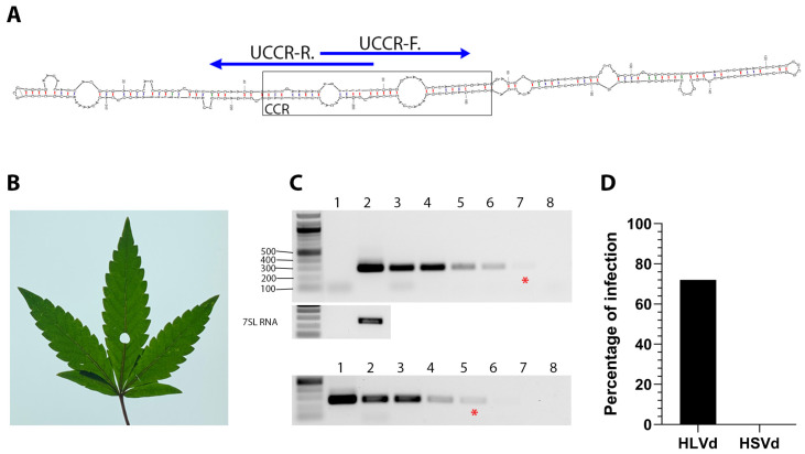
Figure 1.
Diagnostic detection of HLVd in infected hemp plants. (A) The central conserved region (CCR) is the most conserved secondary structure among viroid species. Specific detection primers were developed based on the HLVd upper strand of the CCR (UCCR) and yield 256 bp amplicons. (B) Leaf from HLVd-infected hemp used for total RNA extraction. (C) Ten-fold serial dilutions of RNA extract from HLVd-infected leaves (lanes 2–8, upper panel) and plasmid cDNA copy of HLVd (lanes 1–7, lower panel) were used in RT-PCR and PCR reactions, respectively. The red (*) indicates the lane with the minimal dilution that could be visually detected. Nuclease-free water was used for mock (lane 1 of RNA gel, and lane 8 of DNA gel) treatment. A 1 kb ladder is on the left of each gel and the sizes of several ladder bands are also indicated on the left of the top gel. RT-PCR amplification of the 7SL RNA shown below target band as internal control. (D) Survey of HLVd and hop stunt viroid (HSVd) in 111 hemp samples obtained from a hemp nursery.

