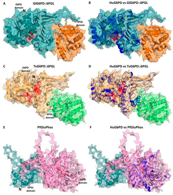
Figure 3.
Structural alignment of the minimized G6PD::6PGL models. Structural alignment of the human G6PD enzyme (PDB entry 2BH9; blue cartoon) and the minimized model of (A) GlG6PD::6PGL from G. lamblia. G6PD domain is shown in blue cartoon; 6PGL domain in orange cartoon (B) Alignment of HuG6PD vs. GlG6PD::6PGL. (C) TvG6PD::6PGL from T. vaginalis. G6PD domain is shown in yellow cartoon; 6PGL domain in green cartoon (D) Alignment of HuG6PD vs. TvG6PD::6PGL. (E) PfGluPhos from P. falciparum. G6PD domain is shown in pink cartoon; 6PGL domain in cyan cartoon (F) Alignment of HuG6PD vs. PfGluPhos.

