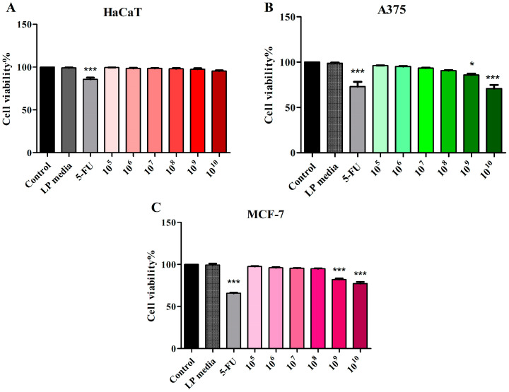
Figure 1.
Cell viability of HaCaT (A), A375 (B), and MCF-7 (C) cells following 48 h treatment with LP 105–1010 CFU/mL and 5-FU (10 μΜ). The results are expressed as viability percentage in comparison with the control group, considered 100% (* p < 0.05 and *** p < 0.001). The data represent the mean values ± SD of three independent experiments performed in triplicate.

