Abstract
This paper reports a case of furuncular myiasis caused by the human bot-fly Dermatobia hominis in a domestic cat from Brazil. A crossbred shorthaired female cat of approximately 3 years old, presented with three boil-like cutaneous lesions at the left cranioventral region of the neck. These were diagnosed as furuncular myiasis. The animal was sedated, and after shaving the fur, bot-fly larvae were removed from the lesion by digital compression. Afterwards, the wounds were treated with 10% iodine solution and also with wound-healing cream containing sulfanilamide, urea and beeswax. Maggots were identified as third-stage larvae of D hominis. Clinical case reports of human bot-fly myiasis in cats are relevant due to its scarce occurrence in feline veterinary practice in some countries.
The human bot-fly Dermatobia hominis (Diptera: Cuterebridae) is distributed throughout neotropical America. Its larvae are obligatory parasites of mammals and its infestation is characterised by primary myiasis. The female fly oviposits on the abdominal region of dipteran vectors that are responsible for carrying the eggs until larval hatching on worm-blooded animals. Larvae penetrate the intact host skin and develop in the subcutaneous tissues within weeks. 1
Because of its low host specificity, there are reports of several domestic animal species naturally parasitized by D hominis in the literature, including cattle, dogs, goats, pigs, horses, cats, rabbits, and even man. 2–4 This fly is considered the major causal agent of myiasis in neotropical America, causing significant economical losses to livestock. 5 Companion animals, especially dogs, have been described as usual hosts in both urban and rural areas. 6,7
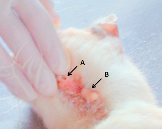
A crossbreed shorthaired female cat, approximately 3 years old, was severely debilitated probably due to feline herpesvirus-1 infection (feline rhinotracheitis virus) and was taken to the laboratory for treatment. This infection is commonly seen in stray animals or multi-cat households, like the one this cat lived in. It also showed mucopurulent discharge typical of the later stages of the disease, caused by a secondary bacterial infection, which aggravated its condition. During physical examination three boil-like cutaneous nodules measuring approximately 1.0 cm of diameter were detected at the left cranioventral region of the neck, resembling furuncular myiasis. The lesions conferred considerable discomfort to the cat and the animal was sedated with ketamine (10 mg/kg intramuscular). After shaving the fur, bot-fly larvae were removed by digital compression (Fig. 1). Subsequently the wounds were treated with 10% iodine solution and wound-healing cream containing sulfanilamide, urea and beeswax. Collected specimens were observed under stereomicroscopy and identified according to a taxonomic key. 1 The animal received support treatment for the respiratory infection and was kept under observation for the following weeks. However, she did not show desirable clinical recovery.
Fig 1.

Cat with boil-like cutaneous lesions (arrows A and B) caused by bot-fly larva in the left cranioventral region of the neck. The bot-fly larvae were removed by digital compression.
Specimens were classified as third-instar D hominis: pyriform larvae with double rows of posteriorly directed dark-coloured spines on its anterior segments and brownish prominent caudal respiratory spiracles. This case along with other published reports, suggest that this specific infestation might be more common in cats from neotropical countries than previously thought. 6,7 This infestation is still considered less frequent in cats than in dogs from the same areas. The cat in this case used to live near a pasture for research cattle. As Dermatobia species larvae periodically parasitize cattle, they are likely to be both the reservoir and the source of infection for this cat and other species. 4
A survey based on a questionnaire reported that this infestation was diagnosed in only eight veterinary clinics out of 227 establishments in Rio de Janeiro Municipality. The majority of cases were caused by a single larva, but in some cases up to three larvae were reported. In general, infested felines were shorthaired crossbreeds. However, the number of cases did not allow us to determine any kind of preference for those animals. Similarly, shorthaired dogs are reportedly to be more affected; shorthair might facilitate the contact of vector flies and the host skin, promoting larval hatching. 7
The feline self-grooming behaviour serves as a basic hygienic purpose. Perhaps, it can promote the removal of recently deposited D hominis larvae, avoiding myiasis' installation. The affected cat was clinically debilitated, which may have compromised this behaviour, possibly facilitating infestation.
Clinical diagnosis can be undertaken by both visualization and palpation of skin nodules that occasionally drains a serous, serosanguineous or seropurulent exudate from a central punctum. 1 The adjacent area is usually edematous and painful.
In temperate countries, the patient's history of residence or travel to the parasites' endemic area can also support diagnosis; imported cases have been reported in Europe and North America. 8
Small animal treatment is based on removal of the larvae and application of wound-healing products to avoid secondary bacterial infections or the installation of secondary myiasis caused by Cochliomyia hominivorax. 9 (In production animals, the treatment is based on chemotherapy.) As preventive measure is recommended to maintain adequate hygienic conditions in the household avoiding anthropozoophilic dipteran species, possible D hominis vectors, and their contact with indoor cats, dogs and people. 7,8
This infestation could be considered less aggressive than other feline myiasis such as C hominivorax. 9–11 It is usually classified as benign, and thus effectively treated once larvae penetrate subcutaneously, in a similar way to infestations by Lucilia eximia, 12 Lucilia sericata, 13,14 Chrysomyia erytrocephala, 15 and Cuterebra species. 16
Infestation by D hominis larvae in tropical countries is of concern due to the high incidence of infestation in various domestic species, including small animals. Clinical case reports of human bot-fly myiasis in cats are relevant because of its infrequent occurrence in feline veterinary practice in different countries. Small animal practitioners should inform their clients about preventive methods and suspicion of infection.
References
- 1.Guimarães J.H., Papavero N., Prado A.P. As miíases da Região Neotropical, Rev Bras Zool 1, 1983, 239–416. [Google Scholar]
- 2.Silva-Júnior V.P., Leandro A., Moya-Borja G.E. Ocorrência do Berne, Dermatobia hominis (Diptera: Calliphoridae) em vários hospedeiros, no Rio de Janeiro, Brasil, Parasitol Dia 22, 1998, 97–101. [Google Scholar]
- 3.Daemon E., Prata M.C.A. Miíase conjuntival por larvas de Dermatobia hominis (Linnaeus Jr, 1781) (Diptera, Cuterebridae) em equino: Descrição de um caso, Rev Bras Med Vet 19, 1997, 81–84. [Google Scholar]
- 4.Verocai G.G., Fernandes J.I., Ribeiro F.A., Melo R.M.P.S., Correia T.R., Scott F.B. Furuncular myiasis caused by the human bot-fly Dermatobia hominis in the domestic rabbit: Case report, J Exot Pet Med 18, 2009, 153–155. [Google Scholar]
- 5.Grisi L., Massard C.L., Moya G.E., Pereira J. Impacto econômico das principais ectoparasitoses em bovinos no Brasil, Hora Vet 21, 2002, 8–10. [Google Scholar]
- 6.Marcial T., Roman E.M., Pivat I.V. Estúdio retrospectivo de doscientos casos de miíasis presentados en el Hospital de Pequeños Animales ‘Dr Daniel Cabello-Mariani’. Facultad de Ciencias Veterinárias, Universidad Central de Venezuela durante los años de 1996 a 1999, Rev Fac Cien Vet UCV 44, 2003, 87–95. [Google Scholar]
- 7.Monteiro HHMS, Cramer-Ribeiro BC, Sanavria A, Souza FS, Rocco FS. Myiasis by Dermatobia hominis in cats (Felis catus) of the northern, southern, western and central zones of Rio de Janeiro City. Anais do XII Congresso Brasileiro de Parasitologia Veterinária; 2002; Rio de Janeiro, Brazil.
- 8.Bowman D.D., Hendrix C.M., Lindsay D.S., Barr S.C. Feline clinical parasitology, 1st edn, 2000, Iowa State University Press/Blackwell Science: Ames. [Google Scholar]
- 9.Mendes-de-Almeida F., Labarthe N., Guerrero J., et al. Cochliomyia hominivorax myiasis in a colony of stray cats (Felis catus Linnaeus, 1758) in Rio de Janeiro, RJ, Vet Parasitol 146, 2007, 376–378. [DOI] [PubMed] [Google Scholar]
- 10.Cramer-Ribeiro B.C., Sanavria A., Oliveira M.Q., Souza F.S., Rocco F.S., Cardoso P.G. Inquérito sobre os casos de miíases por Cochliomyia hominivorax em gatos das zonas norte, sul e oeste e do centro do município do Rio de Janeiro no ano 2000, Braz J Vet Res An Sci 39, 2002, 165–170. [Google Scholar]
- 11.Souza C.P., Verocai G.G., Ramadinha R.H.R. Myiasis caused by the New World screwworm fly Cochliomyia hominivorax (Diptera: Calliphoridae) in cats from Brazil: report of five cases, J Feline Med Surg 12, 2010, 165–170. [DOI] [PMC free article] [PubMed] [Google Scholar]
- 12.Madeira N.G., Silveira G.A.R., Pavan C. The occurrence of primary myiasis caused by Phaenicia eximia (Diptera: Calliphoridae), Mem Inst Oswaldo Cruz 84, 1989, 341. [Google Scholar]
- 13.Marluis J.C., Schnack J.A., Cervinazzo I., Quintana C. Cochliomyia hominivorax (Coquerel, 1858) and Phaenicia sericata (Meigen, 1826) parasiting domestic animals in Buenos Aires and vicinities (Diptera, Calliphoridae), Mem Inst Oswaldo Cruz 89, 1994, 139. [DOI] [PubMed] [Google Scholar]
- 14.Vignau M.L., Arias D.O. Myiasis cutaneo-ulcerosas en pequeños animales, Parasitol Dia 21, 1997, 36–39. [Google Scholar]
- 15.Rodriguez J.M., Perez M. Cutaneous myiasis in three obese cats, Vet Q 18, 1996, 102–103. [DOI] [PubMed] [Google Scholar]
- 16.Slansky F. Feline cuterebriasis caused by lagomorph-infesting Cuterebra spp larva, J Parasitol 93, 2007, 959–961. [DOI] [PubMed] [Google Scholar]


