

Abstract
Practical relevance In cats, three species of demodex mites have been identified as causes of demodicosis, which may manifest as pruritus, miliary dermatitis and/or self-induced alopecia. The condition has been recognized in various countries but does seem to show regional preferences.
Clinical challenges Diagnosis of feline demodicosis can be a challenge as mites are not always readily found within scrapings of the skin examined microscopically. One or more species of demodex mite may be involved in an infestation. Furthermore, the condition can be difficult to treat effectively.
Audience This review is intended as a clinical update for veterinary surgeons in practice who rarely encounter demodicosis.
Feline demodex mites
Feline demodicosis is a parasitic condition affecting cats’ skin. While it is considered a relatively rare parasitism overall, certain areas of the world appear to have an increased incidence of the disorder; it is not uncommon in the Gulf Coast States of the USA, for example. A wide variety of clinical signs are associated with feline demodicosis, and they seem to differ according to the individual species of mite involved. Pruritus may or may not be a feature. Concurrent infestation with different species of mites has also been reported.1–3
Three species of feline demodex mites have been described: Demodex gatoi, Demodex cati and an, as yet unnamed, species.
Demodex gatoi
The mite
D gatoi (Figure 1) appears to be the most common of the three feline demodex mites. It is a short (mean body length 91 µm [male], 108 µm [female]), 3 broad mite that inhabits the superficial skin (stratum corneum).
Figure 1.
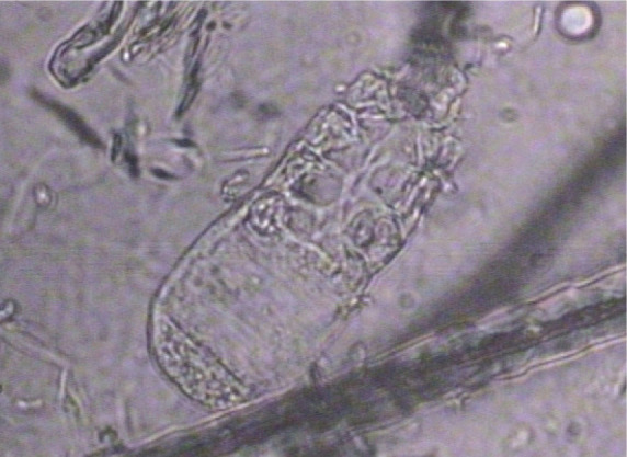
Demodex gatoi mite. Magnification x 100
Clinical signs of infestation
The primary clinical sign associated with infestation is pruritus, which usually manifests as overgrooming. Affected cats rarely have dermatitis; they usually present with self-induced alopecia. Any area can be affected, although the ventral abdomen, inner thighs, flanks and forelimbs appear to be most commonly involved (Figure 2). This is not surprising as these are the easiest areas for the cat to reach and overgroom. Occasionally, cats will present with miliary dermatitis or indolent lip ulcers (possibly associated with overgrooming). 4
Figure 2.

Two-year-old female neutered domestic shorthair cat with broken, barbered hair dorsally and an alopecic ventrum associated with D gatoi infestation
Pathogenesis
D gatoi is quite different from the other species of demodex mites recognized in cats (and dogs). Because this mite dwells superficially, it can be easily removed with overgrooming. Very few mites may be present, yet the affected cat continues to be quite pruritic. This is similar to canine scabies and suggests that a hypersensitivity to the mite may develop in some cases. Further support for this lies in the fact that some cats may have abundant mites present on the skin, yet are not pruritic.
D gatoi infestation is unique in that the mite is transmissible to other cats via casual contact. This has been well documented, and does not appear to be a feature of other Demodex species infestations in cats (and dogs).5–7 In contrast to D cati infestation, this demodicosis is not felt to be associated with underlying immunocompromise in most cases. Although many confirmed cases have a history of previous glucocorticoid administration, it is suspected that in most of these cats the glucocorticoids were administered as a result of the associated pruritus, and were not the cause of the infestation.
Diagnosis
The diagnosis of D gatoi infestation can be difficult as skin scrapings often fail to yield positive results in cats that ultimately respond to treatment. Broad superficial scrapings should be performed, with mineral oil applied to the blade or skin. Multiple areas should be scraped, including non-alopecic areas that the cat would have difficulty reaching. These sites may prove the most productive. Cellophane tape may also be used; it is pressed to the surface of the skin and then examined on a glass slide under a microscope.

When evaluating skin scrapings it is important always to use a cover slip as this helps to keep the mites in the same plane of focus. Lowering the condenser may also be helpful. Scan the slide at x 10 magnification looking for mites and/or eggs. The finding of even a single D gatoi mite is significant.
Other cats in the household should undergo skin scraping as well. Some cats may be unaffected clinically and appear normal, yet mites may be found on scrapings. Mites may also be found on fecal examination due to ingestion associated with overgrooming. Ultimately, the final diagnostic test for D gatoi is response to treatment, much like canine scabies.
Treatment
The treatment of choice for D gatoi infestation is a series of 2% lime sulfur dips (see box on page 211). Unfortunately, this product may not be available in some parts of Europe, including the UK. Oral ivermectin, 0.2–0.3 mg/kg q24h or q48h, has been effective in some cases (see Table 1), but failures have also been reported. Topical selamectin administered on a weekly basis is ineffective. 8 There are anecdotal reports (V Fabok, personal communication) of success with topical moxidectin (Advantage Multi; Bayer Animal Health), but others have found this to be ineffective. 6
Table 1.
Comparison of treatment options for feline demodicosis
| 2% lime sulfur dips | Ivermectin 0.2–0.3 mg/kg PO q24–48h* | Amitraz dips 0.0125–0.025% weekly | Doramectin | Topical moxidectin† | |
|---|---|---|---|---|---|
| D gatoi | Most reliable treatment | Occasional failures, but generally effective. Continue for 2 weeks beyond clinical cure 4 | Reliable treatment. Routine use not recommended given other alternatives and risk of toxicosis | Anecdotal reports of success, but also reports of failures. Not reliable | |
| D cati | Occasional failures, especially in hard to treat (eg, periocular) areas | Effective. Continue until negative scrapings are obtained | Reliable treatment. Routine use is not recommended given other alternatives and risk of toxicosis | 600 ?g/kg SC weekly for 2–3 injections resolved the condition in three cats 14 | |
| Unnamed feline demodex mite | One report of success in six cats in combination with ivermectin | Unsuccessful as a single treatment in one report. Successful in combination with lime sulfur dips | Unsuccessful |
Because use of this medication is off-label, and there is potential for neurotoxicity as well as toxicity associated with propylene glycol (Heinz body anemia), lime sulfur dips are preferred 4
Advantage Multi (Bayer Animal Health)
Environmental treatment is not necessary.

Figure 3.

Cat with D gatoi infestation undergoing a lime sulfur dip
Figure 4.

The same cat as in Figure 2 following a series of six weekly lime sulfur dips
Demodex cati
The mite
D cati is a long (mean body length 182 µm [male], 291 µm [female]), 3 slender mite with a similar morphology to Demodex canis. Like D canis, D cati resides in the hair follicles and sebaceous glands and ducts. This mite provokes inflammation in the hair follicle.
Clinical signs of infestation
Clinical signs may be quite variable and include alopecia, comedones, seborrhea, scale, papules, crusts, miliary dermatitis, erosions and ulceration (Figure 5). The cat may or may not be pruritic. The disease may be generalized in distribution or localized; when localized, it tends to involve the face. D cati may also be the cause of a ceruminous otitis externa; the otodemodicosis may be associated with skin disease, or may occur by itself.
Figure 5.

Furunculosis of the chin of a Siamese cross cat associated with D cati infestation
Depending on the clinical signs present, differential diagnoses may include dermatophytosis, pemphigus foliaceus, bacterial folliculitis, Otodectes otitis/dermatitis, Notoedres cati infestation, drug eruptions, allergic dermatitis or cutaneous lymphoma.
Diagnosis
The diagnosis of D cati infestation is based on history, physical examination findings and demonstration of the oval-shaped mite on microscopic examination of skin scrapings. Given that the mites reside within the hair follicles, scrapings should be deep enough to cause capillary bleeding. Squeezing the skin between your fingers first may be beneficial. Evaluation of trichograms may be useful in areas that are difficult to scrape as mites may be found attached to the hairs. The demonstration of mite eggs (which can be quite abundant) is also diagnostic. On histopathology, mites may be observed within hair follicles. Biopsies may be necessary in ulcerated or very inflamed areas that are difficult to scrape.
Pathogenesis
The pathogenesis of this disorder is unknown. The mite is considered to be part of the naturally occurring microfauna of feline skin. Mite reproduction to the point of causing dermatitis may be associated with an underlying systemic disease.2,4,9–11 However, some patients with D cati infestation have no apparent underlying disease or history of predisposing drug use (much like D canis infestation in the dog).4,11 Patients should be screened for retroviral infections, and concurrent systemic or infectious disease. In the case of localized, very inflamed lesions, biopsy is recommended as D cati infection has been reported to occur in conjunction with squamous cell carcinoma in situ. 12
Treatment
Treatment comprises miticidal therapy. The traditional approach has been whole body dips in 2% lime sulfur. Oral ivermectin, 0.2–0.3 mg/kg q24h or q48h, has also been used successfully in some cases (Table 1); note there is some potential for neurotoxicity with this medication. Both of these treatments may occasionally fail, in which case the use of amitraz dips may be considered. The recommended concentration is 0.0125% amitraz applied weekly. This is half the manufacturer’s recommended dose for dogs, at more frequent intervals. 13 Amitraz should only be used as the last treatment option, as there is potential for toxicity.
Environmental treatment is not necessary.

Case notes
A 5-year-old female spayed domestic shorthair cat residing indoors presented for evaluation of self-induced alopecia that had progressively worsened over a 6-month period. The owner reported that a kitten had been introduced into the household 8 months earlier. The adult cat had no prior history of skin disease and the kitten appeared normal.

History Previous diagnostics in this cat had included skin scrapings with microscopic examination (which did not produce positive findings), and a dermatophyte culture with negative findings. Prior treatments included repositol glucocorticoids (methylprednisolone acetate), which had produced a brief period of improvement, and clomipramine, diazepam and fluoxetine (for presumed behavioral stress-induced alopecia due to the new cat), with no improvement. At the time of examination the cat was receiving no medications other than monthly flea and heartworm control (selamectin).
Physical examination The cat was in good body condition. There was no dermatitis evident, but she had complete alopecia of the ventral abdomen and inner thighs. Alopecia extended onto the flank region and lateral thorax as well as the upper forelegs.
Differential diagnoses The differentials list comprised:
Allergic skin disease (flea allergy, atopic dermatitis, food allergy/intolerance)
Ectoparasites (D gatoi)
Behavioral self-induced alopecia
The fact that the problem began after the introduction of a kitten into the household made either ectoparasites or behavioral alopecia most likely. The latter is a diagnosis of exclusion, in the author’s opinion, and should only be considered once pruritic causes of hair removal have been ruled out.

Multiple D gatoi mites found on superficial scrapings from the asymptomatic housemate of the cat
Diagnostic tests Multiple skin scrapings were performed on the adult cat. Findings were negative. Fecal flotation did not reveal any demodex eggs. Skin scrapings were performed on the kitten and several D gatoi mites were found at each site scraped.
Diagnosis and treatment plan The presumptive diagnosis in this cat was D gatoi infestation, and treatment with a series of six lime sulfur dips, performed at weekly intervals, was commenced. This involved placing both cats in a litter pan filled with 2% lime sulfur solution and continuously sponging the solution over them for a 5-min period. The cats were then placed in an Elizabethan collar to prevent them from ingesting and removing the dip.
Clinical outcome The clinically affected cat had improved hair growth and was showing reduced grooming prior to the third dip. All clinical signs had resolved by the sixth dip. There was no recurrence of signs at a 2-year follow-up.
Funding
The author received no specific grant from any funding agency in the public, commercial or not-for-profit sectors for the preparation of this review article.
Conflict of interest
The author declares that there is no conflict of interest.
References
- 1. Kwochka KW. Demodicosis. In: Griffin CE, Kwochka KW, MacDonald JM. (eds). Current veterinary dermatology. St Louis: Mosby Year Book, 1993, pp 72–84. [Google Scholar]
- 2. Neel JA, Tarigo J, Tater KC, Grindem CB. Deep and superficial skin scrapings from a feline immunodeficiency virus-positive cat. Vet Clin Pathol 2007; 36: 101–104. [DOI] [PubMed] [Google Scholar]
- 3. Lowenstein C, Beck W, Bessmann K, Mueller RS. Feline demodicosis caused by concurrent infestation with Demodex cati and an unnamed species of mite. Vet Rec 2005; 157: 290–292. [DOI] [PubMed] [Google Scholar]
- 4. Morris DO, Beale KM. Demodicosis. In: August JR. (ed). Consultations in feline internal medicine. Vol 5. St Louis, MO: Elsevier Saunders, 2006, pp 247–250. [Google Scholar]
- 5. Saari SA, Juuti KH, Palojarvi JH, Vaisanen KM, Rajaneimi RL, Saifonmaa-Koulumies LE. Demodex gatoi-associated contagious pruritic dermatosis in cats a report from six households in Finland. Acta Vet Scand 2009; 51: 40–49. [DOI] [PMC free article] [PubMed] [Google Scholar]
- 6. Morris DO. Contagious demodicosis in three cats residing in a common household. J Am Anim Hosp Assoc 1996: 32: 350–352. [DOI] [PubMed] [Google Scholar]
- 7. Beale KM. Contagious and occult demodicosis in a family of two cats. Proceedings of the 14th AAVD/ACVD meeting; 1998; San Antonio, p 99. [Google Scholar]
- 8. Beale KM, Rustemeyer-May E. Selamectin in the treatment of feline demodex [abstract]. Vet Derm 2001; 12: 237. [Google Scholar]
- 9. White SD, Carpenter JL, Moore FM, Ogilvie G. Generalized demodicosis associated with diabetes mellitus in two cats. J Am Vet Med Assoc 1987; 191: 448–450. [PubMed] [Google Scholar]
- 10. Chalmers S, Schick RO, Jeffers J. Demodicosis in two cats seropositive for feline immunodeficiency virus. J Am Vet Med Assoc 1989; 194: 256–257. [PubMed] [Google Scholar]
- 11. Morris DO, Beale KM. Feline demodicosis: a retrospective study of 15 cases. Proceedings of the 13th AAVD/ACVD meeting; 1997; Nashville, USA, pp 127–128. [Google Scholar]
- 12. Guaguere E, Olivry T, Delverdier-Poujade E. Demodex cati infestation in association with feline cutaneous squamous cell carcinoma in situ: a report of 5 cases. Vet Dermatol 1999; 10: 61–67. [DOI] [PubMed] [Google Scholar]
- 13. Cowan LA, Campbell K. Generalized demodicosis in a cat responsive to amitraz. J Am Vet Med Assoc 1988; 10: 1442–1444. [PubMed] [Google Scholar]
- 14. Johnstone IP. Doramectin as a treatment for canine and feline demodicosis. Aus Vet Pract 2002; 32: 98–103. [Google Scholar]
- 15. Chesney CJ. An unusual species of demodex mite in a cat. Vet Rec 1988; 123: 671–673. [PubMed] [Google Scholar]
- 16. Lowenstein C, Beck W, Bessman K, Mueller RS. Feline demodicosis caused by concurrent infestation with Demodex cati and an unnamed species of mite. Vet Rec 2005; 157: 290–292. [DOI] [PubMed] [Google Scholar]
- 17. Newbury S, Moriello KA, Steinburg H. An outbreak of Demodex gatoi and an unnamed demodex mite in an open admission animal shelter. Proceedings of the 21st North American Veterinary Dermatology Forum; 2006; Palm Springs, p 188. [Google Scholar]