Abstract
Peritoneopericardial diaphragmatic hernia is a common incidental finding in cats and is rarely symptomatic. The case report described herein presented with dyspnoea secondary to incarceration of hepatic cysts within the pericardial space of a cat with a peritoneopericardial diaphragmatic hernia.
Case report
An 8-year-old, castrated Himalayan cat presented with a 5-week history of respiratory difficulty and occasional coughing. There was no loss of appetite or weight during this period. The cat lived predominantly indoors and had been vaccinated against feline herpesvirus, calicivirus and parvovirus. Physical examination revealed mild tachypnoea (50 breaths per min) and increased respiratory effort, particularly during expiration. Breath sounds were decreased ventrally on both sides of the thoracic cavity and heart sounds were barely audible over the right hemithorax. Jugular pulses were prominent bilaterally.
Blood tests, chest radiography and thoracic ultrasonography were performed. Haematology and biochemistry results were normal.
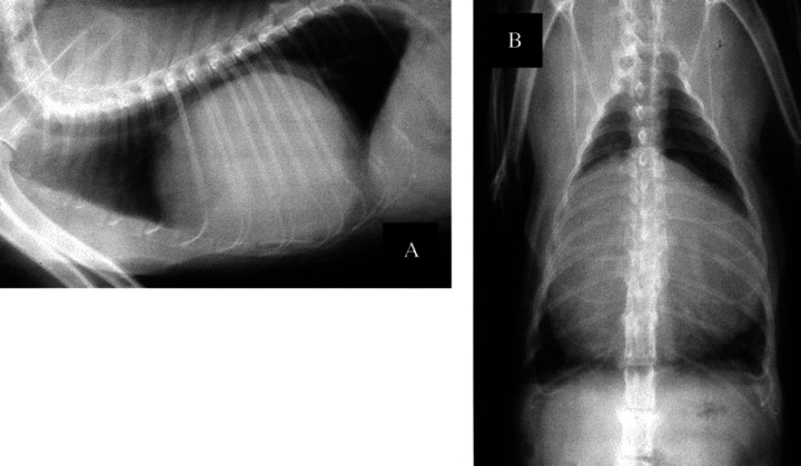
Thoracic radiography [Figs 1(A, B)] revealed generalised cardiomegaly with increased sternal and diaphragmatic contact and caudal displacement of the diaphragm. In the lateral projection, the cardiac silhouette is globular and the trachea is displaced dorsally. In the ventrodorsal projection, the cardiac silhouette is asymmetrical with a spherical bulge on the left side and greater cardiac mass evident on the right.
Fig 1.
Lateral (A) and ventrodorsal (B) thoracic radiographs.
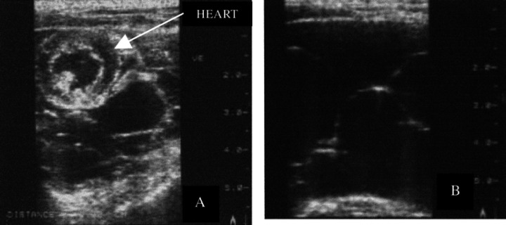
Ultrasonographic examination of the heart and pericardium revealed a loculated hypoechoic mass (2 cm diameter following ultrasound-guided aspiration) is present in the far field and extends to the right thoracic wall [Fig. 2(A)]. In the magnified image [Fig. 2(B)], the intrapericardial mass is characterised by a web of hyperechoic septae separating hypoechoic fluid. Echocardiographic examination was unremarkable. Cytological examination of the aspirate from the cystic pericardial lesion was consistent with a transudate (total protein <10 g/l; nucleated cells 48.9×10⁶/l).
Fig 2.
(A) Ultrasonographic image obtained from the left thoracic wall at the level of the sixth intercostal space. (B) A magnified image of the loculated mass obtained from a right intercostal window.
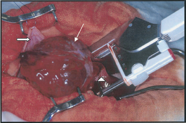
A cranial ventral midline celiotomy-caudal median sternotomy was performed. A cystic mass, approximately 8 cm diameter, was present in the caudal aspect of the pericardium and attached to a segment of the right lateral liver lobe incarcerated in a small peritoneopericardial diaphragmantic hernia (PPDH). A radial incision of the central diaphragm was performed to expose and mobilise the liver lobe and hepatic cyst. The cyst was removed with an automatic stapling device (TA-30V, Tyco Healthcare, Mansfield, MA) (Fig. 3). A chest tube was inserted into the right hemithorax (and removed 6 h post-operatively). The PPDH and diaphragmatic incision were closed with 4–0 polydioxinone (PDS II, Ethicon, Johnson & Johnson, Somerville, NJ) in a simple continuous pattern. The sternotomy-celiotomy was closed routinely.
Fig 3.
The hepatic cyst is being removed with an automatic stapling device (TA-30V, Tyco Healthcare).
The cat recovered uneventfully with resolution of respiratory distress and coughing. Histological examination of the pericardial mass was consistent with a hepatic cyst. The cat remains asymptomatic 6 months postoperatively with no ultrasonographic evidence of recurrence of either the PPDH or intrapericardial cyst.
Discussion
Peritoneopericardial hernias are the most common congenital defect of the diaphragm (Wallace et al 1992, Less et al 2000). The embryological development of the diaphragm is complex and involves fusion of the ventral septum transversum, central caudal mediastinum and dorsolateral pleuroperitoneal membranes (Noden & de Lahunta 1985). The embryogenesis of PPDH is unknown but may include malformation or trauma of the septum transversum and pleuroperitoneal folds (Evans & Biery 1980, Noden & de Lahunta 1985, Wallace et al 1992, Less et al 2000).
Peritoneopericardial diaphragmatic hernia is usually an incidental finding although it may cause gastrointestinal or respiratory signs (Evans & Biery 1980, Wallace et al 1992, Less et al 2000). Respiratory difficulty and coughing in the present case were probably caused by reduced lung capacity and tracheal compression secondary to pericardiomegaly. Umbilical and cranioventral abdominal hernias and sternal defects are commonly associated with PPDH in dogs (Bellah et al 1989, Sisson et al 1993, Simpson et al 1999), however, sternal defects are the only reported concurrent problem in cats (Evans & Biery 1980, Less et al 2000).
The diagnosis of PPDH depends on radiographic, ultrasonographic and surgical findings. Radiographic features typical of PPDH include enlargement of the cardiac silhouette with dorsal elevation of the trachea, overlapping of the diaphragm and caudal aspect of the cardiac silhouette, differential opacities or bowel within the cardiac silhouette, presence of a dorsal peritoneopericardial mesothelial remnant extending from the caudal pericardium to the diaphragm, and absence of radiographic signs associated with cardiac failure (Evans & Biery 1980, Berry et al 1990). In the present case, ultrasound was invaluable for detecting a multiloculated intrapericardial cystic structure and excluding cardiac disease and pericardial effusion as the cause of radiographic cardiomegaly-pericardiomegaly.
An intrapericardial hepatic cyst incarcerated in a small PPDH was confirmed at surgery. Intrapericardial cysts have been reported in two cats: one was an incidental finding (Busalacchi 1993) while the other, similar to the present case, was associated with a PPDH and caused cardiac tamponade and respiratory distress (Less et al 2000). Intrapericardial hepatic cysts probably develop as a result of vascular and lymphatic congestion and fluid retention secondary to constriction of the incarcerated liver lobe (Less et al 2000).
Herniorrhaphy, either through an abdominal or combined abdominal-thoracic approach, is recommended for definitive treatment of PPDH. An abdominal approach, involving closure of the PPDH without entering the pleural space or separating the pericardium from the diaphragm, has been described (Wallace et al 1992). However, a combined median sternotomy-celiotomy was performed in the present case to aid visualisation and resection of the intrapericardial cyst. The prognosis following surgical closure of a PPDH is generally excellent although, in one report, respiratory problems continued in two cats and the PPDH recurred in one cat (Wallace et al 1992). The respiratory problems resolved in the present case and there was no evidence of recurrence 6 months following surgery.
References
- Bellah JR, Whitton DL, Ellison GW, Phillips L. (1989) Surgical correction of concomitant cranioventral abdominal wall, caudal sternal, diaphragmatic, and pericardial defects in young dogs. Journal of the American Veterinary Medical Association 195, 1722–1726. [PubMed] [Google Scholar]
- Berry CR, Koblik PD, Ticer JW. (1990) Dorsal peritoneopericardial mesothelial remnant as an aid to the diagnosis of feline congenital peritoneopericardial diaphragmatic hernia. Veterinary Radiology 31, 239–245. [Google Scholar]
- Busalacchi A. (1993) Hyperthyroidism and a pericardial cyst in a cat. Compendium on the Continuing Education for the Practicing Veterinarian 15, 1250–1254. [Google Scholar]
- Evans SM, Biery DN. (1980) Congenital peritoneopericardial diaphragmatic hernia in the dog and cat: A literature review and 17 additional case histories. Veterinary Radiology 21, 108–116. [Google Scholar]
- Less RD, Bright JM, Orton EC. (2000) Intrapericardial cyst causing cardiac tamponade in a cat. Journal of the American Animal Hospital Association 36, 115–119. [DOI] [PubMed] [Google Scholar]
- Noden DM, de Lahunta A. (1985) Respiratory system and partitioning of body cavities. In: The Embryology of Domestic Animals: Developmental Mechanisms and Malformations, Noden DM, de Lahunta A. (eds). Williams & Wilkins, Baltimore, pp. 279–291. [Google Scholar]
- Simpson DJ, Hunt GB, Church DB, Beck JA. (1999) Benign masses in the pericardium of two dogs. Australian Veterinary Journal 77, 225–229. [DOI] [PubMed] [Google Scholar]
- Sisson D, Thomas WP, Reed J, Atkins CE, Gelberg HB. (1993) Intrapericardial cysts in the dog. Journal of Veterinary Internal Medicine 7, 364–369. [DOI] [PubMed] [Google Scholar]
- Wallace J, Mullen HS, Lesser MB. (1992) A technique for surgical correction of peritoneal pericardial diaphragmatic hernia in dogs and cats. Journal of the American Animal Hospital Association 28, 503–510. [Google Scholar]


