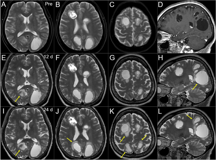
Figure 5. Magnetic resonance images during the 15-fr radiosurgery and subsequent WBRT.
The images show T2-WIs (A-C, E-L); a CE T1-WI (sagittal T2-WIs before WBRT were unavailable) (D); axial images (A-C, E-G, I-K); sagittal images (D, H, L); three days before the initiation of WBRT (pre) (A-D); 12 days (d) after the WBRT initiation (at the 8 fr of WBRT) (E-H); and 24 days after the WBRT initiation (at the completion of SRS boost) (I-L).
(A-H) These images are shown at the same magnification and coordinates under co-registration and fusion based on the pre-SRS images. (E-H) There was no obvious change in the tumor configuration of most lesions. The largest lesion was slightly enlarged (arrow in H). The perilesional edema aggravated in some lesions (arrow in E). (I-L) There is still no obvious change in the tumor configuration of almost all the lesions. The expansion of the largest lesion has not progressed. The perilesional edemas appear or aggravate in multiple lesions (arrows in I-L).
WIs: weighted images; CE: contrast-enhanced; WBRT: whole-brain radiotherapy; SRS: stereotactic radiosurgery; fr: fraction

