Abstract
We developed and applied transposon-based transformation vectors for molecular manipulation and analysis of spotted fever group rickettsiae, which are obligate intracellular bacteria that infect ticks and, in some cases, mammals. Using the Epicentre EZ::TN transposon system, we designed transposons for simultaneous expression of a reporter gene and a chloramphenicol acetyltransferase (CAT) resistance marker. Transposomes (transposon-transposase complexes) were electroporated into Rickettsia monacensis, a rickettsial symbiont isolated from the tick Ixodes ricinus. Each transposon contained an expression cassette consisting of the rickettsial ompA promoter and a green fluorescent protein (GFP) reporter gene (GFPuv) or the ompB promoter and a red fluorescent protein reporter gene (DsRed2), followed by the ompA transcription terminator and a second ompA promoter CAT gene cassette. Selection with chloramphenicol gave rise to rickettsial populations with chromosomally integrated single-copy transposons as determined by PCR, Southern blotting, and sequence analysis. Reverse transcription-PCR and Northern blots demonstrated transcription of all three genes. GFPuv transformant rickettsiae exhibited strong fluorescence in individual cells, but DsRed2 transformants did not. Western blots confirmed expression of GFPuv in R. monacensis and in Escherichia coli, but DsRed2 was expressed only in E. coli. The DsRed2 gene, but not the GFPuv gene, contains many GC-rich amino acid codons that are rare in the preferred codon suite of rickettsiae, possibly explaining the failure to express DsRed2 protein in R. monacensis. We demonstrated that our vectors provide a means to study rickettsia-host cell interactions by visualizing GFPuv-fluorescent R. monacensis associated with actin tails in tick host cells.
Rickettsiae occupy an obligate intracellular niche in eukaryotic hosts and live free in the cytoplasm, in contrast to other genera of obligate intracellular pathogenic bacteria (e.g., Anaplasma, Chlamydia, Coxiella, and Ehrlichia), which are restricted to intracytoplasmic vesicles (63). Rickettsiae are adapted to arthropods, in which they are often maintained by transtadial and transovarial transmission (5), but some are transmitted to vertebrates by blood-feeding arthropods. Pathogenic rickettsial infections in humans include endemic and epidemic typhus and Rocky Mountain spotted fever, which are caused, respectively, by Rickettsia typhi, Rickettsia prowazekii, and Rickettsia rickettsii. Typhus rickettsiae and spotted fever group rickettsiae (SFGR) occur worldwide and are important emerging or reemerging pathogens (5, 51).
In addition, ticks harbor symbiotic rickettsiae that have little or no pathogenicity in laboratory animals (6, 9) but play a role in tick physiology and affect the population dynamics and transmission of pathogenic rickettsiae (10, 11). We isolated a symbiont, Rickettsia monacensis, from Ixodes ricinus ticks collected in an urban locale in which rickettsiae have never been associated with a spotted fever outbreak, and we confirmed its nonpathogenic character by inoculation of hamsters, which developed high immunoglobulin G (IgG) antibody titers but no evidence of illness (58). The recently cultured R. monacensis, Rickettsia peacockii (57), and other symbiotic SFGR from ticks (61) offer an opportunity to study rickettsiae without the hazards of working with pathogenic rickettsiae that require biosafety level 3 facilities. We have begun to enhance the potential of symbionts as models of rickettsial host interactions through transformation with reporter genes and a selectable marker.
Selection of transformant rickettsiae through use of a robust drug-selectable marker is complicated by the limited number of effective antibiotics (54) and the fact that the obvious candidates, tetracyclines, are the drugs of choice in treatment of rickettsiosis. Until recently, patients for whom tetracyclines were considered contraindicated, primarily children and pregnant women, were treated with chloramphenicol (CHL), despite serious potential side effects, including aplastic anemia, leukemia, and sudden death, as well as a much-increased risk of death from the rickettsiosis due to treatment failure (15, 32, 59). The Centers for Disease Control and Prevention and the American Academy of Pediatrics now recommend doxycycline as the drug of choice in treatment of all patients, including pregnant women and children regardless of age (21, 26, 42, 45). Effective alternatives in some circumstances include rifampin with erythromycin or josamycin alone (7, 12, 50).
Versatile rickettsial transformation vectors allowing facile insertion and expression of genes under regulation of effective promoters and containing a robust drug-selectable marker have not been available. In this report, we describe transformation of R. monacensis with EZ::TN transposon vectors designed to express a chloramphenicol acetyltransferase (CAT) resistance gene and a green fluorescent protein (GFP) reporter gene (GFPuv) or a red fluorescent protein reporter gene (DsRed2) under regulation of the R. rickettsii outer membrane protein ompA and ompB gene promoters (43). Transformant populations containing chromosomally integrated transposons were obtained by CAT selection and were stable over long-term passage after removal of selection. GFPuv transformants exhibited strong epifluorescence that was easily visualized by microscopy, but DsRed2 transformants did not. Western blots showed that R. monacensis did not express DsRed2 protein, while Escherichia coli transformed with the same vector did. The GFPuv gene closely matched the preferred AT-rich codon suite of Rickettsia, but the DsRed2 gene codon suite was highly biased towards GC-rich codons that occur rarely in Rickettsia. To demonstrate the utility of transformant rickettsiae in investigation of rickettsia-host cell interactions, we showed association of GFPuv-fluorescent R. monacensis with host cell actin tail structures.
MATERIALS AND METHODS
Growth and preparation of rickettsiae.
R. monacensis, a SFGR isolated from I. ricinus ticks (58), was grown in the Ixodes scapularis ISE6 cell line or, to demonstrate actin tail formation, in the IDE8 cell line (35). Tick cell cultures were maintained in L-15B300 medium as described previously (37). To prepare purified rickettsiae, infected cells were recovered by centrifugation at 2,500 × g and 4°C for 10 min, resuspended in 4 ml of cold SPG buffer, and lysed by vortexing with 3-mm-diameter glass beads (47). Lysates were centrifuged as described above, and rickettsiae in the supernatant not destined for electroporation were collected by centrifugation at 18,400 × g and 4°C for 5 min. Supernatants with R. monacensis destined for electroporation were layered on Percoll (Sigma, St. Louis, Mo.) cushions prepared by mixing 2.1 ml of SPG buffer with 0.9 ml of 90% Percoll-10% CMRL medium (Life Technologies, Rockville, Md.) and centrifuged at 3,000 × g and 4°C for 30 min. Rickettsial pellets were resuspended twice in 6 ml of 250 mM sucrose and repelleted at 18,400 × g and 4°C for 5 min. Washed rickettsiae were resuspended in 250 mM sucrose, an aliquot was mixed with live-dead stain (BacLight bacterial viability kit; Molecular Probes, Eugene, Oreg.), and rickettsiae were counted with a Petroff-Hauser chamber by using epifluorescence microscopy. Rickettsiae were adjusted to 3 × 10⁶ cells/μl.
Plaque purification of rickettsiae.
Serial dilutions of purified rickettsiae in SPG buffer were inoculated onto 75% confluent ISE6 cells in 35-mm-diameter tissue culture plates with medium removed. The plates were rocked for 30 min at room temperature, overlaid with 3 ml of L15B (37°C) mixed with 1 ml of 1% agarose in H2O cooled to 56°C, and incubated in candle jars at 34°C. After 10 days, individual isolated plaques were removed as agar cores in sterile Pasteur pipettes and inoculated onto fresh ISE6 cells in sealed flasks.
Preparation of rickettsial genomic DNA.
Purified rickettsiae were lysed in 300 μl of buffer containing 10 mM Tris-HCl (pH 7.8), 0.5 mM EDTA, 0.5% sodium dodecyl sulfate (SDS), 40 μg of RNase A (Promega, Madison, Wis.) per ml, and 250 μg of proteinase K (Fisher, Pittsburgh, Pa.) per ml at 55°C for 2 to 16 h. Lysates were phenol-chloroform extracted, and DNA was ethanol precipitated and resuspended in nuclease-free H2O (Life Technologies).
Enzymes, buffers, and primers used in nucleic acid procedures.
All enzymes were obtained from Life Technologies unless stated otherwise. Electrophoresis and blot transfer buffers were prepared as described previously (31) unless stated otherwise. All primers were from Invitrogen (Carlsbad, Calif.) or Integrated DNA Technologies (Coralville, Iowa). Primer sequences are shown in Table 1.
TABLE 1.
PCR and sequencing primers
| Primer | Sequence (5′ to 3′)a |
|---|---|
| ompAPromF | AGTCGGATCCGCAACAAGGCCAACACCGC |
| ompAPromRC | CTGAGCCGGCATATGTAAAACCTTAATCAAAATT |
| ompBPromF | AGTCGGATCCGCGGTCAAGCCAC |
| ompBPromRC | CTGAGCCGGCATATGTTTGGTCCTATATTTAAGTTAAAATTT |
| ompBPrommutRC | CTACGGGGTGAGCATACTATATATTG |
| ompATerF | CTGAGCCGGCTAAGCAACCGTTCTATAATCATAAAAAAAGC |
| ompATerRC | AGTCGGATCCTCGCAATGACGCTTCTC |
| CATF | CCCCCATATGGAGAAAAAAATCACTGGATATACC |
| CATRC | AACCGCTTAGCCCGCCCCGCCCTGCCACTCATC |
| DsRed2F | CCCCCATATGGCCTCCTCCGAGAACGTC |
| DsRed2RC | AACCGCTTAGCCCAGGAACAGGTGGTGGCGG |
| GFPuvF | GTCGACTCTAGAGGATCCCCGGGTACCGGTAGAACATATGAGTAAAGGAG |
| GFPuvRC | GGGGTCTAGACGCTTAGCCTTATTTGTAGAGCTCATCCATGCCATG |
| Rm658R1 | CTAACCAACTGAGCTAATCACCC |
| InvPCR658#1 | GAGCCAATATGCGAGAACACCCGAGAA |
| InvPCR658#4 | GTAGTTTTGAGAAGCGTCATTGCG |
| InvPCR658#7 | TTAAGCATTCTGCCGACATGGAAGC |
| InvPCR658#8 | CGGTTGCTTATTAGCCCGCCC |
| InvPCR601#2 | GCGTTATGCCTTATCAGTGTCATCC |
| InvPCR601#3 | ACAATATATAGTATGCTCAGCCCG |
Single underlining indicates BamHI (GGATCC), BpuI 102I (GCTAAGC), and NdeI (CATATG) restriction enzyme sites. Double underlining indicates BpuI 102I (GCTTAGC) and NgoAIV (GCCGGC) sites. Boldface indicates ribosome binding site consensus sequences.
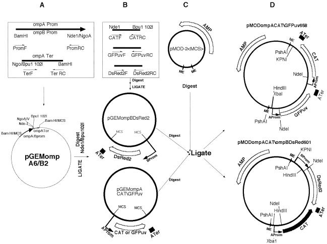
Cloning of the ompA promoter and transcription terminator and ompB promoter (Fig. 1A).
FIG. 1.
Construction of rickettsial transposon vectors. (A) PCR amplification of R. rickettsii Hlp#2 ompA and ompB promoters with forward (PromF) and reverse complement (PromRC) primers and of the ompA transcription terminator with TerF and TerRC primers, all containing end-terminal restriction endonuclease sites (Table 1). Amplicons were digested with BamHI and NgoAIV and ligated into the pGem3Z BamHI site within the MCS to obtain pGEMompA6 and -ompB2. (B) PCR amplification of CAT, GFPuv, and DsRed2 gene open reading frames with forward (F) NdeI and reverse complement (RC) BpuI 102I end-terminal primers. Amplicons and pGEMompA6 and -B2 plasmids were digested with the same enzyme pair and ligated to produce pGEMompACAT or -GFPuv and pGEMompBDsRed2. (C) The Epicentre pMOD-2-<MCS> transposon vector was cleaved at restriction sites within the MCS and ligated with two rickettsial gene expression cassettes (ompACAT and ompAGPPuv or ompBDsRed2) removed (see text) from the plasmids shown in panel B. (D) Final vectors, pMODompACAT\GFPuv658 (pMOD658) and pMODompACAT\ompBDsRed601 (pMOD601), showing relative positions of 19-nucleotide mosaic element (ME) sites recognized by EZ::TN transposase, ompA and -B promoters (Aprom and Bprom), the transcription terminators (ATer), and the CAT, Discoma red fluorescent protein (DsRed2), green fluorescent protein (GFPuv), and ampicillin resistance (AMP) genes, with directions of transcription indicated by arrows. Lines indicate positions of restriction enzyme sites involved in Southern blot experiments, plasmid rescue cloning, and excision of the transposons (PshA1) from the vectors for incubation with EZ::TN transposase to form transposome complexes for electroporation.
The ompA and ompB promoters are from genes encoding the abundant SFGR major outer membrane proteins OmpA and OmpB (1, 23). Both promoters have been well characterized, including by primer extension analysis and quantitative evaluation of their activities in transformed E. coli clones (43), and are among the better described of known rickettsial promoters. To clone ompA and -B promoter sequences, we used DNA extracted from R. rickettsii Hlp#2, a strain of low virulence originally isolated from the rabbit tick Haemaphysalis leporis-palustris (41). The promoters and ompA transcription terminator were PCR amplified from Hlp#2 DNA and cloned into vector pGEM-3Z (Promega). The 5′ ompA promoter primer, ompAPromF, contained a BamHI site followed by 19 nucleotides corresponding to positions −110 to −92 relative to the R. rickettsii ompA transcription start site (43). The 3′ reverse complementary primer (ompAPromRC) corresponded to nucleotides +10 to +34 and included the ribosome binding site consensus and an NdeI site containing the translation start codon preceded by an overlapping NgoAIV site. A similar pair of primers corresponding to positions −134 to −116 (ompBPromF) and +134 to +102 (ompBPromRC) was used to amplify the ompB promoter (43). The ompB promoter contained a problematic BpuI 102I site beginning within the −10 TATA box and ending at a CCC tract resembling the TTTTT pyrimidine tract at the same relative position within the ompA promoter. To remove that BpuI 102I site, we used standard PCR mutagenesis techniques and an antisense primer (ompBPrommutRC) to introduce a G-to-C transversion at position −4, lengthening the pyrimidine tract to CCCC. The 5′ transcription terminator primer, ompATerF, contained an NgoAIV site overlapping a BpuI 102I site followed by nucleotides 6822 to 6847 immediately downstream of the R. rickettsii ompA termination codon (1). The 3′ reverse complement primer, ompATerRC, contained a BamHI site followed by nucleotides 6914 to 6895 of the ompA sequence. We obtained 164-bp ompA promoter and 115-bp terminator PCR amplicons and a 281-bp ompB promoter amplicon from 50-μl reaction mixtures containing 10 pmol of each primer, 40 ng of template DNA, and 1.5 U of Taq polymerase in PCR buffer (Promega) cycled in a Robocycler (Stratagene, La Jolla, Calif.) as follows: 1 cycle at 95°C for 5 min; 35 cycles at 94°C for 30 s, 59°C for 30 s, and 72°C for 1 min; and a final 5-min cycle at 72°C. The amplicons were recovered after electrophoresis through 1.5% agarose-Tris-acetate-EDTA gels with Qiagen (Valencia, Calif.) Gel Pure kit spin columns. After digestion with BamHI and NgoAIV, the promoter and terminator amplicons were ligated into pGEM-3Z digested with BamHI and were transformed into XL-1 Blue cells (Stratagene) to obtain recombinant vectors pGEMompA6 and pGEMompB2. The vectors contained a rickettsial promoter and transcription terminator separated by a restriction site linker allowing insertion of PCR-amplified cDNAs beginning with an NdeI site at the start codon and ending with a BpuI 102I site at the stop codon (Fig. 1A).
Cloning of CAT, DsRed2, and GFPuv genes into pGEMrompA6 and pGEMrompB2 (Fig. 1B).
The CAT, DsRed2, and GFPuv genes were PCR amplified (with reaction conditions as described above) from 0.5 μg of pCAT3 (Promega) and pDsRed2 (4) or pGFPuv (Becton Dickson, Palo Alto, Calif.) vector as the template. Forward primers CATF, DsRed2F, and GFPuvF contained NdeI sites beginning at start codons, and reverse complement primers CATRC, DsRed2RC, and GFPuvRC contained BpuI 102I sites followed by stop codons and 3′-terminal nucleotides of the genes. PCR amplicons were purified as described above, digested with BpuI 102I, end filled with Klenow enzyme, digested with NdeI, and ligated into pGEMompA6 or -ompB2 digested with the same enzymes to obtain pGEMompACAT, pGEMompBDsRed2, and pGEMompAGFPuv (Fig. 1B).
Cloning of ompACAT and GFPuv and ompBDsRed2 expression cassettes into the pMOD transposon vector and production of transposomes (Fig. 1C and D).
Expression cassettes were removed from pGEMompA6 and -ompB2 vectors by restriction digestion within the vector multiple cloning site (MCS) and ligated into the pMOD-2<MCS> transposon construction vector (Epicentre, Madison, Wis.) (Fig. 1C). The pGEMompAGFPuv vector was digested with KpnI downstream of the transcription terminator, blunt ended with mung bean nuclease (Stratagene), and digested with XbaI upstream of the ompA promoter to release the GFPuv expression cassette. The pGEMompACAT vector was digested with HindIII upstream of the ompA promoter, end filled with Klenow enzyme, and digested with EcoRI downstream of the terminator to release the CAT expression cassette. The cassettes were purified as described above and ligated in head-to-tail orientation into the pMOD vector digested with EcoRI and XbaI to obtain pMODompACAT\GFPuv 658 (pMOD658) (Fig. 1D). Expression cassette orientation and integrity of the CAT and GFPuv open reading frames (ORFs) were confirmed by DNA sequence analysis with an ABI 377 automated sequencer (Advanced Genetic Analysis Center, University of Minnesota). A similar strategy was used to construct and confirm the integrity of pMODompACAT\ompBDsRed601 (pMOD601) containing the R. rickettsii Hlp#2 ompB promoter and the DsRed2 fluorescent reporter gene in opposite orientation to the ompACAT expression cassette (Fig. 1D). The pMOD658 and -601 transposons were separated from the pMOD vector by digestion with PshAI and purified on a 0.8% agarose-Tris-acetate-EDTA gel. Transposon bands were recovered as described above and reacted with EZ::TN transposase to form transposomes (transposon-transposase complexes) according to the instructions of the manufacturer (Epicentre).
Electroporation of rickettsiae with transposomes.
Prior to execution, the University of Minnesota Institutional Biosafety Committee approved our research plan involving transformation of R. monacensis with a CAT gene. A 100-μl aliquot (3 × 10⁸ cells) of R. monacensis in 250 mM sucrose at 4°C mixed with 1 μl of transposome in a 0.2-cm-gap electroporation cuvette was pulsed once (4 to 5 ms, 2.5 kV, 200 Ω, 25 μF) with a Gene Pulser II (Bio-Rad, Hercules, Calif.). Control rickettsiae were electroporated with no-DNA transposome mixes. Rickettsiae were mixed with 1 ml of ice-cold SPG buffer, transferred to a monolayer of ISE6 cells in a 25-cm2 tissue culture flask without culture medium, and rocked for 1 h at room temperature before addition of 5 ml of medium and transfer to 34°C. For selection of transformants, cultures were fed after 24 h with medium containing 2.5 μg of CHL (Roche, Indianapolis, Ind.) per ml. After 5 days, the CHL concentration was raised to 5 μg/ml, and the medium was changed every 4 to 5 days thereafter.
PCR and Southern blot detection of transposons in rickettsial genomic DNA.
CAT, DsRed2, and GFPuv genes within the transposons were amplified from rickettsial DNA by PCR with JumpStart Taq DNA polymerase (Sigma) and the manufacturer's buffer conditions, using forward (F) and reverse complement (RC) primer combinations (Table 1). Cycling conditions were 1 cycle at 95°C for 1 min; 30 cycles at 95°C for 1 min, 55°C for 1 min, and 72°C for 1 min; and 1 cycle at 72°C for 10 min. Amplicons were electrophoresed on 1% agarose gels stained with ethidium bromide (EtBr). For Southern blots, 100 ng of DNA was digested overnight with HindIII, KpnI, or NdeI; electrophoresed on 1% agarose gels; and transferred overnight onto Zeta Probe GT genomic membrane (Bio-Rad) in 0.4 M NaOH. The blots were rinsed in 3× SSC buffer (1× SSC is 0.15 M NaCl plus 0.015 M sodium citrate), baked at 80°C for 30 min, prehybridized at 65°C for 2 h in 2× block buffer (22), and hybridized overnight at 65°C with CAT, DsRed2, or GFPuv digoxigenin-labeled probes prepared with the PCR DIG Probe Synthesis kit (Roche) and end-terminal primers as described above. Blots were washed twice in 2× SSC-0.1% SDS for 5 min at 22°C, twice in 0.5× SSC-0.1% SDS for 15 min at 65°C, and once in 0.1× SSC-0.1% SDS for 15 min at 65°C; developed with the DIG Wash and Block Buffer Set and CDP-Star detection reagent according to the protocol of the manufacturer (Roche); and exposed to Kodak X-OMAT AR film.
Cloning and sequencing of transposon genome integration sites.
We cloned and sequenced transposon integration sites by inverse PCR amplification or by plasmid rescue cloning. First-round inverse PCR cloning of the pMOD658 CAT gene end integration site from 0.5 μg of R. monacencis DNA digested with AseI (cleaving within the ompA promoter), primer pair InvPCR658#4 and InvPCR658#8 (Table 1), and Hot Start Pfu polymerase (Stratagene) with the manufacturer's buffer conditions used 1 cycle at 95°C for 1 min; 40 cycles at 95°C for 1 min, 53°C for 1 min, and 72°C for 2 min; and a final cycle at 72°C for 10 min. A 1,300-bp amplicon was gel purified for sequencing from a second-round inverse PCR with 1 μl of first-round reaction product as the template, primer pair InvPCR658#1 and InvPCR658#7, with Taq polymerase (Promega) with the manufacturer's buffer conditions and 1 cycle at 95°C for 2 min; 35 cycles at 95°C for 30 s, 56°C for 30 s, and 72°C for 2 min; and a final single cycle at 72°C for 10 min. Inverse PCR cloning of the pMod601 DsRed gene end integration site from 0.5 μg of AseI-digested DNA, primer pair InvPCR601#2 and InvPCR658#3, and Jumpstart Taq DNA polymerase in the manufacturer's buffer conditions used 1 cycle at 95°C for 5 min; 40 cycles at 95°C for 30 s, 48°C for 1 min, and 72°C for 2 min; and a final cycle at 72°C for 10 min. A 2,000-bp amplicon was gel purified for sequencing. To clone the pMOD601 CAT gene side integration site, we ligated KpnI-digested Rmona601DNA into the pGEM-3Z vector for transformation of E. coli Stabl-2 cells (Invitrogen), followed by selection on Luria-Bertani agarose plates containing 25 μg of CHL per ml. The pMOD658 GFP gene side integration site was cloned from KpnI-digested Rmona658 DNA ligated into the pSMART-cDNA vector for transformation of Replicator electrocompetent cells according to the protocol of the manufacturer (Lucigen, Madison, Wis.) with CHL selection as described above. Drug-resistant colonies were cultured in LB medium with 170 μg of CHL per ml to prepare plasmid DNA for sequencing.
RT-PCR and Northern blot detection of CAT, DsRed2, and GFPuv mRNAs.
Fifty-nanogram aliquots of RNA prepared from bacteria with the RiboPure Kit (Ambion) according to the manufacturer's protocol, including DNase I treatment, were used as templates in Access System (Promega) reverse transcription-PCRs (RT-PCRs) according to the manufacturer's protocol with F and RC primer pairs (Table 1). Reaction products were electrophoresed in 1% agarose gels stained with EtBr or SYBR Green (Molecular Probes) for UV visualization. For Northern blots, RNA prepared from bacteria in RNAprotect Bacteria Reagent and recovered from RNeasy kit spin columns according to the protocols of the manufacturer (Qiagen) was electrophoresed on 1% agarose-formaldehyde gels in 1× MOPS (morpholinepropanesulfonic acid) running buffer and subjected to rapid alkaline downward transfer (34) onto a positively charged nylon membrane (Roche). Blots were hybridized with probes used for Southern blots (above) as described previously (22) but were washed three times in wash buffer 1 for 20 min at 60°C before development and exposure to film as described above.
Western immunoblot analysis of GFPuv and DsRed2 expression.
Expression of GFPuv and DsRed2 proteins by pMOD658- and pMOD601-transformant R. monacensis was analyzed by Western immunoblotting. Rickettsiae and control E. coli expressing GFPuv and DsRed2 from pMOD658 and -601 plasmids and DsRed2-transformant rhesus monkey endothelial cells (RF/6A) (M. Herron, unpublished data) were lysed by being boiled in 5 volumes of Tris buffer-2% SDS-660 mM β-mercaptoethanol-bromophenol blue tracking dye. Ten microliters of rickettsial lysate (108 cells), 5 μl of E. coli lysate (106 cells), or 20 μl of RF/6A lysate (105 cells) was loaded per lane of a GeneMate Express mini-SDS-polyacrylamide gradient gel (8 to 16%; ISC BioExpress, Kaysville, Utah). Protein bands, including SeeBlue Plus 2 molecular mass markers (Invitrogen), were resolved by electrophoresis at 150 V for 60 min in a discontinuous Tris-glycine buffer system (30), blotted onto Immobilon-P membranes (Millipore, Bedford, Mass.) at 100 mA for 1 h, and washed in PBS. Blots were blocked with 10% nonfat dry milk in PBS for 2 h and were incubated with goat anti-GFP IgG (Rockland Immunochemicals, Gilbersville, Pa.) or rabbit polyclonal or mouse monoclonal anti-DsRed IgG (Becton Dickson) diluted according to the manufacturer's instructions in PBS with 3% bovine serum albumin for 1 h at room temperature and then washed in PBS. GFP- and DsRed-specific bands were detected with anti-goat, -mouse, or -rabbit IgG conjugated to horseradish peroxidase (Pierce, Rockford, Ill.) and the ICN detection system (Kirkegaard and Perry Laboratories, Gaithersburg, Md.).
Chloramphenicol acetyltransferase assay of protein extracts.
To prepare protein extracts for assay of CAT activity, E. coli cells (107) and R. monacensis cells (108) were resuspended in 200 and 100 μl of CelLytic reagent (Sigma), respectively, containing 0.2 μg of lysozyme (Fisher, Pittsburgh, Pa.) per μl and 0.02 U of DNase I (Ambion, Austin, Tex.) per μl. After 15 min at room temperature, the cells were vortexed (top speed, 2 min), followed by three freeze-thaw cycles in liquid N2 and 37°C water baths. Lysates were clarified by centrifugation at 21,000 × g and 4°C for 1 min, diluted 10-fold in H2O, and stored at −80°C. Protein concentrations were determined with the Bradford protein assay kit II (Bio-Rad). To assay CAT activity, 100 μl of lysate was heated at 60°C for 10 min, placed on ice, and mixed with 5 μl of n-butyryl-coenzyme A (Sigma), prepared as a lithium salt at 5 mg/ml in 10 mM NaOAc (pH 5.0), and 0.15 μCi of 14C-labeled CHL in 20 μl (MP Biomed, Irvine, Calif.). The lysate mixtures were incubated for 3 to 5 h at 37°C and then extracted sequentially once in xylene and twice in Tris-HCl (pH 7.8) according to the pCAT3 reporter vector protocol (Promega). A 180- to 200-μl aliquot of the final xylene phase was mixed with scintillant, and radioactivity was counted in a Beckman LS3801 liquid scintillation counter.
Estimation of chloramphenicol MIC50.
Four-microliter aliquots of R. monacensis (4 × 10⁵ cells) in L-15B300 medium were inoculated in triplicate onto monolayers of 3 × 10⁵ ISE6 cells in 96-well plates containing100 μl of culture medium per well. After 4 h at 34°C in candle jars to allow infection of host cells, the medium was replaced with CHL diluted in medium at 0, 0.5, 1, 2, 4, 6, 8, and 16 μg/ml. After 3 days cells were fed fresh CHL medium, and after 6 days they were spun onto slides and Giemsa stained. The percentage of cells infected was determined by microscopy (approximately 300 cells were counted per replicate).
Visualization of GFP-fluorescent rickettsiae and association with actin tails.
Rhesus monkey RF/6A cells grown in L-15B300 (38) and tick IDE8 cells were infected with rickettsiae at a multiplicity of infection of 0.1 and grown in candle jars on 12-mm-diameter coverslips in 24-well plates for 12 days at 34°C. Association of rickettsiae with actin tails was demonstrated by using rhodamine-conjugated phalloidin (Molecular Probes) as described previously (36). Coverslips were mounted on slides in Vectashield mounting medium (Vector Labs, Burlingame, Calif.) and examined on a Nikon Eclipse E400 microscope with epifluorescent illumination and a dual-color fluorescein isothiocyanate-Texas Red filter set. Images were collected with a DXM 1200 digital camera and the ACT-1 imaging program (Nikon, Melville, N.Y.).
RESULTS
Rickettsial transformation vectors.
We chose the ompA and -B promoters because they drive expression of the abundant OmpA and -B outer membrane proteins in SFGR and have been well characterized (1, 23, 43, 44). PCR amplification with primers containing end-terminal restriction enzyme recognition sites allowed cloning of pGEM3Z recombinant vectors (Fig. 1A) that could accept insertion of reporter genes between a rickettsial promoter and terminator. We chose two fluorescent reporter protein genes with widely separated excitation and emission spectra: DsRed2, optimized for reduced aggregation and faster maturation compared to DsRed1 (4), and GFPuv, optimized for short-wavelength excitation and bacterial cytoplasmic solubility (14). We chose the CAT gene as a robust selectable marker because CHL is active against rickettsiae but is no longer recommended for treatment of pregnant women and children (see the introduction), while use of alternatives in transformation of typhus group rickettsiae has been complicated by high rates of spontaneous resistance mutants (47, 48, 56). Fluorescent reporter and CAT expression cassettes constructed in the pGEM vector (Fig. 1B) were ligated into the pMOD vector (Fig. 1C) to generate transposon vectors pMODompACAT/GFPuv658 (pMOD658) and pMODompACAT/ompBDsRed (pMOD601) (Fig. 1D).
Isolation of transformant R. monacensis.
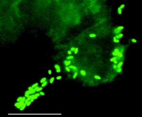
Rickettsiae electroporated with pMOD658 (one cuvette) or pMOD601 (2 cuvettes) transposomes, or control rickettsiae without a transposome (two cuvettes), were inoculated onto ISE6 cells at a multiplicity of infection of 100 and were initially exposed to 2.5 μg of CHL per ml 24 h after inoculation. After 5 days, extracellular rickettsiae remained in both control and transposome samples, and some cytopathic effect was evident. CHL was increased to 5 μg/ml, and by 10 days extracellular rickettsiae were absent and host cells appeared healthy. At 21 days, a few rickettsiae were visible in fewer than 5% of Giemsa-stained control host cells, but as many as 30 pMOD658-electroporated rickettsiae were visible in approximately 50% of host cells. In the two cultures inoculated with pMOD601-electroporated rickettsiae, rickettsial numbers were similar to those of the pMOD658 culture in one culture but were comparable to controls in the second. Abundant rickettsiae and extensive cytopathic effect were apparent in the pMOD658 and the first pMOD601 cultures after 1 month but were absent in the second pMOD601 and control cultures. CHL-selected rickettsiae were serially passaged by inoculation of 0.1 ml of infected culture onto new ISE6 cell cultures maintained with 5 μg of CHL per ml and were designated R. monacensis pMODompACAT/GFPuv line 658 (Rmona658) and pMODompACAT/ompBDsRed line 601 (Rmona601). Under epifluorescence microscopic observation, strong GFPuv fluorescence was visible in Rmona658 rickettsiae (Fig. 2). The Rmona658 line, currently at the 26th passage and removed from CHL selection at the 6th passage, has continued to express GFPuv fluorescence, but DsRed fluorescence was never visible in Rmona601 rickettsiae grown under the same conditions as Rmona658 rickettsiae. Removal of drug selection did not result in DsRed fluorescence, nor did growth of cultures at temperatures of as low as 26°C, storage of cultures at 4 or 22°C, or cultivation in the mammalian RF/6A cell line.
FIG. 2.
GFPuv-fluorescent R. monacensis transformant cells (Rmona658) in tick ISE6 cells. Bar, 10 μm. Magnification, ×1,000, with a fluorescein isothiocyanate filter.
Detection of transposons in pMOD658 and -601 rickettsial DNA.
We detected pMOD658 and -601transposons in DNA extracts of CHL-resistant rickettsia by PCR with primers (Table 1) designed to produce full-length amplicons of the CAT, DsRed2, and GFPuv genes. The predicted 720-bp GFPuv amplicon was obtained from Rmona658 DNA (Fig. 3A, lane 3) but not from R. monacensis control DNA (lane 2). The predicted 660-bp CAT amplicon was obtained from Rmona658 (lane 5) and Rmona601 (lane 6) DNA but not from control DNA (lane 4). Despite an absence of DsRed fluorescence in Rmona601, we obtained the predicted 670-bp DsRed2 amplicon from Rmona601 (lane 8) but not from control DNA (lane 7). With primers corresponding to the ompA and -B promoters and the ompA terminator as well as reporter genes, additional amplicons were obtained from Rmona658 and -601 DNAs that verified the integrity of the transposons (data not shown). DNA sequence analysis confirmed the integrity of the CAT, DsRed2, and GFPuv ORFs.
FIG. 3.
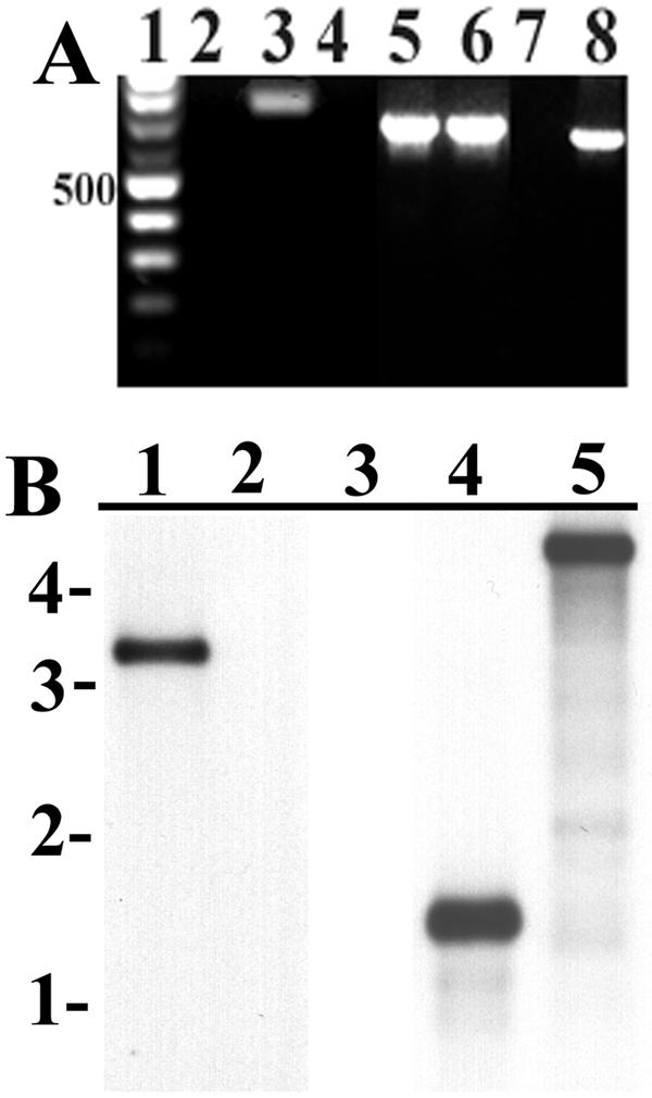
Detection of transposons in Rmona658 and Rmona601 DNA extracts. (A) PCR amplicons electrophoresed on a 1% agarose gel stained with EtBr. Lane 1 contains a 100-bp marker ladder, with the 500-bp band indicated at left. GFP primer amplicons from untransformed control R. monacensis (lane 2) and Rmona658 (lane 3); CAT primer amplicons from controls (lane 4), Rmona658 (lane 5), and Rmona601 (lane 6); DsRed2 primer amplicons from controls (lane 7) and Rmona601 (lane 8). (B) Southern blots with gel migration positions of a 1-kb marker ladder indicated at left. HindIII-digested DNA from Rmona658 (lane 1) or untransformed controls (lane 2) hybridized with a GFPuv probe; NdeI-digested DNA from controls (lane 3) or Rmona601 (lane 4) and KpnI-digested DNA from Rmona601 (lane 5) hybridized with a DsRed 2 probe.
We confirmed genomic integration of intact transposons by Southern blot analysis of Rmona658 and -601 DNAs. Digestion of Rmona658 DNA with HindIII, cleaving the pMOD658 transposon once near its 3′ terminus (Fig. 1D), and hybridization with GFPuv probe produced a single strongly hybridizing band at approximately 3.3 kb (Fig. 3B, lane 1), while no hybridization was observed with untransformed Rmona control DNA (lane 2). The result was consistent with single-site genomic integration of a pMOD658 transposon approximately 1.3 kb downstream of a genomic HindIII site. Because we were unable to detect DsRed fluorescence in Rmona601 cells, we rigorously verified chromosomal integration of an intact transposon. We hybridized a DsRed2 probe with Rmona601DNA digested with NdeI, cleaving the pMOD601 transposon twice at the translation initiation codons of the opposed DsRed2 and CAT genes (Fig. 1D), and obtained the predicted 1.4-kb band (Fig. 3B, lane 4), while no hybridization was observed with control DNA (lane 3). The result was consistent with genomic integration of an intact DsRed2- and CAT-coding portion of the pMOD601 transposon. The same probe hybridized to KpnI-digested Rmona601 DNA, cleaving the pMOD601 transposon once near its 5′ terminus, produced a single band at approximately 4.7 kb (lane 5), consistent with single-site genomic integration of an intact transposon 2.7 kb upstream of a genomic KpnI site. Supporting results were obtained by hybridization of the DsRed2 probe with HindIII-digested Rmona601 DNA, cleaving within the ompB promoter and again near the 3′ end of the transposon, resulting in the predicted 1.6-kb band (data not shown). Southern blots of single-plaque isolates of the Rmona658 (passage 20) and Rmona601 (passage 10) lines were consistent with those described above, indicating that the Rmona658 and -601 lines were clonal and derived from single-copy transposon integrations.
Genome insertion sites of the pMOD658 and -601 transposons.
We identified transposon chromosomal insertion sites by sequencing inverse PCR- and plasmid rescue-cloned R. monacensis DNA flanking the transposons followed by alignment of the cloned sequences to the complete genomes of R. conorii and R. prowazekii (GenBank accession no. AE006914 and AJ235269, respectively). Both transposons integrated with intact mosaic element sequences at each end flanked by 9-bp insertion site duplications typical of EZ::TN transposase (CAAGAACTG for pMOD658 and AGCAAGGAG for pMOD601). The CAT gene of the pMOD658 transposon was flanked by approximately 400 bp carrying a unique open reading frame followed by a 75-bp sequence that was 92 and 90% similar to tRNA-Phe genes of R. conorii and R. prowazekii, respectively. Extension of the R. monacensis sequence by using a primer (Rm658R1 [Table 1]) annealing downstream of the tRNA-Phe sequence reached a second region of 110 bp that was 90 and 78% similar to cytidylate kinase genes in the R. conorii and R. prowazekii genomes, respectively. The opposite side of the transposon was flanked by unique DNA sequence. The results were consistent with the comparative genome map (http://igs-server.cnrs-mrs.fr/mgdb/Rickettsia/) of R. conorii and R prowazekii, showing a colinear arrangement of the tRNA-Phe and cytidylate kinase genes just upstream of regions carrying unique ORFs followed by unique noncoding sequences, and they indicated an R. monacensis transposon insertion corresponding approximately to R. conorii position 716300. Analysis of the pMOD601 transposon integration site in the R. monacensis chromosome showed that it inserted at position 433496 relative to the R. conorii genome within a Rickettsia palindromic element 5 sequence overlapping the 3′ terminus of a 507-nucleotide ORF. Alignment of 916 bp of R. monacensis sequence with R. conori sequence in that region indicated 93% identity.
Detection of CAT and fluorescent reporter gene mRNAs.
We confirmed expression of CAT and fluorescent reporter mRNAs in transformant R. monacensis by RT-PCR and Northern blot analyses (Fig. 4). RT-PCRs with Rmona658 RNA yielded predicted amplicons of 650 bp with CAT primers (Fig. 4A, lane 4) and 720 bp with GFPuv primers (lane 6), but untransformed control reactions (lanes 5 and 7) and no-RT control reactions with Rmona658 RNA (lanes 2 and 3) did not. Rmona601 RNA yielded predicted CAT (Fig. 4B, lane 3) and DsRed2 (lane 5) amplicons, but untransformed control (lanes 2 and 4) and no-RT control (lanes 6 and 7) reactions did not.
FIG. 4.
Detection of CAT, GFPuv, and DsRed2 mRNAs in Rmona658 and Rmona601 RNA extracts by RT-PCR and Northern blotting. (A) Rmona658 RT-PCR analysis. Lane 1: 100-bp marker ladder, with the 700-bp band indicated at left (asterisk). Rmona658 no-RT control reactions with CAT (lane 2) and GFPuv (lane 3) primers, respectively. Rmona658 (lane 4) and untransformed control (lane 5) RT-PCRs with CAT primers. Rmona658 (lane 6) and untransformed control (lane 7) RT-PCRs with GFPuv primers. (B) Rmona601 RT-PCR analysis. Lane 1: 100-bp marker as described above. Untransformed control (lane 2) and Rmona601 (lane 3) RT-PCRs with CAT primers. Untransformed control (lane 4) and Rmona601 (lane 5) RT-PCRs with DsRed2 primers. Rmona601 no-RT control reactions with CAT (lane 6) and DsRed2 (lane 7) primers. (C) Northern blot. Lane 1: 0.5- and 1.0-kb RNA markers. Rmona658 (lane 2) and untransformed control (lane 3) RNA extracts hybridized with GFPuv probe. Rmona658 (lane 4), untransformed control (lane 5), and Rmona601 (lane 6) RNA extracts hybridized with CAT probe. Untransformed control (lane 7) and Rmona601 (lane 8) RNA extracts hybridized with DsRed2 probe.
We confirmed full-length mRNA expression with Northern blots, particularly because the transcription terminator in our vectors was predicted on the basis of an inverted repeat followed by an A-rich sequence immediately downstream of the ompA open reading frame but was not confirmed by functional assay (1). We predicted minimum mRNA lengths of 750 (CAT), 810 (GFPuv), and 860 (DsRed2) nucleotides based on promoter transcription start sites and untranslated mRNA sequences (1, 23, 43) as well as transcription of 70 nucleotides of terminator to form mRNA hairpins and stop transcription. Northern blotting showed that the GFPuv probe hybridized to Rmona658 RNA in a single band (Fig. 4C, lane 2) at approximately 900 nucleotides relative to marker RNA (lane 1) but did not hybridize to untransformed control RNA (lane 3). The CAT probe hybridized to Rmona658 and -601 RNAs (lanes 4 and 6, respectively) in single bands at approximately 750 nucleotides but did not hybridize to control RNA (lane 5). The DsRed2 probe hybridized to Rmona601 RNA in two bands of roughly equivalent intensity corresponding to the predicted 860 nucleotides and an unexpected 400 to 600 nucleotides (lane 8), but it did not hybridize to control RNA (lane 7). The results confirmed expression of full-length mRNAs and were consistent with a functional transcription terminator.
Western immunoblot analyses of GFPuv and DsRed2 expression.
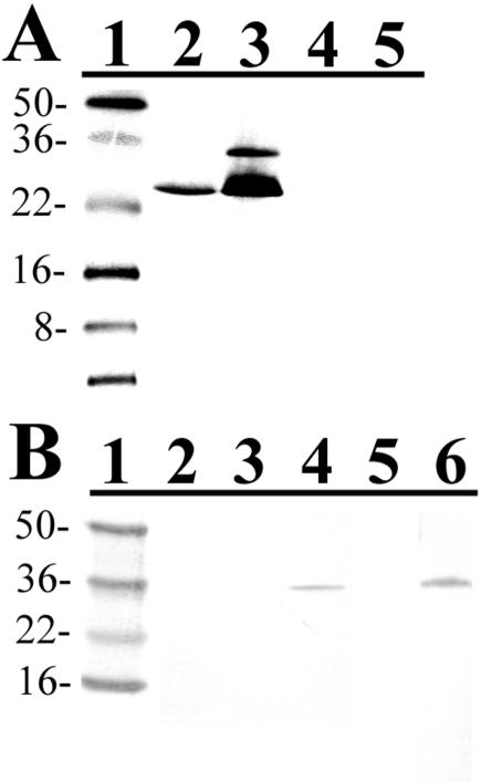
We studied expression of fluorescent proteins because Rmona658 cells exhibited strong GFPuv fluorescence but Rmona601 cells, despite expressing DsRed2 mRNA and in contrast to E. coli cells transformed with the pMOD601 plasmid, were not fluorescent. Extracts of Rmona658 (Fig. 5A, lane 2) or E. coli transformed with pMOD658 plasmid (lane 3) probed with polyclonal anti-GFP antibody produced prominent protein bands consistent with the predicted GFPuv molecular mass of 26 kDa. A 30-kDa band was also present in E. coli 658 extract, but proteins in R. monacensis and E. coli control extracts (lanes 4 and 5) were not recognized by antibody. Extracts of DsRed2-transformant RF/6A cells (Fig. 5B, lane 4) and E. coli transformed with pMOD601 (lane 6), but not E. coli controls (lane 5), probed with polyclonal anti-DsRed antibody produced a single band migrating just below the 36-kDa marker, as predicted (55). R. moncensis control (lane 2) or Rmona601(lane 3) proteins were not recognized by anti-DsRed antibody. Monoclonal anti-DsRed antibody produced identical results except that a 36-kDa band was not detected in RF/6A cell extracts (data not shown). E. coli therefore expressed both GFPuv and DsRed2 proteins, but R. monacensis expressed only GFPuv protein.
FIG. 5.
Western immunoblots of R. monancensis, E. coli, and RF/6A cell protein extracts. (A) GFP immunoblot. Lane 1: SeeBlue Plus 2 markers, with sizes in kilodaltons indicated at left. Lane 2: Rmona658 extract. Lane 3: pMOD658 transformant E. coli extract. Lane 4: untransformed R. monacensis extract. Lane 5: untransformed E. coli extract. (B) DsRed immunoblot. Lane 1: SeeBlue Plus 2 markers. Lane 2: Rmona601 extract. Lane 3: untransformed R. monacensis extract. Lane 4: DsRed2 transformant RF/6A extract. Lane 5: untransformed E. coli extract. Lane 6: pMOD601 transformant E. coli extract.
Expression of CAT and determination of CHL MIC50.
We assayed CAT activity to confirm CAT protein expression in Rmona658 and -601 cells. Protein extracts of fourth-passage preparations of Rmona658 and -601 cells had 57- and 338-fold, respectively, the CAT activity of R. monacensis control extracts (Table 2). Extracts of E. coli transformed with pMOD658 and -601 plasmids had 254- and 490-fold, respectively, the activity of control extracts. Log plots (not shown) of [CHL] versus percentage of infected host cells from infection inhibition assays indicated MIC50s of 1.5 and 6.0 μg/ml for control rickettsiae and Rmona658 cells, respectively (Rmona601 rickettsiae were not analyzed).
TABLE 2.
Chloramphenicol acetyltransferase activities of R. monacensis and E. coli protein extracts
| Bacterial extract | Activitya (mean ± SD) | Fold increaseb |
|---|---|---|
| R. monacensis pMOD658 | 497.7 ± 16.2 | 57 |
| R. monacensis control | 8.7 ± 0.5 | |
| R. monacensis pMOD601 | 1959.3 ± 55.6 | 338 |
| R. monacensis control | 5.8 ± 0.8 | |
| E. coli pMOD658 | 3687.3 ± 63.5 | 254 |
| E. coli pMOD601 | 7115.7 ± 246.5 | 491 |
| E. coli control | 14.5 ± 1.0 |
Activity (means of three replicates) expressed as counts per minute of 14C incorporated per microgram of protein extract.
Activity of transformant versus untransformed control.
Visualization of GFPuv-fluorescent rickettsiae with host cell actin tails.
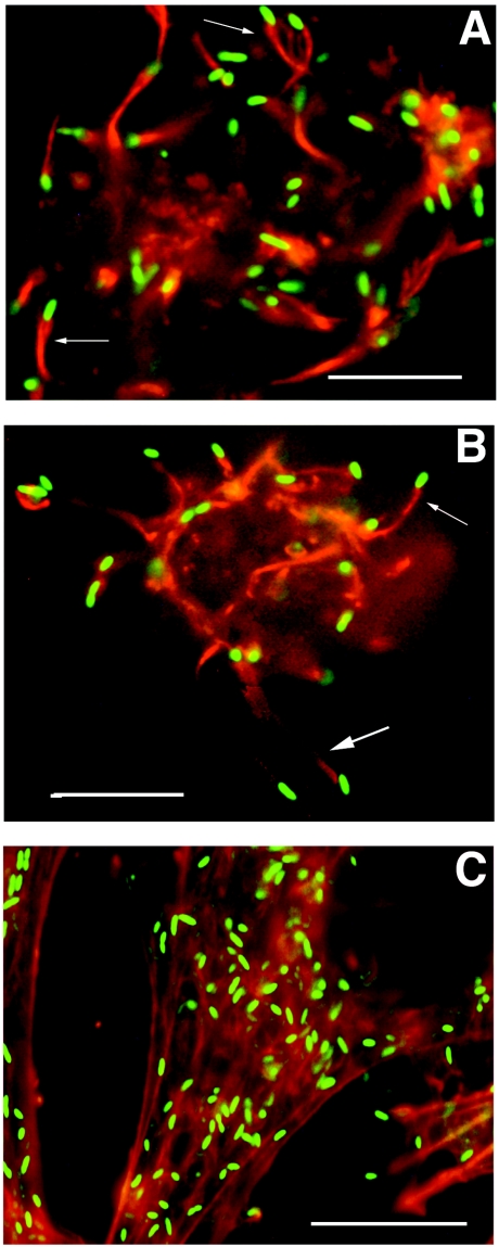
To demonstrate the utility of transformant rickettsiae for study of host-rickettsia interactions, we used Rmona658 cells to confirm previous double-immunolabeling studies that showed an association of R. monacensis with host cell F-actin tail structures (58). Visualization of rhodamine-labeled actin and Rmona658 rickettsiae in tick IDE8 cells showed rickettsiae in areas of high actin content and a specific association with primarily cytosolic actin tail structures (Fig. 6A) or within pseudopodia and possibly leaving host cells on tails up to 10 μm in length (Fig. 6B). In vertebrate RF6A cells, rickettsiae concentrated in areas of high actin content, but association with actin tails was equivocal (Fig. 6C).
FIG. 6.
Visualization of GFPuv-fluorescent rickettsiae associated with rhodamine-labeled (red) host cell actin structures. (A and B) Rickettsiae (green rods) in tick IDE8 cells associated with actin tail structures (arrows) that were primarily cytosolic (A) or in pseudopodia (B). (C) Rickettsiae in vertebrate RF6A cells. Bar, 10 μm.
DISCUSSION
Development of molecular techniques for study of rickettsiae has been hindered by their slow obligate intracellular growth and the lack of characterized mutants or a reliable transformation system (65). The recently sequenced R. prowazekii and R. conorii (SFGR) genomes (3, 39) and demonstrations of rickettsial transformation have ameliorated that impediment. R. prowazekii was first transformed by electroporation and homologous recombination of DNA carrying an rpoB allele conferring resistance to rifampin (47). R. typhi GFPuv transformants generated by a similar strategy were unstable and not visibly fluorescent (60). Rifampin selection is compromised by spontaneous resistance mutations (56), and a subsequent effort to transform and select erythromycin-resistant R. prowazekii was again problematic due to spontaneous resistance (48). Improved rifampin selection was recently achieved by electroporation and transformation of R. prowazekii with a rifampin-ribosylation resistance factor gene in the EZ::TN transposon vector (46).
We developed EZ::TN transposon vectors containing two rickettsial promoter expression cassettes for transformation with genes of interest linked to a robust selectable marker and demonstrated visible expression of GFPuv in individual cells, a result not achieved in previous GFP rickettsial transformations (52, 60). We obtained clonal transformant populations, overcoming a further limitation described in previous studies (47, 48, 52, 60). Our transformants were the result of transposition events rather than recombination and have remained stable over long-term passage even without selection.
We recovered transformant R. monacensis from two of three electroporations of 3 × 10⁸ cells each, indicating a transformation efficiency of 10−8. The EZ::TN transposon system produces single-site integrations in bacterial cells (18), including R. prowazekii (46), but reports of rickettsial transformation have not included efficiency estimations, due to difficulty in quantifying rickettsiae. Our use of live-dead stain allowed fast, accurate quantification. The transformation efficiency of R. monacensis was 100-fold lower than that of the facultative intracellular pathogen Bartonella henselae, generated by electroporation with EZ::TN transposomes and isolated by extracellular growth on selective media (53). The relative inefficiency of R. monacensis transformation may extend to other rickettsiae due to factors related to their obligate intracellular growth and lability outside host cells.
Plaque assay estimates of CHL MIC50s for SFGR range from 0.25 to 1.0 μg/ml (49, 64), in contrast to our Giemsa stain-based estimate of 1.5 μg/ml for R. monacensis. Expression of CAT activity by Rmona658 rickettsiae increased the MIC50 to 6 μg/ml, a value below the 8-μg/ml interpretive standard limit for classification as susceptible (13) and the approximate human therapeutic dosage (20 μg/ml of serum). Rmona658 rickettsiae cultured in tick cells expressed sufficient CAT activity to be selectable but were not CHL resistant. Moreover, CHL is toxic and is no longer recommended for treatment of rickettsioses (see the introduction). Its use as a selectable marker in transformation of rickettsiae confined to the laboratory will facilitate advances in the study of rickettsial physiology and pathogenesis analogous to those obtained by application of the same technology to other bacteria, many of which are pathogens.
Rmona658 cells expressed GFPuv protein, but Rmona601 cells did not express DsRed2 protein despite expressing mRNA with an intact ORF. Codon usage effects on translation may explain failed DsRed2 expression in R. monacensis versus successful expression in E. coli. The concentrations of stable RNA and ribosomes in R. prowazekii and E. coli are similar (40), but low-GC rickettsial genomes have a strong AT bias in third-base codon positions and higher and lower frequencies of AT-coded and GC-coded amino acids, respectively, relative to E. coli (2). E. coli expresses 46 tRNAs (20) from 86 genes (8), but rickettsiae have only 33 single-copy tRNA genes (3, 39). Codon bias of the mRNA pool is likely reflected in relative abundances of the cognate tRNAs (20, 27).
The DsRed2 gene GC content of 63.8%, with G or C in 100% of third-base codon positions, contrasts with that of the GFPuv gene and rickettsial genomes (Table 3). Over 71% of DsRed2 codons, versus 16.5% of GFPuv and 33.4% of CAT codons, occur rarely or very rarely in rickettsiae, but only 12.9% and 14.2% of DsRed2 codons occur rarely or very rarely in E. coli. Codon bias extremes represented by the DsRed2 gene and rickettsial genomes may have disrupted translation of DsRed2 mRNA in R. monacensis due to low levels of appropriate tRNAs. Studies of E. coli showed that rare codons (frequencies of 1% or less) could cause amino acid misincorporation, frameshifts, and premature termination (28, 29, 33), especially when clustered or near the 5′ end of mRNA (24, 28). Considering only the 59 more restrictively defined very rare rickettsial codons (Table 3) in the DsRed2 gene, 16 occur as doublets, 9 occur as triplets, 4 occur as a quadruplet, and 17 occur within the first 200 nucleotides. Of 13 very rare rickettsial codons in the GFPuv gene, only 2 occur as a doublet and 1 occurs within the first 200 nucleotides, while 4 of 17 in the CAT gene occur as doublets and 7 occur within the first 200 nucleotides. Those comparisons imply compromised translation of DsRed2 by R. monacensis due to “hungry” codons calling for rare or deacylated cognate tRNAs that resulted in stalled ribosomes (17, 29). Ribosomes stalled at rare codons introduced into the E. coli ompA gene triggered premature termination of transcription (reference 16 and references therein). Our Northern blots showed similar amounts of approximately half- and full-length DsRed2 mRNAs in Rmona601 extracts versus predominantly full-length CAT and GFPuv mRNAs, consistent with possible premature termination of DsRed2 transcription. However, other possible explanations for failed DsRed2 expression in R. monacensis include reduced DNA melting capacity and GC-rich template activity of R. prowazekii RNA polymerase versus that of E. coli (19).
TABLE 3.
GC content and codon bias in DsRed2, CAT, and GFPuv genes versus rickettsia and E. coli genomesa
| Gene or genome | % GC | Codon third-base % GC | Total codons (103) | % Codonsb
|
||
|---|---|---|---|---|---|---|
| Rare | Very rare | Total rare + very rare | ||||
| R. prowazekii | 29.0 | 18.4 | 280 | 7.4 | 4.8 | 12.2 |
| R. conorii | 32.4 | 23.5 | 340 | 9.8 | 3.3 | 13.1 |
| GFPuv | 41.0 | 35.2 | 0.238 | 10.8 | 5.7 | 16.5 |
| CAT | 45.1 | 51.9 | 0.218 | 24.6 | 8.8 | 33.4 |
| DsRed2 | 63.8 | 100 | 0.225 | 47.1 | 24.4 | 71.5 |
| E. coli | 50.7 | 55.8 | 1,400 | 11.4 | 1.5 | 12.9 |
| DsRed2 versus E. coli | 63.8 | 100 | 0.225 | 14.2 | 0 | 14.2 |
The R. prowazekii, R. conorii, and E. coli genomes contain, respectively, 834, 1,374, and 4,289 protein-coding sequences.
We define rare and very rare codons as those occurring at frequencies of 0.51 to 1.0% and of 0.5% or less in the rickettsial and E. coli protein coding sequences, respectively. Data are drawn from (http://gpath.ym.edu.tw:8090/kegg/codon_table/). Very rare rickettsial codons include CUC, CUG, UCC, UGC, CCC, CAC, CGC, CGG, AGG, GUC, and GCG. In E. coli, UCC, UGC, CCC, and CAC are rare codons.
Development of transformation and gene expression vectors for intracellular bacteria should be guided by genomic sequence analyses. Anaplasma marginale has a 50% GC genome, and its 967 ORFs (K. Brayton [Washington State University, Pullman], personal communication) contain fewer rare codons than those of E. coli, a hopeful prospect for DsRed2 expression in Anaplasma. Other arthropod-adapted intracellular bacteria, including Ehrlichia (Cowdria) (www.sanger.ac.uk/Projects/C_ruminantium/), Buchnera, Wigglesworthia, and Wolbachia species (25, 62), share genome characteristics with Rickettsia. Expression of potentially problematic GC-rich genes in such bacteria might require coexpression of rare or absent tRNAs and aminoacyl synthetases. Genetic manipulation of intracellular bacteria remains in its infancy but holds great promise. Our vectors provide a new tool for study of rickettsial molecular biology and physiology and may be useful for transformation of other arthropod-borne intracellular bacteria as well.
Acknowledgments
This research was supported by NIH grant RO1 AI49424 to U.G.M.
REFERENCES
- 1.Anderson, B. E., G. A. McDonald, D. C. Jones, and R. L. Regnery. 1990. A protective protein antigen of Rickettsia rickettsii has tandemly repeated, near-identical sequences. Infect. Immun. 58:2760-2769. [DOI] [PMC free article] [PubMed] [Google Scholar]
- 2.Andersson, S. G. E., and P. M. Sharp. 1996. Codon usage and base composition in Rickettsia prowazekii. J. Mol. Evol. 42:525-536. [DOI] [PubMed] [Google Scholar]
- 3.Andersson, S. G. E., A. Zomorodipour, J. O. Andersson, T. Sicheritz-Ponten, U. C. M. Alsmark, R. M. Podowski, A. K. Naslund, A. Eriksson, H. H. Winkler, and C. G. Kurland. 1998. The genome sequence of Rickettsia prowazekii and the origin of mitochondria. Nature 396:133-140. [DOI] [PubMed] [Google Scholar]
- 4.Anonymous. 2001. Living colors DsRed2. Clonetechniques 16:2-3. [Google Scholar]
- 5.Azad, A. F., and C. B. Beard. 1998. Rickettsial pathogens and their arthropod vectors. Emerg. Infect. Dis. 4:179-186. [DOI] [PMC free article] [PubMed] [Google Scholar]
- 6.Bell, J. E., G. M. Kohls, H. G. Stoenner, and D. B. Blackman. 1963. Nonpathogenic rickettsias related to the spotted fever group from ticks, Dermacentor variabilis and Dermacentor andersoni from eastern Montana. J. Immunol. 90:770-781. [PubMed] [Google Scholar]
- 7.Bella, F., B. Font, S. Uriz, T. Munoz, E. Espejo, J. Traveria, J. A. Serrano, and F. Segura. 1990. Randomized trial of doxycycline versus josamycin for Mediterranean spotted fever. Antimicrob. Agents Chemother. 34:937-938. [DOI] [PMC free article] [PubMed] [Google Scholar]
- 8.Blattner, F. R., G. I. Plunkett, C. A. Bloch, et al. 1997. The complete genome sequence of Escherichia coli K12. Science 277:1453-1474. [DOI] [PubMed] [Google Scholar]
- 9.Burgdorfer, W., D. J. Sexton, R. K. Gerloff, R. L. Anacker, R. N. Philip, and L. A. Thomas. 1975. Rhipicephalus sanguineus: vector of a new spotted fever group rickettsia in the United States. Infect. Immun. 12:205-210. [DOI] [PMC free article] [PubMed] [Google Scholar]
- 10.Burgdorfer, W., S. F. Hayes, and A. J. Mavros. 1981. Nonpathogenic rickettsiae in Dermacentor andersoni: a limiting factor for the distribution of Rickettsia rickettsii, p. 585-594. In W. Burgdorfer and R. L. Anacker (ed.), Rickettsiae and rickettsial diseases. Academic Press, New York, N.Y.
- 11.Childs, J. E., and C. D. Paddock. 2002. Passive surveillance as an instrument to identify risk factors for fatal Rocky Mountain spotted fever: is there more to learn? Am. J. Trop. Med. Hyg. 66:450-457. [DOI] [PubMed] [Google Scholar]
- 12.Cohen, J., Y. Lasri, and Z. Landau. 1999. Mediterranean spotted fever in pregnancy. Scand. J. Infect. Dis. 31:202-203. [DOI] [PubMed] [Google Scholar]
- 13.Conte, J. E., and S. L. Barriere. 1992. Antibiotics and infectious diseases, 7th ed., p. 117-137. Lea and Febiger, London, United Kingdom.
- 14.Crameri, A., E. A. Whitehorn, E. Tate, and W. P. Stemmer. 1996. Improved green fluorescent protein by molecular evolution using DNA shuffling. Nat. Biotechnol. 14:315-319. [DOI] [PubMed] [Google Scholar]
- 15.Dalton, M. J., M. J. Clarke, R. C. Holman, J. W. Krebs, D. B. Fishbein, J. G. Olson, and J. E. Childs. 1995. National surveillance for Rocky Mountain spotted fever, 1981-1992: epidemiologic summary and evaluation of risk factors for fatal outcome. Am. J. Trop. Med. Hyg. 52:405-413. [DOI] [PubMed] [Google Scholar]
- 16.Deana, A., R. Ehrlich, and C. Reiss. 1998. Silent mutations in the Escherichia coli ompA leader peptide region strongly affect transcription and translation in vivo. Nucleic Acids Res. 26:4778-4782. [DOI] [PMC free article] [PubMed] [Google Scholar]
- 17.Del Tito, B. J., J. M. Ward, J. Hodgson, C. J. Gershater, H. Edwards, L. A. Wysocki, F. A. Watson, G. Sathe, and J. F. Kane. 1995. Effects of a minor isoleucyl tRNA on heterologous protein translation in Escherichia coli. J. Bacteriol. 177:7086-7091. [DOI] [PMC free article] [PubMed] [Google Scholar]
- 18.Derbyshire, K. M., C. Takacs, and J. Huang. 2000. Using the EZ::TN transposome for transposon mutagenesis in Mycobacterium smegmatis. Epicentre Forum 7:1-4. [Google Scholar]
- 19.Ding, H. F., and H. H. Winkler. 1993. Characterization of the DNA-melting function of the Rickettsia prowazekii RNA polymerase. J. Biol. Chem. 268:3897-3902. [PubMed] [Google Scholar]
- 20.Dong, H., L. Nilsson, and C. G. Kurland. 1996. Co-variation of tRNA abundance and codon usage in Escherichia coli at different growth rates. J. Mol. Biol. 260:649-663. [DOI] [PubMed] [Google Scholar]
- 21.Donovan, B. J., D. J. Weber, J. C. Rublein, and R. H. Raasch. 2002. Treatment of tick-borne diseases. Ann. Pharmacother. 36:1590-1597. [DOI] [PubMed] [Google Scholar]
- 22.Engler-Blum, G., M. Meir, J. Frank, and G. A. Muller. 1993. Reduction of background problems in nonradioactive Northern and Southern blot analyses enables higher sensitivity than 32P-based hybridizations. Anal. Biochem. 210:235-244. [DOI] [PubMed] [Google Scholar]
- 23.Gilmore, R. D., Jr., W. Cieplak, Jr., P. F. Policastro, and T. Hackstadt. 1991. The 120 kilodalton outer membrane protein (rOmpB) of Rickettsia rickettsii is encoded by an unusually long open reading frame: evidence for protein processing from a large precursor. Mol. Microbiol. 5:2361-2370. [DOI] [PubMed] [Google Scholar]
- 24.Goldman, E., A. H. Rosenberg, G. Zubay, and F. W. Studier. 1995. Consecutive low-usage leucine codons block translation only when near the 5′ end of a message in Escherichia coli. J. Mol. Biol. 245:467-473. [DOI] [PubMed] [Google Scholar]
- 25.Herbeck, J. T., D. P. Wall, and J. J. Wernegreen. 2003. Gene expression level influences amino acid usage, but not codon usage, in the tsetse fly endosymbiont Wigglesworthia. Microbiology 149:2585-2596. [DOI] [PubMed] [Google Scholar]
- 26.Holman, R. C., C. D. Paddock, A. T. Curns, J. W. Krebs, J. H. McQuiston, and J. E. Childs. 2001. Analysis of risk factors for fatal Rocky Mountain spotted fever: evidence for superiority of tetracyclines for treatment. J. Infect. Dis. 184:1437-1444. [DOI] [PubMed] [Google Scholar]
- 27.Ikemura, T. 1981. Correlation between the abundance of Escherichia coli transfer RNAs and the occurrence of the respective codons in its protein genes. J. Mol. Biol. 146:1-21. [DOI] [PubMed] [Google Scholar]
- 28.Kane, J. F. 1995. Effects of rare codon clusters on high-level expression of heterologous proteins in Escherichia coli. Curr. Opin. Biotechnol. 6:494-500. [DOI] [PubMed] [Google Scholar]
- 29.Kurland, C., and J. Gallant. 1996. Errors of heterologous protein expression. Curr. Opin. Biotechnol. 7:489-493. [DOI] [PubMed] [Google Scholar]
- 30.Laemmli, U. K. 1970. Cleavage of structural proteins during the assembly of the head of bacteriophage T4. Nature 227:680-685. [DOI] [PubMed] [Google Scholar]
- 31.Maniatis, T., E. F. Fritsch, and J. Sambrook. 1982. Molecular cloning: a laboratory manual. Cold Spring Harbor Laboratory, Cold Spring Harbor, N.Y.
- 32.Masters, E. J., G. S. Olson, S. J. Weiner, and C. D. Paddock. 2003. Rocky Mountain spotted fever: a clinician's dilemma. Arch. Intern. Med. 163:769-774. [DOI] [PubMed] [Google Scholar]
- 33.McNulty, D. E., B. A. Claffee, M. J. Huddleston, and J. F. Kane. 2003. Mistranslational errors associated with the rare arginine codon CGG in Escherichia coli. Protein Expr. Purif. 27:365-374. [DOI] [PubMed] [Google Scholar]
- 34.Munch, S. 1994. Nonradioactive Northern blots in 1.5 days with substantially increased sensitivity through “alkaline blotting.” Biochemica 11:29-31. [Google Scholar]
- 35.Munderloh, U. G., Y. Liu, M. Wang, C. Chen, and T. J. Kurtti. 1994. Establishment, maintenance and description of cell lines from the tick Ixodes scapularis. J. Parasitol. 80:533-543. [PubMed] [Google Scholar]
- 36.Munderloh, U. G., S. F. Hayes, J. Cummings, and T. J. Kurtti. 1998. Microscopy of spotted fever rickettsia movement through tick cells. Microsc. Microanal. 4:115-121. [Google Scholar]
- 37.Munderloh, U. G., S. D. Jauron, V. Fingerle, L. Leitritz, S. F. Hayes, J. M. Hautman, C. M. Nelson, B. W. Huberty, T. J. Kurtti, G. G. Ahlstrand, B. Greig, M. A. Mellen, and J. L. Goodman. 1999. Invasion and intracellular development of the human granulocytic ehrlichiosis agent in tick cell culture. J. Clin. Microbiol. 37:2518-2524. [DOI] [PMC free article] [PubMed] [Google Scholar]
- 38.Munderloh, U. G., M. J. Lynch, M. J. Herron, A. T. Palmer, T. J. Kurtti, R. D. Nelson, and J. L. Goodman. 2004. Infection of endothelial cells with Anaplasma marginale and A. phagocytophilum. Vet. Microbiol. 101:53-64. [DOI] [PubMed] [Google Scholar]
- 39.Ogata, H., S. Audic, P. Renesto-Audiffren, P. E. Fournier, V. Barbe, D. Samson, V. Roux, P. Cossart, J. Weissenbach, J. M. Claverie, and D. Raoult. 2001. Mechanisms of evolution in Rickettsia conorii and R. prowazekii. Science 293:2093-2098. [DOI] [PubMed] [Google Scholar]
- 40.Pang, H., and H. H. Winkler. 1994. The concentrations of stable RNA and ribosomes in Rickettsia prowazekii. Mol. Microbiol. 12:115-120. [DOI] [PubMed] [Google Scholar]
- 41.Parker, R. R., E. G. Pickens, D. B. Lackman, E. J. Bell, and F. B. Thraikill. 1951. Isolation and characterization of rocky mountain spotted fever rickettsiae from the rabbit tick Haemaphysalis lepori-palustris Packard. Public Health Rep. 66:455-463. [PMC free article] [PubMed] [Google Scholar]
- 42.Pickering, L. K. (ed.). 2000. 2000 red book: report of the Committee on Infectious Diseases, 25th ed., p. 491-493. American Academy of Pediatrics, Elk Grove Village, Ill.
- 43.Policastro, P. F., and T. Hackstadt. 1994. Differential activity of Rickettsia rickettsii ompA and ompB promoter regions in a heterologous reporter gene system. Microbiology 140:2941-2949. [DOI] [PubMed] [Google Scholar]
- 44.Policastro, P. F., U. G. Munderloh, E. R. Fischer, and T. Hackstadt. 1997. Rickettsia rickettsii growth and temperature-inducible protein expression in embryonic tick cell lines. J. Med. Microbiol. 46:839-845. [DOI] [PubMed] [Google Scholar]
- 45.Purvis, J. J., and M. S. Edwards. 2000. Doxycycline use for rickettsial disease in pediatric patients. Pediatr. Infect. Dis. J. 19:871-874. [DOI] [PubMed] [Google Scholar]
- 46.Qin, A., A. M. Tucker, A. Hines, and D. O. Wood. 2004. Transposon mutagenesis of the obligate intracellular pathogen Rickettsia prowazekii. Appl. Environ. Microbiol. 70:2816-2822. [DOI] [PMC free article] [PubMed] [Google Scholar]
- 47.Rachek, L. I., A. M. Tucker, H. H. Winkler, and D. O. Wood. 1998. Transformation of Rickettsia prowazekii to rifampin resistance. J. Bacteriol. 180:2118-2124. [DOI] [PMC free article] [PubMed] [Google Scholar]
- 48.Rachek, L. A., A. Hines, A. M. Tucker, H. H. Winkler, and D. O. Wood. 2000. Transformation of Rickettsia prowazekii to erythromycin resistance encoded by the Escherichia coli ereB gene. J. Bacteriol. 182:3289-3291. [DOI] [PMC free article] [PubMed] [Google Scholar]
- 49.Raoult, D., P., Roussellier, G. Vestris, and J. Tamalet. 1987. In vitro antibiotic susceptibility of Rickettsia rickettsii and Rickettsia conorii: plaque assay and microplaque colorimetric assay. J. Infect. Dis. 155:1059-1062. [DOI] [PubMed] [Google Scholar]
- 50.Raoult, D., and M. Drancourt. 1991. Antimicrobial therapy of rickettsial diseases. Antimicrob. Agents Chemother. 35:2457-2462. [DOI] [PMC free article] [PubMed] [Google Scholar]
- 51.Raoult, D., and V. Roux. 1997. Rickettsioses as paradigms of new or emerging infectious diseases. Clin. Microbiol. Rev. 10:694-719. [DOI] [PMC free article] [PubMed] [Google Scholar]
- 52.Renesto, P., E. Gouin, and D. Raoult. 2002. Expression of green fluorescent protein in Rickettsia conorii. Microb. Pathog. 33:17-21. [DOI] [PubMed] [Google Scholar]
- 53.Rieb, T., B. Anderson, A. Fackelmayer, I. B. Autenrieth, and V. A. J. Kempf. 2003. Rapid and efficient transposon mutagenesis of Bartonella henslae by transposome technology. Gene 313:103-109. [DOI] [PubMed] [Google Scholar]
- 54.Rolain, J. M., M. Maurin, G. Vestris, and D. Raoult. 1998. In vitro susceptibilities of 27 rickettsiae to 13 antimicrobials. Antimicrob. Agents Chemother. 42:1537-1541. [DOI] [PMC free article] [PubMed] [Google Scholar]
- 55.Sachetti, A., V. Subramaniam, T. M. Jovin, and S. Alberti. 2002. Oligomerization of DsRed is required for the generation of a functional red fluorescent chromophore. FEBS Lett. 525:13-19. [DOI] [PubMed] [Google Scholar]
- 56.Severinov, K., M. Soushko, A. Golfarb, and V. Nikiforov. 1993. New rifampicin-resistant and streptolydigin-resistant mutants in the B subunit of Escherichia coli RNA polymerase. J. Biol. Chem. 268:14820-14825. [PubMed] [Google Scholar]
- 57.Simser, J. A., A. T. Palmer, U. G. Munderloh, and T. J. Kurtti. 2001. Isolation of a spotted fever group rickettsia, Rickettsia peacockii, in a Rocky Mountain wood tick, Dermacentor andersoni, cell line. Appl. Environ. Microbiol. 67:546-552. [DOI] [PMC free article] [PubMed] [Google Scholar]
- 58.Simser, J. A., A. T. Palmer, V. Fingerle, B. Wilske, T. J. Kurtti, and U. G. Munderloh. 2002. Rickettsia monacensis sp. nov., a spotted fever group rickettsia, from ticks (Ixodes ricinus) collected in a European city park. Appl. Environ. Microbiol. 68:4559-4566. [DOI] [PMC free article] [PubMed] [Google Scholar]
- 59.Smilock, J. D., W. R. Wilson, and F. R. Cockerill III. 1991. Tetracyclines, chloramphenicol, erythromycin and metronidazole. Mayo Clin. Proc. 66:1270-1280. [DOI] [PubMed] [Google Scholar]
- 60.Troyer, J. M., S. Radulovic, and A. F. Azad. 1999. Green fluorescent protein as a marker in Rickettsia typhi transformation. Infect. Immun. 67:3308-3311. [DOI] [PMC free article] [PubMed] [Google Scholar]
- 61.Weller, S. J., G. D. Baldridge, U. G. Munderloh, H. Noda, J. Simser, and T. J. Kurtti. 1998. Phylogenetic placement of rickettsiae from the ticks Amblyomma americanum and Ixodes scapularis. J. Clin. Microbiol. 36:1305-1317. [DOI] [PMC free article] [PubMed] [Google Scholar]
- 62.Wernegreen, J. 2002. Genome evolution in bacterial endosymbionts of insects. Nat. Rev. 3:850-861. [DOI] [PubMed] [Google Scholar]
- 63.Winkler, H. H. 1990. Rickettsia species (as organisms). Annu. Rev. Microbiol. 44:131-153. [DOI] [PubMed] [Google Scholar]
- 64.Wisseman, C. L., and S. V. Ordonez. 1986. Actions of antibiotics on Rickettsia rickettsii. J. Infect. Dis. 153:626-627. [DOI] [PubMed] [Google Scholar]
- 65.Wood, D. O., and A. F. Azad. 2000. Genetic manipulation of rickettsiae: a preview. Infect. Immun. 68:6091-6093. [DOI] [PMC free article] [PubMed] [Google Scholar]


