

Abstract
Introduction
Reconstructing large defects of the columella and upper lip is an interesting challenge in facial reconstruction due to the high visibility of this aesthetic subunit and the difficulties posed by the unique characteristics of the skin in these areas, which differs from that of the surrounding regions. Among the various techniques proposed, the use of local flaps remains the most commonly used and effective method in this type of reconstruction.
Presentation of the case
A 47-year-old man in good clinical condition presented with a nodular lesion on the columella and upper lip. The lesion was excised (revealing it to be a squamous cell carcinoma) and reconstructed using two opposing nasogenian flaps, resulting in an optimal aesthetic and functional restoration.
Discussion
The use of local flaps remains the most effective technique for columella defect reconstruction. However, many described techniques require multiple surgical stages or result in visible scarring. Additionally, they do not guarantee effective reconstruction in cases involving the upper lip. On the other hand, the use of free flaps, while more expensive and requiring expert teams, may not ensure optimal color and skin texture matching.
Conclusions
The use of opposing nasogenian flaps allows for a rapid and effective reconstruction of defects involving the columella and upper lip, leading to a swift return to normal life for the patient.
Keywords: Spinocellular carcinoma, columella reconstruction, Nasogenian flaps, Nose reconstruction, Case report
Highlights
-
•
Reconstructing defects of the columella and the upper lip still presents a challenge today.
-
•
Numerous techniques have been described in the literature.
-
•
In this work, we use two opposing nasogenian flaps for a simple and effective reconstruction of this type of defect.
1. Introduction
The columella represents one of the aesthetic subunits of the nose [1]. Despite its small size, any alteration to it can lead to significant changes both aesthetically (as it is an important pillar in the projection and shape of the tip, and also defines the nasolabial angle the relationship between the nasal base and alar rims) and functionally (as it can disrupt the airflow during inspiration [2]). Alterations to the columella can be caused by surgical excision of skin neoplasms, trauma, or iatrogenic factors ischaemic injuries, vascular malformations and congenital malformation of nasal anatomy [3]. Over time, various techniques have been proposed for its reconstruction, ranging from methods based on the use of full thickness skin grafts [4] or composite grafts [5] to those based on the use of local flaps (such as nasal vestibule flaps [6] and nasolabial flap [7]) or regional flaps (such as the forehead flap [8]), and even involving the use of free flaps [9]. Reconstruction of the columella remains a significant challenge today due to its aesthetic importance, central position within the face and long-term results are usually unsatisfactory. The development of new techniques for this surgery continues to characterize modern times. In this work (reported in line with the SCARE criteria [10]), we present our experience in the reconstruction of this area using the interposition of two nasogenian flaps, a technique which provide satisfactory reconstruction even with large defect, with a simple procedure which can be performed even from novitiate surgeons.
2. Case report
A 47-year-old male patient visited our department with a nodular growth on his columella, which had been present for several years and had gradually increased in size over time. The patient had no other underlying health conditions and no prior history of skin cancer. The lesion, measuring approximately 3 cm in its largest dimension, was causing nasal airway obstruction and altering the appearance of the nose (see Fig. 1). We proceeded with the surgical removal of the skin neoformation with a scalpel, leaving 5 mm of healthy skin margins. With the excision of the neoformation were included the entire columellar base and body and both the soft triangles. The lateral margins of the incision included about 3 mm of the nasal vestibule mucosa, the upper incision includes half of the infratip lobule while the lower margin stopped about 5 mm from the vermilion edge of the upper lip. A strip of 2 mm of caudal septum was included in the excision as well as both the medial crus of the lower lateral cartilages. The lesion spared the anterior nasal spine. After a careful hemostasis, the two opposing nasogenian flaps were set up. This technique was chosen to ensure coverage of both the columella and the upper lip, as both areas were affected by the growth.
Fig. 1.

Preoperative drawing.
The flap, which has a random blood supply, was drawn with a width of about 1.5 cm and a length of about 3 cm at the level of the nasogenic groove bilaterally, to hide the scar and have an optimal aesthetic result. The dissection was advanced at the subcutaneous plane up to about the projection of the lateral canthus bilaterally to allow the movement of the flap. Two discharge incisions were drawn on a line parallel to the edge of the vermilion. Then the flaps were transposed to cover the defect. The donor site was closed with a simple detached stitch suture using 3-0 Vycril™ plus Ethicon and 4-0 Prolene™ Ethicon (see Fig. 2). The two opposing nasogenian flaps were sutured together at the midline region of the upper lip and at the level of the columellar region. The subcutaneous tissue was sutured with 4-0 Monocryl™ Ethicon. Three stiches with 4-0 Monocryl™ Ethicon were used to suture the remaining caudal septum to the subcutaneous tissue of the flaps to rebuild the nasolabial angle. The skin was sutured with simple detached stitch using 4-0 Prolene™ Ethicon. The dressing was carried out with tulle gauze and sterile gauze. Subsequent histological examination of the excised lesion confirmed it as squamous cell carcinoma with disease-free margins, with infiltration of the cartilaginous tissue. The patient achieved an excellent functional recovery, and after the sutures were removed (after ten days from the surgery), he could resume chewing without any issues and also experienced improved breathing (see Figs. 3 and 4). The follow-up after one year from the surgery (see Figs. 5, 6 and 7) shows that the aesthetic and functional result is satisfactory.
Fig. 2.
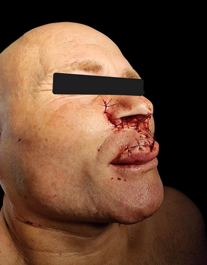
Immediate postoperative.
Figs. 3, 4.

Postoperative follow up at 6 weeks.
Figs. 5, 6, 7.

Postoperative follow-up at 1 year.
3. Discussion and conclusions
Columellar reconstruction remains a significant challenge today. Reconstructing a highly visible facial area with skin characteristics different from the surrounding tissues remains a challenge. Among the various reconstruction techniques reported in the literature, the use of local flaps is the most commonly chosen option by various authors [10]. The use of skin grafts does not ensure optimal coverage in cases of non-superficial defects, and it also does not guarantee an optimal aesthetic outcome. As a result, it is a limited-use reconstructive technique in patients with significant comorbidities [11]. On the other hand, the use of free flaps is burdened by both long operating times and procedural complexity (not accessible to all surgeons and in all settings), as well as the presence of excessive bulking in the reconstruction, which may require further interventions to improve aesthetics and function [12,13].
Numerous local flaps have been suggested for addressing these defects. For instance, some authors [14] have recommended the utilization of an alar rim flap. This technique can deliver a satisfactory reconstruction in terms of color and skin texture. However, it's most effective for small defects and can lead to distortion of the nasal ala.
Other techniques described in the literature include the use of two nasal vestibule flaps [15] (which allows for the reconstruction of extensive portions of superficial columellar defects but not those involving the upper lip), the use of nasal sill advancement flaps [16] (which require a two-step procedure and cartilage grafts for adequate reconstruction), the utilization of philtrum flaps [17] (which, by their nature, are not applicable in cases of defects involving the upper lip) and the use of a peri-alar hatchet flap [18] (which also requires a two-step procedure).
The utilization of two opposing nasogenial flaps provides a highly effective method for reconstructing not only the columella but also a portion of the upper lip. This technique offers the advantage of concealing scars within the nasogenial sulcus and the phyltrum of the upper lip. Importantly, it does not result in any functional deficits at the donor site. Furthermore, by enabling reconstruction in a single surgical procedure, it significantly reduces patient discomfort and minimizes the healthcare costs associated with additional surgeries. This approach allows for optimal reconstruction through a straightforward procedure, minimal alteration to the patient's facial appearance, and results in fewer surgical interventions, reduced recovery time, and a subsequent reduction in economic impact.
Consent
Written informed consent was obtained from the patient for publication and any accompanying images. A copy of the written consent is available for review by the Editor-in-Chief of this journal on request.
Ethical approval
The work does not require approval by an ethics committee, as it does not constitute a modification of the standard of care (which always remains the use of surgical exclusion of squamous cell carcinoma and subsequent reconstruction).
Funding
We have no funding to disclosure.
Author contribution
Faenza M conceptualization, project administration, surgery.
Molle M conceptualization, writing, data curation.
Mazzarella V writing, data curation.
Crisci E writing.
Pieretti G data curation.
Nicoletti Maria Maddalena writing.
Guarantor
Faenza M.
Molle M.
Research registration number
N/A.
Conflict of interest statement
We have no conflict of interest.
References
- 1.Gonzalez-Ulloa M. Restoration of the face covering by means of selected skin in regional aesthetic units. Br. J. Plast. Surg. Oct 1956;9(3):212–221. doi: 10.1016/s0007-1226(56)80036-2. (PMID: 13374260) [DOI] [PubMed] [Google Scholar]
- 2.Smith V., Papay F.A. Surgical options in columellar reconstruction. Otolaryngol. Head Neck Surg. Jun 1999;120(6):947–951. doi: 10.1016/S0194-5998(99)70347-5. (PMID: 10352460) [DOI] [PubMed] [Google Scholar]
- 3.Pan B.S., Vu A.T., Rapp S.J., Saenger N.J. Reconstruction of the isolated columellar defect: a novel 2-stage technique and review of the literature. J. Oral Maxillofac. Surg. Apr 2017;75(4):822–827. doi: 10.1016/j.joms.2016.11.023. (Epub 2016 Dec 10. PMID: 28012842) [DOI] [PubMed] [Google Scholar]
- 4.Scott A.R., Boahene K.D. A novel approach to columellar reconstruction in a child. Laryngoscope. Dec 2018;128(12):2718–2720. doi: 10.1002/lary.27229. (Epub 2018 May 4. PMID: 29729007) [DOI] [PubMed] [Google Scholar]
- 5.Chang C.S., Swanson J.W., Wilson A., Low D.W., Bartlett S.P. Columellar reconstruction following nasal continuous positive airway pressure injury. Plast. Reconstr. Surg. Jan 2018;141(1):99e–102e. doi: 10.1097/PRS.0000000000003978. (PMID: 28938361) [DOI] [PubMed] [Google Scholar]
- 6.Mavili M.E., Akyürek M. Congenital isolated absence of the nasal columella: reconstruction with an internal nasal vestibular skin flap and bilateral labial mucosa flaps. Plast. Reconstr. Surg. Aug 2000;106(2):393–399. doi: 10.1097/00006534-200008000-00022. (PMID: 10946938) [DOI] [PubMed] [Google Scholar]
- 7.Chang C.S., Swanson J.W., Wilson A., Low D.W., Bartlett S.P. Columellar reconstruction following nasal continuous positive airway pressure injury. Plast. Reconstr. Surg. Jan 2018;141(1):99e–102e. doi: 10.1097/PRS.0000000000003978. (PMID: 28938361) [DOI] [PubMed] [Google Scholar]
- 8.Baker S.R., Swanson N.A. Oblique forehead flap for total reconstruction of the nasal tip and columella. Arch. Otolaryngol. Jul 1985;111(7):425–429. doi: 10.1001/archotol.1985.00800090039003. (PMID: 4015492) [DOI] [PubMed] [Google Scholar]
- 9.Maruccia M., Elia R., Nacchiero E., Giudice G. Microsurgical reconstruction of the isolated columellar defect with a prelaminated radial forearm free flap. A case report and a review of the literature. Microsurgery. Feb 2020;40(2):241–246. doi: 10.1002/micr.30472. (Epub 2019 May 21. PMID: 31112632) [DOI] [PubMed] [Google Scholar]
- 10.Sohrabi C., Mathew G., Maria N., Kerwan A., Franchi T., Agha R.A. The SCARE 2023 guideline: updating consensus Surgical CAse REport (SCARE) guidelines. Int. J. Surg. Lond. Engl. 2023;109(5):1136. doi: 10.1097/JS9.0000000000000373. [DOI] [PMC free article] [PubMed] [Google Scholar]
- 11.Nowicki J., Abbas J.R., Sudbury D., Anari S. Nasal columella reconstruction - a comprehensive review of the current techniques. J. Plast. Reconstr. Aesthet. Surg. May 2020;73(5):815–827. doi: 10.1016/j.bjps.2020.01.016. (Epub 2020 Jan 21. PMID: 32146115) [DOI] [PubMed] [Google Scholar]
- 12.Sherris D.A., Fuerstenberg J., Danahey D., Hilger P.A. Reconstruction of the nasal columella. Arch. Facial Plast. Surg. Jan-Mar 2002;4(1):42–46. doi: 10.1001/archfaci.4.1.42. (PMID: 11843678) [DOI] [PubMed] [Google Scholar]
- 13.Benito-Ruiz J., Raigosa M., Yoon T.S. Columella reconstruction using a free flap from the first web space of the foot. Ann. Plast. Surg. Sep 2012;69(3):279–282. doi: 10.1097/SAP.0b013e31822af8b9. (PMID: 21862919) [DOI] [PubMed] [Google Scholar]
- 14.Saad M.N., Barron J.N. Reconstruction of the columella with alar margin flaps. Br. J. Plast. Surg. Oct 1980;33(4):427–429. doi: 10.1016/0007-1226(80)90108-3. (PMID: 7426824) [DOI] [PubMed] [Google Scholar]
- 15.Vecchione T.R. Columella reconstruction using internal nasal vestibular flaps. Br. J. Plast. Surg. Oct 1980;33(4):399–403. doi: 10.1016/0007-1226(80)90101-0. (PMID: 7426819) [DOI] [PubMed] [Google Scholar]
- 16.Pan B.S., Vu A.T., Rapp S.J., Saenger N.J. Reconstruction of the isolated columellar defect: a novel 2-stage technique and review of the literature. J. Oral Maxillofac. Surg. Apr 2017;75(4):822–827. doi: 10.1016/j.joms.2016.11.023. (Epub 2016 Dec 10. PMID: 28012842). [DOI] [PubMed] [Google Scholar]
- 17.Di Santo D., Trimarchi M., Galli A., Bussi M. Columella reconstruction with an inferiorly-based philtral advancement flap in a cocaine abuser. Indian J. Plast. Surg. Jan-Apr 2017;50(1):96–99. doi: 10.4103/ijps.IJPS_163_16. (PMID: 28615819; PMCID: PMC5469245) [DOI] [PMC free article] [PubMed] [Google Scholar]
- 18.Gupta D.K. Peri-alar hatchet flap for the reconstruction of the columella and nasal tip. Eur. J. Plast. Surg. 2015;38:55–60. doi: 10.1007/s00238-014-1042-5. [DOI] [Google Scholar]