



Abstract
Background
The role of deep brain stimulation in the treatment of dystonia has been widely documented. However, there is limited literature on the outcome of lesioning surgery in unilateral dystonia.
Objective
We restrospectively reviewed our cases of focal and hemidystonia undergoing unilateral Pallidotomy at our institute to evaluate the short‐term and long‐term outcome.
Methods
Patients who underwent radiofrequency lesioning of GPi for unilateral dystonia between 1999 and 2019 were retrospectively reviewed. All patients were evaluated using the Burke–Fahn–Marsden Dystonia Rating Scale (BFMDRS) and Dystonia Disability Scale (DDS) preoperatively at the short term follow‐up (<1 year) and at long‐term follow‐up (2–7.5 years). Video recordings performed at these time points were independently reviewed by a blinded movement disorders specialist.
Results
Eleven patients were included for analysis. The preoperative, short‐term, and long‐term follow‐up motor BFMDRS and DDS scores were 15.5 (IQR [interquartile range]: 10.5, 23.75) and 10.5 (IQR: 6.0, 14.5); 3.0 (IQR: 1.0, 6.0, P = 0.02) and 3.0 (IQR: 3.0, 8.0, P = 0.016); and 14.25 (IQR: 4.0, 20.0, P = 0.20) and 10.5 (IQR: 2.0, 15.0, P = 0.71) respectively. For observers B, the BFMDRS scores at the same time points were 19 (IQR: 12.5, 27.0), 7.5 (IQR: 6.0, 15.0, P = 0.002), and 21 (IQR: 7.0, 22.0, P = 0.65) respectively. The improvement was statistically significant for all observations at short‐term follow‐up but not at long‐term follow‐up.
Conclusion
Pallidotomy is effective for hemidystonia or focal dystonia in the short term. Continued benefit was seen in the longer term in some patients, whereas others worsened. Larger studies may be able to explain this in future.
Keywords: hemidystonia, lesioning, pallidotomy
Dystonia is described as sustained contraction of muscle causing repetitive twisting and abnormal posture. 1 Based on the etiology, dystonia can be classified as primary, where the cause is genetic or unknown, and secondary, if it is due to a known or unknown secondary insult. As this review spans over a long period starting from 1999 onward, we have continued to use the old classification. “Hemidystonia” is referred to dystonia occurring in one half of the body. Lesional surgeries have been offered to medically refractory patients. 2 , 3 , 4 , 5 In 1976, Irving Cooper presented a large series of thalamotomy for dystonia, with favorable outcomes. 3 Later in 1983, Andrew et al. analyzed the results of 55 thalamotomy procedures in different types of dystonia and found that the hemidystonia group with unilateral brain damage obtained most benefit. 4 However, these results have failed to be consistently replicated, and several centers have had to perform a second surgery on a different target. 3 , 6 , 7 Thalamotomy also had a higher incidence of complications, especially dysarthria, as compared to pallidotomy. 8 , 9 , 10 During the same period other researchers tried subthalamic nucleus, pulvinar, centromedian, and sensory ventrolateral thalamic lesion. 3 , 4 , 7 , 11 , 12 From the 1990s onward, the globus pallidus internus (GPi) became a favored target for lesions surgery in dystonia. 13 , 14 There is however little literature on long‐term outcomes after pallidotomy for hemidystonia. We have been practicing pallidotomy for focal or hemidystonia patients since 1999, and we present short‐term and long‐term outcomes in a cohort of these patients.
Patients and Methods
We retrospectively reviewed the medical records and pre‐ and postoperative video recordings of all the patients who underwent unilateral pallidotomy surgery for dystonia at Jaslok Hospital and Research Centre between 1999 and 2019. Only patients with focal dystonia (excluding task‐specific dystonia) and hemidystonia were included in this study. Patient records were analyzed for the data pertaining to demography, etiology, video recordings, and Burke–Fahn–Marsden Dystonia Rating Scale (BFMDRS) and Dystonia Disability Scale (DDS) scores. 15 All surgeries were done by one neurosurgeon (P.K.D.). The study was approved by the institutional review board of our institute.
Patient Selection
Patients with stable dystonia for at least 1 year were selected for the surgery (Table 1). Task‐specific dystonia (though a focal dystonia) has been excluded from this report as it is subjected to thalamotomy and has been reported previously. 16
TABLE 1.
Demography and follow‐up details of dystonia patients
| Case | Age/sex | Side of dystonia | Etiology | Duration of symptoms (yr) | Follow‐up duration |
|---|---|---|---|---|---|
| Long term (yr) | |||||
| 1 | 25/M | R hemi | Secondary (encephalitis, missense mutation in ITPR3 gene) | 24 | 3 |
| 2 | 18/F | Lt. hemidystonia | Secondary (bacterial meningitis) | 2.5 | 7.5 |
| 3 | 31/F | L hemidystonia | Primary (idiopathic) | 3 | 5 |
| 4 | 60/M | L hemidystonia | Secondary (stroke) | 3 | 3 |
| 5 | 32/M | Lt. hemidystonia | Secondary (encephalitis) | 28 | 6 |
| 6 | 57/F | L hemidystonia | Secondary (viral meningitis) | 10 | 7.5 |
| 7 | 56/M | R focal (upper limb) | Primary (idiopathic) | 2.5 | 8.5 |
| 8 | 39/M | L focal (upper limb) | Secondary (hypoplastic splenium) | 10 | 5.5 |
| 9 | 31/M | R focal (forearm and hand) | Primary (idiopathic) | 9 | 2 |
| 10 | 60/M | L hemidystonia | Secondary (stroke) | 6 | Not available |
| 11 | 18/M | L hemidystonia | Secondary (meningitis) | 15 | 7 |
Abbreviations: M, male; R, right; F, female; L, left.
Evaluation
All patients underwent BFMDRS and DDS scoring preoperatively, at short‐term (12 months) and last/long‐term (2–7.5 years) follow‐up, by unblinded local observer (observer A). Video recordings were also performed at all these time points. Observers B, K.B., and E.M. together, performed blinde"d evaluations of all the videos that were anonymized and randomized. These evaluations were then compared with the results of the authors’ team. As the DDS involved an assessment of activities of daily living, a blinded evaluation was not possible, and therefore author B performed BFMDRS rating only.
Pallidotomy Technique
Pallidotomy was performed using targeting based on magnetic resonance imaging (MRI) fused with stereotactic computed tomography (CT) scan. We performed thin‐slice T2‐weighted and inversion recovery coronal MRI, perpendicular to the anterior commissure (AC)–posterior commissure (PC) plane. On the slice passing through the mammillary body, the pallidal target was chosen just above the lateral one third of the optic tract. This would usually fall 2 mm anterior to the mid‐AC‐PC point. This target was confirmed to be within the gray matter of postero‐ventero‐lateral GPi on the proton density axial MRI and T2 MRI. The entry point was planned just anterior to the coronal suture, 2.5–3.0 cm lateral to the midline. It was ensured that the trajectory point at the level of AC–PC was within posterior one third of the GPi and 2 to 3 mm lateral to the internal capsule; if not, the laterality of the target or the trajectory would need to be adjusted to achieve that. We performed microelectrode recording (MER) to define the boundaries of globus pallidus externus and GPi. The recording was usually started 10 mm above the target and was stopped 1 mm above the target. This was to avoid any damage to the optic tracts by sharp microelectrodes. A typical MER would demonstrate the globus pallidus externus, a silent zone (external laminae), and the GPi. Macrostimulation was performed to look for improvement and side effects. We performed three tract recordings, central, posterior, and medial, which helped us to define the posterior and medial boundaries of GPi. Once we confirmed the target, we used a 2 × 2‐mm exposed tip Radionics electrode to make lesions starting 4 mm above the target and extending up to the target. The lesioning electrode was advanced at 2‐mm increments. Usually three lesions were made. Initially a test lesion was performed at 45°C for 60 seconds. If there was no significant deficit, a permanent lesion was performed at 70°C for 60 seconds. After lesioning at each station, the findings were documented. Assessments were made for improvement in dystonia, reduction in pain, and feelings of relaxation. The patient was also evaluated for any motor deficit.
The visual fields were evaluated using confrontational methods in the postoperative period for evaluating any gross visual field deficits.
Postoperative Imaging
Postoperative CT scan or MRI was performed to rule out hemorrhage in all patients (Fig. 1). The postoperative CT scan was merged with the preoperative MRI, and the lesions were found to be in the intended target region (Fig. 2). Postoperative imaging data were available for 8 patients. Further analysis of the volume of the lesion revealed that the average volume for patients with good long‐term outcome was 209 mm3 and for those with poor long‐term outcome was 160 mm3 (P = 0.25) (Table S1). The average coordinates were 18.95 lateral and 2.5 mm anterior to mid‐commissural point at the AC–PC plane; once again the difference between good and poor outcome patients was insignificant.
FIG. 1.
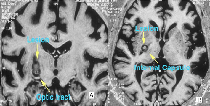
MRI (magnetic resonance imaging) T1‐weighted (A) coronal and (B) axial sequence showing lesion in the right pallidum. The lesion borders the inferior margin of the optic tract (A) and the medial margin of the internal capsule (B).
FIG. 2.

Postoperative plot of lesion sites at the AC–PC (anterior commissure–posterior commissure) plane, with the x and y coordinates scaled to the Schaltenbrand Wahren atlas AC–PC length. The variability in the lesion sites is due to the individual variability in the GPi (globus pallidus internus) and therefore cannot be reproduced effectively here. However, each individual lesion when evaluated was within the GPi. On the left side, the star, plus, and square represent cases 1, 4, and 6. On the right side, the star, triangle, diamond, circle, and square represent cases 3, 5, 8, 9, and 11, respectively.
Statistics
We estimated the median and interquartile range for the scores. We used the Wilcoxon matched pair signed‐rank test to assess the equality of matched pairs. As this study included observations of the involved (unblinded, observer A) and the noninvolved (blinded movement disorders specialists from different countries, observers B) teams, we used the Bland–Altman plots to visualize the difference in measurements between the blinded and unblinded reviewers. A P‐value <0.05 was considered statistically significant.
Results
Of the 13 patients who underwent pallidotomy, follow‐up was available in 11 cases. Eight patients had hemidystonia (7 secondary to lesions and 1 idiopathic) and 3 had focal dystonia (2 idiopathic and 1 with a hypoplastic splenium of unknown cause) (Table 1).
Blinded scoring was possible at all but three‐time points (one short‐term and two long‐term) because of inadequate video quality or missing video data. At short‐term follow‐up, there was a 12.1 (80%) and 7.5 (49%) point improvement in (median) BFMDRS scores, for observer A and observers B, respectively. This improvement was statistically significant for both observers; 4 of 11 patients had complete/near‐complete resolution of dystonia (BFMDRS 0–2). At long‐term follow‐up, the improvement in the median scores was zero, and it increased by 6.5 points, for Observer A and Observers B. This was statistically insignificant for observer A (Table 2).
TABLE 2.
BFMDRS scores of the patients
| Observer A | Observers B | Observer A | Observers B | Observer A | Observers B | |
|---|---|---|---|---|---|---|
| Preoperative | Preoperative | Postoperative <1 yr | Postoperative <1 yr | Last follow‐up as in December 2020 | Last follow‐up as in December 2020 | |
| Patient number | BFMDRS—motor score | BFMDRS—motor score | BFMDRS—motor score | BFMDRS—motor score | BFMDRS—motor score | BFMDRS—motor score |
| 1 | 15 | 10 | 6 | 6 | 4 | 6 |
| 2 | 19.5 | 15 | 6 | 9.5 | 4.5 | 11 |
| 3 | 12 | 24 | 7 | 7 | 2 | 7 |
| 4 | 12 | 12 | 3 | 8 | 13.5 | 21 |
| 5 | 32 | 40 | 6 | 25.5 | 15 | 22 |
| 6 | 9 | 14 | 0 | 2 | 16 | 24.5 |
| 7 | 9 | 13 | 1 | 7 | 20 | 27 |
| 8 | 6 | 6 | 0 | 2 | 0 | 2 |
| 9 | 17 | 29 | 2 | 15 | 15 | |
| 10 | 16 | 23 | 16 | 15 | 20 | 22 |
| 11 | 36 | 2 | 28 | |||
| Median | 15.0 | 14.5 | 3.0 | 7.5 | 15.0 | 21.0 |
| IQR | 9.0, 19.5 | 12.0, 24.0 | 1.0, 6.0 | 6.0, 15.0 | 4.0, 20.0 | 7.0, 22.0 |
| P‐value | 0.002 | 0.002 | 0.18 | 0.65 |
Abbreviations: BFMDRS, Burke–Fahn–Marsden Dystonia Rating Scale; IQR, interquartile range.
DDS scores were assessed only by observer A. There was a 6‐point (67%) improvement in short‐term follow‐up, which was completely lost at long‐term follow up (Table 3).
TABLE 3.
Dystonia Disability Scale scores for observer A
| Case number | Preoperative | Postoperative <1 yr | Last follow‐up as in December 2020 |
|---|---|---|---|
| Dystonia Disability Scale | Dystonia Disability Scale | Dystonia Disability Scale | |
| 1 | 5 | 2 | |
| 2 | 13 | 8 | 5 |
| 3 | 4 | 2 | 2 |
| 4 | 9 | 3 | 19 |
| 5 | 20 | 19 | 15 |
| 6 | 15 | 0 | 16 |
| 7 | 8 | 3 | 11 |
| 8 | 2 | 0 | 1 |
| 9 | 12 | 12 | 12 |
| 10 | 7 | 7 | 10 |
| 11 | 15 | 3 | 13 |
| Median | 9.0 | 3.0 | 11.0 |
| IQR | 5.0, 15.0 | 2.0, 8.0 | 2.0, 15.0 |
| P‐value | 0.01 | 0.69 |
Abbreviation: IQR, interquartile range.
Analyzing the data for primary (3 cases) and secondary dystonia (8 cases) revealed that at short‐term follow‐up there was 83% and 71% improvement in primary dystonia and 70% and 40% improvement in secondary dystonia, for observer A and observers B, for BFMDRS scores, respectively. This improvement was lost at long‐term follow‐up for observer A, whereas observers B continued to see an improvement of about 30% in the case of primary dystonia. For secondary dystonia, on long‐term follow‐up, observer A found almost no change, and observers B documented worsening. These observations are overall in line with the observations of the entire cohort, except the long‐term follow‐up improvement seen in primary dystonia, by observers B (Tables S2 and S3). As regards the DDS, the scores improved by 73% and 0% in cases of secondary dystonia and 63% and ‐37% for primary dystonia, at short‐term and long‐term follow‐up (Tables S4 and S5).
A comparison of interobserver variability between the blinded and unblinded assessors found that both groups were coherent as far as statistical significance was concerned. The Wilcoxon signed‐rank test comparing the percentage improvement between the two groups at short‐term and long‐term follow‐up showed no significant difference between the two groups. The interobserver variability data analyzed using Bland–Altman plots also showed good agreement (Fig. 3).
FIG. 3.

(A) Preoperative Bland–Altman plot with good coherence between the two observers. (B) Postoperative (short‐term) Bland–Altman plot with good coherence between the two observers. (C) Postoperative (long‐term) Bland–Altman plot with good coherence between the two observers.
Complications
Three patients experienced minor postoperative complications (2 pneumocephalus, 1 transient weakness), all of which resolved spontaneously within the first postoperative week. There were no visual field defects or gross cognitive changes.
Discussion
Long‐term follow‐up data after unilateral pallidotomy for focal dystonia and hemidystonia are limited. We present the long‐term follow‐up of these patients. Independent review of the videos and scoring of the same by movement disorders specialists (K.B. and E.M.), not associated with the surgical team, provide further strength to the observations of this study.
Between 1990 and 1998, after the resurgence in the interest in pallidotomy, there were several papers on pallidotomy for primary dystonia and a limited number of papers on secondary dystonia, 12 most reports comprising single case studies 5 , 17 , 18 , 19 and limiting their analysis to short‐term follow‐up. Between 1996 and 2007, 29 cases of pallidotomy for primary and 40 cases for secondary dystonia were reported in the literature, but only a small number underwent evaluation using standardized measures. 20 Lin et al. reported 18 patients who underwent bilateral contemporaneous or staged pallidotomy for generalized dystonia and found that 44% of patients showed improvement at 12‐month follow‐up 19 ; average BFMDRS score improved by 13%. Seven patients developed adverse effects, including hemiparesis and visual field defects, a few of them with more than one adverse effect.
Horisawa et al. recently published the outcome of 69 patients undergoing unilateral pallidotomy in primary dystonia. They found that the total BFMDRS score improved from 11.2 ± 14.7 preoperatively to 5.4 ± 7.6 at last‐available follow‐up (51.8% improvement, P < 0.001). 21 The mean follow‐up was 15.3 ± 10.9 months. All these patients suffered from bilateral and axial dystonia, and there was statistically significant improvement in the axial symptoms also. However, the ipsilateral side had only minor benefit. Long‐term outcome of these improvements in axial symptoms needs to be confirmed. Another study conducted by Eltahawy et al. found only 10.6% improvement in 6 patients with secondary dystonia undergoing pallidotomy or pallidal DBS. They did not find any difference in outcome between either procedure in their short follow‐up. 22 Our study had similar improvement in the BFMDRS and DDS scores, but this was lost on long‐term follow‐up in a few cases. Contrary to other reported series, 19 , 21 , 22 , 23 we noted most of the short‐term improvements in the immediate postoperative period. One reason could be the difference in the patient groups, as all the reported series was for generalized dystonia.
Is the Long‐Term Outcome Definitely Poor?
There is only one case report on the long‐term outcome of unilateral pallidotomy for secondary dystonia, which showed symptom progression over the long term. We analyzed our long‐term outcome in detail. We found symptom progression in 3 cases in the same body segments (no new parts involved, cases 4, 6, and 7) as their long‐term BFMDRS scores were worse than preoperative scores (Table 2) (Video 1). If we exclude them, the improvement will be 41% and 53% for observers A and B, respectively (Video 2). Both these observations were statistically significant. This is in line with the other reported series. 21 , 23 Loher et al. noted that though there was sustained long‐term improvement after unilateral GPi DBS in hemidystonia patients, some of the symptoms worsened after 2 years. 24 A similar observation has been noted after thalamotomy for tremors. 25 Chuang et al. 26 reviewed the natural history of 33 cases of secondary hemidystonia. They noted that dystonia could progress from one part to the other over a period of time, ranging from 2 months to 23 years. Lee et al. also noted that dystonia could progress over several years, ranging from several months to years, after head trauma. 27 This could be one explanation for worsening in 3 cases. The other explanation could be inadequate lesion; as mentioned earlier, there are several reports in the literature to suggest that. 3 , 7 , 26 However, in the present series this was not the case, as there was no significant difference in the site of the lesion nor the volume of the lesions between patients who had favorable and not‐so‐favorable long‐term outcome. Also the total volume of the lesions was similar to those reported earlier. 28 , 29 Therefore, the site and adequacy of the lesion were not the reasons for differential outcome in the present series. The third explanation, for which we do not have much evidence or support, is that this is the long‐term side effect of the lesion.
Video 1.
This video shows preoperative, short‐term, and long‐term follow‐up (8.5 years) of case 7, which had a poor outcome after pallidotomy at all three time points.
Video 2.
This video shows preoperative, short‐term, and long‐term follow‐up (7.5 years) of case 2 suffering from left hemidystonia, which had a good outcome after pallidotomy at all three‐time points.
This series has shown that there is a definite role for pallidotomy in the management of focal dystonia or hemidystonia. All patients experienced short‐term benefit, and the majority of the other patients also experienced long‐term relief (if we exclude those patients with progression). In the majority of patients, the quality‐of‐life improvement continued even at long‐term follow‐up, and in a few where there was progression, surgery did provide a relatively symptom‐free/reduced symptom period. The outcome after secondary dystonia has always been variable, with some patients experiencing remarkable improvement, either with lesioning or DBS, and some not having so much benefit, which again is reflected in our experience. A larger series may be helpful in defining the cohort of patients who may benefit in the long term. Future prospective studies should also include responder analysis to evaluate the benefits of pallidotomy. Pallidotomy is safe and has minimal morbidity in experienced hands.
Author Roles
(1) Research project: A. Conception, B. Organization, C. Execution; (2) Statistical analysis: A. Design, B. Execution, C. Review and critique; (3) Manuscript preparation: A. Writing of the first draft, B. Review and critique.
P.K.D.: 1A, 1B, 1C, 2A, 3A, 3B
M.B.: 1B, 1C, 3B
Eoin Mulroy : 1C, 3B
J.K.K.: 3B
K.B.: 1A, 1C, 3B
Disclosures
Ethics Compliance Statement: Jaslok Hospital and Research Centre ethics and scientific committee has approved this work. Informed patient consent was obtained in detail for the surgery, review of records and videos, and publication of their video on any platform. We confirm that we have read the Journal's position on issues involved in ethical publication and affirm that this work is consistent with those guidelines.
Funding Sources and Conflicts of Interest: No specific funding was received for this work. The authors declare that there are no conflicts of interest relevant to this work.
Financial Disclosure for previous 12 months: Paresh K. Doshi and Manish Baldia have no additional disclosures to report. Eoin Mulroy is funded by the Edmond J. Safra Foundation. He also receives research support from the National Institute for Health Research University College London Hospitals Biomedical Research Centre. Joachim K. Krauss received fees for consulting from Boston, Medtronic, Aleva, and Inomed, and grant support from the Deutsche Forschungsgemeinschaft. Kailash Bhatia received speaker honoraria from Ipsen, Merz, and MDS; personal compensation for scientific advisory board from Mitsubishi (Neuroderm), Jazz Pharma, and Ipsen; receives royalties from the publication of Oxford Specialist Handbook of Parkinson's Disease and Other Movement Disorders (Oxford University Press, 2008), Cambridge Press; and receives editorial work stipend from MDS for MDCP journal.
Supporting information
Table S1. Table showing the site of the lesion, volume of the lesion, and postoperative status at long‐term follow‐up.
Table S2. BFMDRS (Burke–Fahn–Marsden Dystonia Rating Scale) scores at short‐term and long‐term follow‐up in patients with secondary dystonia.
Table S3. BFMDRS (Burke–Fahn–Marsden Dystonia Rating Scale) scores at short‐term and long‐term follow‐up in patients with primary dystonia.
Table S4. DDS (Dystonia Disability Scale) scores at short‐term and long‐term follow‐up in patients with secondary dystonia.
Table S5. DDS (Dystonia Disability Scale) scores at short‐term and long‐term follow‐up in patients with primary dystonia.
Acknowledgments
We thank the Jaslok Hospital and Research Centre for the support provided for this work. We thank Mrs. Bharati Karkera and Mr. Pankaj Khadye for the support provided to collate patient information and data and Dr. Maninder Setia for his help with statistics. We thank all the neurologists who have supported this work, especially Dr. Pettarusp Wadia and Dr. Mohit Bhatt who have been the movement disorders specialists on the team during the past two decades.
References
- 1. Fahn S. Concept and classification of dystonia. Adv Neurol 1988;50:1–8. [PubMed] [Google Scholar]
- 2. Centen LM, Oterdoom DLM, Tijssen MAJ, Lesman‐Leegte I, van Egmond ME, van Dijk JMC. Bilateral pallidotomy for dystonia: a systematic review. Mov Disord 2021;36:547–557. 10.1002/mds.28384. [DOI] [PMC free article] [PubMed] [Google Scholar]
- 3. Cooper IS. 20‐year followup study of the neurosurgical treatment of dystonia musculorum deformans. Adv Neurol 1976;14:423–452. [PubMed] [Google Scholar]
- 4. Andrew J, Fowler CJ, Harrison MJ. Stereotaxic thalamotomy in 55 cases of dystonia. Brain 1983;106(Pt 4):981–1000. [DOI] [PubMed] [Google Scholar]
- 5. Alkhani A, Bohlega S. Unilateral pallidotomy for hemidystonia. Mov Disord 2006;21:852–855. 10.1002/mds.20838. [DOI] [PubMed] [Google Scholar]
- 6. Yoshor D, Hamilton WJ, Ondo W, Jankovic J, Grossman RG. Comparison of thalamotomy and pallidotomy for the treatment of dystonia. Neurosurgery 2001;48:818–824; discussion 824‐826. 10.1097/00006123-200104000-00025. [DOI] [PubMed] [Google Scholar]
- 7. Cardoso F, Jankovic J, Grossman RG, Hamilton WJ. Outcome after stereotactic thalamotomy for dystonia and hemiballismus. Neurosurgery 1995;36:501–507; discussion 507–508. 10.1227/00006123-199503000-00009. [DOI] [PubMed] [Google Scholar]
- 8. Bertrand C, Molina‐Negro P, Martinez SN. Stereotactic targets for dystonias and dyskinesias: relationship to corticobulbar fibers and other adjoining structures. Adv Neurol 1979;24:395–399. [Google Scholar]
- 9. Hariz MI, De Salles AA. The side‐effects and complications of posteroventral pallidotomy. Acta Neurochir Suppl 1997;68:42–48. 10.1007/978-3-7091-6513-3_8. [DOI] [PubMed] [Google Scholar]
- 10. Krack P, Vercueil L. Review of the functional surgical treatment of dystonia. Eur J Neurol 2001;8:389–399. [DOI] [PubMed] [Google Scholar]
- 11. Richardson RR. Rehabilitative neurosurgery: posttraumatic syndromes. Stereotact Funct Neurosurg 1989;53:105–112. 10.1159/000099525. [DOI] [PubMed] [Google Scholar]
- 12. Krauss JK, Mohadjer M, Braus DF, Wakhloo AK, Nobbe F, Mundinger F. Dystonia following head trauma: a report of nine patients and review of the literature. Mov Disord 1992;7:263–272. 10.1002/mds.870070313. [DOI] [PubMed] [Google Scholar]
- 13. Iacono RP, Kuniyoshi SM, Lonser RR, Maeda G, Inae AM, Ashwal S. Simultaneous bilateral pallidoansotomy for idiopathic dystonia musculorum deformans. Pediatr Neurol 1996;14:145–148. 10.1016/0887-8994(96)00003-3. [DOI] [PubMed] [Google Scholar]
- 14. Krauss JK, Desaloms JM, Lai EC, King DE, Jankovic J, Grossman RG. Microelectrode‐guided posteroventral pallidotomy for treatment of Parkinson's disease: postoperative magnetic resonance imaging analysis. J Neurosurg 1997;87:358–367. 10.3171/jns.1997.87.3.0358. [DOI] [PubMed] [Google Scholar]
- 15. Burke RE, Fahn S, Marsden CD, Bressman SB, Moskowitz C, Friedman J. Validity and reliability of a rating scale for the primary torsion dystonias. Neurology 1985;35:73–77. 10.1212/wnl.35.1.73. [DOI] [PubMed] [Google Scholar]
- 16. Doshi PK, Ramdasi RV, Karkera B, Kadlas DB. Surgical interventions for task‐specific dystonia (Writer's dystonia). Ann Indian Acad Neurol 2017;20:324–327. 10.4103/aian.AIAN_15_17. [DOI] [PMC free article] [PubMed] [Google Scholar]
- 17. Valálik I, Jobbágy A, Bognár L, Csókay A. Effectiveness of unilateral pallidotomy for meige syndrome confirmed by motion analysis. Stereotact Funct Neurosurg 2011;89:157–161. 10.1159/000323341. [DOI] [PubMed] [Google Scholar]
- 18. Tripathi M, Sharan S, Mehta S, et al. Gamma knife Radiosurgical pallidotomy for dystonia: not a fallen angel. Neurol India 2019;67:1515–1518. 10.4103/0028-3886.273644. [DOI] [PubMed] [Google Scholar]
- 19. Lin JJ, Lin SZ, Chang DC. Pallidotomy and generalized dystonia. Mov Disord 1999;14:1057–1059. 10.1002/1531-8257(199911)14:6<1057::aid-mds1034>3.0.co;2-1. [DOI] [PubMed] [Google Scholar]
- 20. Gross RE. What happened to posteroventral pallidotomy for Parkinson's disease and dystonia? Neurotherapeutics 2008;5:281–293. 10.1016/j.nurt.2008.02.001. [DOI] [PMC free article] [PubMed] [Google Scholar]
- 21. Horisawa S, Fukui A, Takeda N, Kawamata T, Taira T. Safety and efficacy of unilateral and bilateral pallidotomy for primary dystonia. Ann Clin Transl Neurol 2021;8:857–865. 10.1002/acn3.51333. [DOI] [PMC free article] [PubMed] [Google Scholar]
- 22. Eltahawy HA, Saint‐Cyr J, Giladi N, Lang AE, Lozano AM. Primary dystonia is more responsive than secondary dystonia to pallidal interventions: outcome after pallidotomy or pallidal deep brain stimulation. Neurosurgery 2004;54:613–619; discussion 619–621. 10.1227/01.neu.0000108643.94730.21. [DOI] [PubMed] [Google Scholar]
- 23. Ondo WG, Desaloms JM, Jankovic J, et al. Pallidotomy for generalized dystonia. Mov Disord 1998;13:693–698. 10.1002/mds.870130415. [DOI] [PubMed] [Google Scholar]
- 24. Loher TJ, Hasdemir MG, Burgunder JM, Krauss JK. Long‐term follow‐up study of chronic globus pallidus internus stimulation for posttraumatic hemidystonia. J Neurosurg 2000;92:457–460. 10.3171/jns.2000.92.3.0457. [DOI] [PubMed] [Google Scholar]
- 25. Dallapiazza RF, Lee DJ, Vloo PD, et al. Outcomes from stereotactic surgery for essential tremor. J Neurol Neurosurg Psychiatry 2019;90:474–482. 10.1136/jnnp-2018-318240. [DOI] [PMC free article] [PubMed] [Google Scholar]
- 26. Chuang C, Fahn S, Frucht SJ. The natural history and treatment of acquired hemidystonia: report of 33 cases and review of the literature. J Neurol Neurosurg Psychiatry 2002;72:59–67. 10.1136/jnnp.72.1.59. [DOI] [PMC free article] [PubMed] [Google Scholar]
- 27. Lee MS, Rinne JO, Ceballos‐Baumann A, Thompson PD, Marsden CD. Dystonia after head trauma. Neurology 1994;44:1374–1378. 10.1212/wnl.44.8.1374. [DOI] [PubMed] [Google Scholar]
- 28. Lim JY, De Salles AAF. Sequential postoperative appearance of radiofrequency pallidotomy lesions on MRI. Stereotact Funct Neurosurg 1997;69:46–53. 10.1159/000099851. [DOI] [PubMed] [Google Scholar]
- 29. Cohn MC, Hudgins PA, Sheppard SK, et al. Pre‐ and postoperative MR evaluation of stereotactic pallidotomy. AJNR Am J Neuroradiol 1998;19:1075–1080. [PMC free article] [PubMed] [Google Scholar]
Associated Data
This section collects any data citations, data availability statements, or supplementary materials included in this article.
Supplementary Materials
Table S1. Table showing the site of the lesion, volume of the lesion, and postoperative status at long‐term follow‐up.
Table S2. BFMDRS (Burke–Fahn–Marsden Dystonia Rating Scale) scores at short‐term and long‐term follow‐up in patients with secondary dystonia.
Table S3. BFMDRS (Burke–Fahn–Marsden Dystonia Rating Scale) scores at short‐term and long‐term follow‐up in patients with primary dystonia.
Table S4. DDS (Dystonia Disability Scale) scores at short‐term and long‐term follow‐up in patients with secondary dystonia.
Table S5. DDS (Dystonia Disability Scale) scores at short‐term and long‐term follow‐up in patients with primary dystonia.