Abstract
Schaumann bodies are the inclusion bodies usually seen in sarcoidosis, but can also be found in other conditions like tuberculosis, chronic beryllium diseases and Crohn’s diseases. Histopathologically, these bodies appear as round to oval shell-like basophilic calcifications usually considered to be as a residuum of lysosomal organelles activity.
Keywords: Basophilic, calcified structure, inclusion body, sarcoidosis, Schaumann body
Schaumann bodies, also known as conchoidal bodies, are round or oval, laminated calcified structure most commonly found in sarcoidosis, tuberculosis, Crohn’s disease, chronic beryllium disease and lymphogranuloma inguinale.[1] The first documentation of the Schaumann bodies in the literature was described by Jorgen Nilsen Schaumann in 1941.[2]
They are basically the inclusion bodies that are composed of two basic elements, namely calcium carbonate crystals with a high refractive index and concentric, laminated and conchoidal bodies.[3] These inclusions are birefringent to polarized light.[4]
They are composed of a protein matrix, impregnated with calcium salts, phosphate or carbonate or both. Both components arise in epithelioid cells or Langhans giant cells.[3] Johnson FB labelled the central dark isotropic void structure in the refractile bodies as a ’kern’.[5]
Giant cells contain crystalline inclusions, which serves as a serve as a nidus for the deposition of calcium leading to the formation of Schaumann bodies.[4] Macrophages in turn elaborate a calcium binding muco-glycoprotein along with platelet-activating factor present in macrophages increase calcium ion concentration. Additionally, damaged cell membrane barriers might allow influx of calcium ions.[6,7,8] These inclusions are believed to be non-specific end-products of the active metabolism and secretion that takes place in the giant cells.[9]
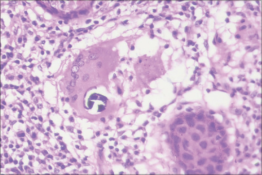
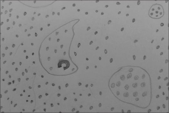
They are mostly found in the cytoplasm of multinucleated giant cells (MGCs) but not in epithelioid cells. These bodies are stained pink, brown, dark blue or black with routine haematoxylin and eosin.[10] [Figures 1 and 2] Electron-microscopically, Schaumann bodies are complex, concentrically stratified concretions, up to 150 micrometre in diameter, that often enclose haematoxyphilic mineralized components or birefringent crystalline material.[9]
Figure 1.

Histopathological image showing non-caseating granuloma with giant cells containing calcified basophilic Schaumann bodies in oral sarcoidosis. (H&E, x400)
Figure 2.

Hand-drawn illustration showing presence of Schaumann bodies in multinucleated giant cells
Financial support and sponsorship
Nil.
Conflicts of interest
There are no conflicts of interest.
REFERENCES
- 1.Purushothaman S, Budeda H, Vinnakoti A, Satyanarayana VV. Lupus vulgaris with Schaumann bodies–An atypical histopathological finding:Lupus vulgaris with Schaumann bodies. Journal of Pakistan Association of Dermatologists. 2021;31:514–7. [Google Scholar]
- 2.Kulkarni M, Agrawal T, Dhas V. Histopathologic bodies:An insight. J Int Clin Dent Res Organ. 2011;3:43–7. [Google Scholar]
- 3.Jones Williams W. The nature and origin of Schaumann bodies. J Pathol Bacteriol. 1960;79:193–201. doi: 10.1002/path.1700790126. [DOI] [PubMed] [Google Scholar]
- 4.Žák F. Contribution to the origin, development and experimental production of laminated calcinosiderotic Schaumann bodies. Acta Medica Scandinavica. 1964;176:21–4. [PubMed] [Google Scholar]
- 5.Reid JD, Andersen ME. Calcium oxalate in sarcoid granulomas:with particular reference to the small ovoid body and a note on the finding of dolomite. American journal of clinical pathology. 1988;90:545–58. doi: 10.1093/ajcp/90.5.545. [DOI] [PubMed] [Google Scholar]
- 6.James EM, Williams WJ. Fine structure and histochemistry of epithelioid cells in sarcoidosis. Thora×. 1974;29:115–20. doi: 10.1136/thx.29.1.115. [DOI] [PMC free article] [PubMed] [Google Scholar]
- 7.Farber JL. Biology of disease:Membrane injury and calcium homeostasis in the pathogenesis of coagulative necrosis. Lab Invest. 1982;47:114–23. [PubMed] [Google Scholar]
- 8.Conrad GW, Rink TJ. Platelet activating factor raises intracellular calcium ion concentration in macrophages. J Cell Biol. 1986;103:439–50. doi: 10.1083/jcb.103.2.439. [DOI] [PMC free article] [PubMed] [Google Scholar]
- 9.Kirkpatrick CJ, Curry A, Bisset DL. Light- and electron-microscopic studies on multinucleated giant cells in sarcoid granuloma:New aspects of asteroid and Schaumann bodies. Ultrastruct Pathol. 1988;12:581–97. doi: 10.3109/01913128809056483. [DOI] [PubMed] [Google Scholar]
- 10.Engle RL., Jr The association of iron-containing crystals with Schaumann bodies in the giant cells of granulomas of sarcoid type. Am J Pathol. 1951;27:1023–35. [PMC free article] [PubMed] [Google Scholar]


