



Abstract
Despite therapeutic advances, overall survival in glioblastoma is dismal. To optimize progress, a more detailed understanding of glioma’s molecular, cellular, and intercellular pathophysiology is needed. Recent investigation has revealed a vital role for exosomes in inter-cellular signaling, tumor cell support, and regulation of the tumor microenvironment. Exosomes carry miRNAs, lncRNAs, mRNAs, proteins, immune regulatory molecules, nucleic acids, and lipids; however, the composition of exosome cargo is variable depending on the cell of origin. Specific exosomal miRNA contents such as miR-21, miR-301a, miR-151a, miR-148a, and miR-5096 are altered in high-grade glioma. Unique proteomic, genomic, and miRNA signatures of tumor exosomes have been associated with disease pathobiology, temozolomide resistance, immunosuppression, and tumor proliferation. Exosomes hold promise for tissue diagnostic glioma diagnosis and monitoring response to therapy. This review summarizes the current understanding of exosomes, their crucial role in glioma pathology, and future directions for their use in diagnosis and treatment.
Graphical Abstract

Summary of exosome origin and scope of effects on glioma cells, microenvironment, and overall therapeutic response. Affects include immunosuppression, angiogenesis, tumor proliferation, and temozolomide resistance.
1. Introduction
Current methods of glioma diagnosis and surveillance include invasive and non-invasive tests. Non-invasive testing involves magnetic resonance imaging (MRI), including diffuse weighted restriction, gadolinium contrast enhancement, functional and MR spectroscopy, and clinical examination for focal deficits [1]. Invasive testing consists primarily of surgical lesion biopsy, either in the form of resection with biopsy or stereotactic needle biopsy. Tissue biopsy provides critical histologic, molecular, and genetic information to determine an accurate diagnosis and the most appropriate treatment regimen [2]. Unfortunately, neurosurgery carries significant morbidity, especially in the setting of older patients or those with multiple chronic conditions.
Both molecular tumor characteristics and effects of radiation and chemotherapy can complicate the interpretation of MRIs, often leading to pseudoprogression or radiation necrosis which mimics tumor growth [3]. Much research has been dedicated to developing less invasive, and hence less morbid, diagnostic approaches to glioma diagnosis and recurrence. Despite advances in surgery, chemotherapy, and radiotherapy, overall glioblastoma (GBM) survival is dismally low at 12–15 months. To optimize progress in this devastating disease, a greater understanding of the molecular, cellular, and intercellular pathophysiology of glioma is needed.
The glioma tumor microenvironment (TME) has been extensively investigated. The TME is heterogeneous and includes endothelial cells, immune cells, glioma stem cells, and astrocytes, though environment composition varies among specific tumor subtypes. GBM has a TME with significant areas of hypoxia, which drive malignancy, enhance therapy resistance and angiogenesis, and correlate with poor outcomes [4]. Communication between cells within the TME is accomplished by direct cell-cell contacts or through secreted soluble mediators like cytokines, chemokines, growth factors, and metabolites. In recent decades, intercellular communication via the release of extracellular vesicles such as exosomes has been revealed under normal and pathologic states. Initially thought to function only to release cellular debris, exosomes have been isolated from various biofluids, including serum, cerebrospinal fluid (CSF), and urine, and have been demonstrated to communicate with neighboring and distant cells via transfer of cargo containing RNA, DNA, lipids, and proteins. Exosomes have become an area of interest for possible diagnostic and therapeutic roles.
Extracellular vesicles (EVs), first described in 1946 by Erwin Chargaff and Randolf West in the setting of coagulation [5, 6], range from 30–100 nm and include microvesicles and exosomes [5]. “Microvesicle” describes EVs released directly from the outward budding of the plasma membrane, as opposed to “exosomes” which are secreted via the endocytic pathway [7, 8]. In 1991 Rose Johnstone and colleagues described exosomes as a cellular waste disposal mechanism [9]; however, modern investigation implicates exosomes in cancer cell-environment communication. Based on their physical characteristics as well as phenotype, EVs have been classified into four general subtypes: microvesicles, membrane particles, apoptotic vesicles, and exosomes [10]. Other nanoparticles such as supermeres and exomeres have been recently identified during the exosome isolation, though comprise a separate category of particles. While these nanoparticles carry molecules involved in intercellular communication, their structure does not contain a lipid membrane. This property differentiates nonvesicular extracellular particles from exosomes [11]. Exosomes are involved in a multitude of processes such as immune regulation, tissue regeneration, and tissue maintenance [12]. A recent investigation into the role of exosomes has revealed their involvement in inter-cellular signaling, supporting tumor cells and their microenvironment [13]. Interestingly, exosome cargo is variable at different stages of cancer development [14].
1.1. Exosome Synthesis
Exosome synthesis is initiated with the invagination of the plasma membrane. This membrane contains components from the outer component and retains them within the new vesicle. Once multiple new small vesicles are formed, termed intraluminal vesicles (ILV), they are sequestered within late endosomal compartments, termed multivesicular bodies (MVB) [15]. MVBs subsequently fuse with the plasma membrane and release the formed intraluminal vesicles, now termed exosomes (Fig.1a) [10]. The exosomes travel to other cells where they are endocytosed, and their cargo is delivered. Their contents include a lipid bilayer membrane, encasing bioactive compounds such as cytoplasmic and membrane proteins, mRNA, miRNA, and lipids (Fig.1b) [16]. Exosome membranes have a higher concentration of lipids associated with lipid rafts, including cholesterol, ceramides, and sphingomyelin, than typical plasma membranes [7]. Exosome proteins include transport proteins, signaling proteins, GTPases, annexins, and integrins, proteins from their origin MVB (Tsg101 and ALIX), heat shock proteins (Hsp90 and Hsp70), as well as tetraspanins (CD9, CD63, CD81, and CD82). Tetraspanins, each with a particular palmitoylation site, are contained in high concentrations and are considered specific exosome surface biomarkers [17, 18].
Figure 1:
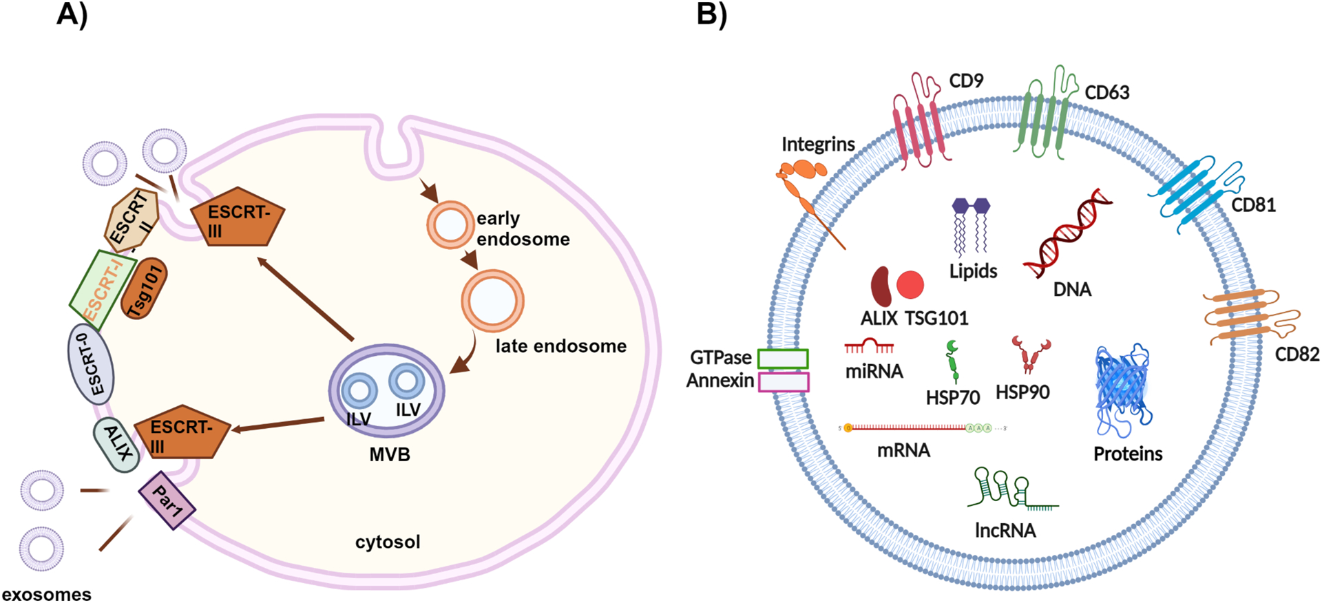
a).Schematic representation of exosome biogenesis and their general composition. The scheme depicts exosome synthesis beginning with invagination of the plasma membrane, which forms new small vesicles called early endosomes. They are then transferred to late endosomal compartments, termed multivesicular bodies (MVB) once intraluminal vesicles (ILVs) are formed. MVBs fuse with the plasma membrane and release the intraluminal vesicles, now called exosomes. b). An example of exosome structure is shown with common membrane proteins CD9, CD63, CD81, CD81, Integrins, GTPase, and Annexin. Common cargo includes mRNA, miRNA, lncRNA, proteins, DNA, lipids, HSP70, HSP90, ALIX, and TSG101. mRNA, messenger RNA; miRNA, microRNA; lncRNA, long noncoding RNA; HSP70, Heat Shock Protein 70 (HSP70); HSP90 Heat Shock Protein 90; ALIX, Apoptosis Linked Gene 2-Interacting Protein; TSG101, Tumor Susceptibility Gene 101.
Endosomal Sorting Complexes Required for Transport (ESCRT) determine which bioactive components are contained within each exosome. The ESCRT complex pathway is a group of 5 complexes, ESCRT-0, -I, -II, -III, and multiple accessory proteins involved in the recognition and packaging of cargo, membrane formation, and MVB biogenesis. Cargos are clustered via an ESCRT-0 complex that initiates the MVB formation pathway. First, invagination of the plasma membrane is triggered by ESCRT-I and ESCRT-II complexes, with resulting vesicles cleaved by ESCRT-III [7]. Early ESCRT complexes, ESCRT-0, -I, and -II, are involved in sorting ubiquitinated molecules via ubiquitin-binding domains. ESCRT-III and the accessory protein complex ATPase Associated with various cellular Activities (AAA ATPase ) Vacuolar protein sorting-associated protein 4 (Vps4) are involved in cargo isolation of ILVs after formation and vesicle shedding [19]. A subunit of ESCRT-I, Tsg101, is a major regulatory factor in exosomal secretion and, as mentioned previously, is often secreted within the final exosomes. Another protein within final exosomes is ALIX, which interacts with ESCRTs. ALIX binds ESCRT III, which sequesters cargo and drives inward budding, as well as the G-protein coupled receptor PAR1. PAR1 then sorts itself into the cargo of an MVB without ubiquitination, a post-translation modification where ubiquitin molecules are attached to protein substrates for protein degradation, required for many other components to prevent degradation during transport to other cells [20, 21]. In addition, ALIX also plays a crucial role in sorting and including syntenin, syndecan, and CD63 into exosomes without ubiquitination [19]. The molecular pathways of early endosome, late endosome, MVB, ILV, and exosome formation have been revealed in exquisite detail, though they are outside the scope of this review [19] (Fig 1A).
1.2. Exosome cargo
Exosomes from normal and malignant cells contain micro ribonucleic acids (miRNAs), long noncoding ribonucleic acids (lncRNAs), messenger ribonucleic acids (mRNAs), proteins, immune regulatory molecules, nucleic acids such as deoxyribonucleic acid (DNA), organelle fragments, lipids, and metabolites. Exosomal RNA cargo content generally reflects the origin cell’s cytosolic composition, with miRNAs loaded via cell-activation-dependent alterations in miRNA targets in the parent cell [22]. The exact mechanisms of molecule selection for exosome cargo is still under investigation. The concentration of each component varies based on the cell of origin and hence affects intercellular communication.
Generally, exosomes contain lipids such as diglycerides, phospholipids, and cholesterol [23]. They generally contain a 2–3 fold enrichment of sphingomyelin, glycophospholipids, cholesterol, and phosphatidylserine, with lower concentrations of phosphatidylinositol and phosphatidylcholine in exosomes. The specific lipid profile in each exosome also varies depending on the type of cell from which it originates. For example, exosomes released from prostate cancer PC-3 cells contain less ceramide and sphingomyelin compared to exosomes released from Oli-neu cells, an oligodendrocyte cell line. Astrocytes shed exosomes with elevated levels of ceramide in Alzheimer’s disease [24, 25]. Exosome lipid profiles as biomarkers, i.e., ‘lipidomics’ have been extensively studied in pancreatic, non-small cell lung, and ovarian cancer; however, less is known about glioma-specific exosome lipid profiles [5].
A significant number of miRNAs play a role in the malignant transformation and progression of glioma, such as miR-21, miR-301a, and miR-148a, which will be discussed in more detail [26]. MiRNAs influence mRNA expression, affecting intercellular communication [27]. Upon interaction with RNA-binding proteins, miRNAs are transported into MVBs and subsequently loaded into exosomes[28]. Exosomal miRNAs are transported to bind and affect mRNA transcription in other cells at distant sites. Generally, miRNA binding to mRNA leads to decreased target gene expression, not complete silencing [29]. In addition to miRNA, single and double-stranded DNA molecules are carried and transferred via exosomes as well. Multiple subtypes of DNA are observed in exosomes, including mitochondrial, ribosomal, nuclear, cell-free, cytoplasmic, genomic, retrotransposon elements, and micro-nuclear. Cancer cells often carry DNA tumor-specific alterations, which can be distributed to other cancer cells and/or non-cancerous cells. DNA molecules isolated from GBM-derived exosomes include the Nanog Homeobox gene (NANOG), Sry-box transcription factor 2 (SOX2), epidermal growth factor receptor mutation vIII (EGFRvIII), O-6-methylguanine-DNA methyltransferase (MGMT), isocitrate dehydrogenase 1 and 2 (IDH1 and IDH2), and the proto-oncogene c-Myc amongst others [30]. MGMT promotor methylation status is a significant prognostic factor for GBM response to chemotherapy with temozolomide (TMZ) and IDH mutation status is a significant positive overall prognostic factor. In 2021, Maire et al. demonstrated accurate detection of MGMT promotor methylation status and IDH mutation status via GBM-derived exosomes from human GBM stem-like cells in culture [31].
2. Exosomes within the Physiologic Central Nervous System
The essential role of exosome-mediated intercellular communication has been explored in various tissue types. Within the central nervous system (CNS), this form of communication is implicated between neurons, glial cells, and microglia. Neuron-derived exosomes isolated from a culture medium contain abundant miRNA and small RNAs [32]. MiR124–3p, essential for neuronal function and identity, is at high levels. Neuron-specific miRNAs can be transferred to astrocytes and other glial cells via exosome communication. In 2013, Morel et al. demonstrated cortical neuron-derived exosomes containing miR-124a resulted in the upregulation of the predominant astroglial glutamate transporter GLT1. Astroglial GLT1 levels are dynamic during neuronal development and pathologic states and are noted to be significantly reduced in mouse models of ALS [32]. As another example of miRNA driving normal physiology, miR-219 is required to produce myelinating oligodendrocytes [32]. Other miRNA transfers have been implicated in the promotion of myelination as well as in the maintenance of brain vascular integrity. Neuron-derived exosomes transfer miR-132 into endothelial cells to regulate vascular integrity. Knockdown or mutation of miR-132 leads to disruption of the brain vasculature via reduced expression of vascular endothelial cadherin, an adherent junction protein, often leading to subsequent hemorrhage [33].
Neurons are not the only cell type in the CNS to communicate via exosomes. In addition to neurons, microglia, phagocytic immune regulatory cells, secrete exosomes during antigen presentation as well as during independent exosome release [34]. Guo et al. demonstrated intercellular communication between neural cells and microglia via the rat pheochromocytoma PC12 cell line with neuroblastic characteristics. Exosomes isolated from PC12 medium containing miR-21–5p promoted M1 polarization of microglia [35]. Microglial exosome levels of miR-124–3p increase from the acute to the chronic phase of neural injury, correlating with a transition from M1 pro-inflammatory M2 anti-inflammatory polarization. This transition inhibits neuronal inflammation and promotes neuritis outgrowth [36]. The CNS utilizes exosome-mediated intercellular communication in normal homeostasis, inquiry response, and pathologic states.
3. Exosomes in Glioma
Exosomes can provide specific and quantitative information correlated with the parent glioma cells from which they are derived. In contrast to free serum RNA, DNA, and proteins, exosomes provide a vehicle for these elements to be transported without degradation, thereby serving as the surrogates of a genomic, proteomic, and transcriptomic profile of the cell of origin. Isolation of exosomes from serum provides a unique opportunity to detect specific tumor-derived metabolites and markers.
In glioma specifically, several tumor-derived exosomal proteins and miRNAs have been discovered. Packaging miRNAs and proteins into exosomes is a selective process that is altered in tumorigenesis. Isolation of glioma-specific exosomal miRNAs and proteins has been demonstrated in CSF and serum.
3.1. Exosomal miRNA in gliomagenesis
Many miRNAs appear to contribute to the pathogenesis of glioma (Table 1). MiR-21 is a well-known miRNA up-regulated in almost all cancer types, which targets Programmed Cell Death 4 (PDCD4), Tissue Inhibitor of Metalloproteinase 3 (TIMP3), and Reversion Inducing Cysteine Rich Protein With Kazal Motifs (RECK), leading to tumor cell proliferation, metastasis, and invasion [37]. Glioma cells in hypoxic conditions transfer radiation resistance to normoxic glioma cells via exosomal miRNA. For example, exosomal miR-301a directly targets and downregulates the expression of TCEAL7, which functions as a tumor suppressor. This leads to persistent activation of the Wnt/beta-catenin pathway and increased translocation of beta-catenin from the cytoplasm to the nucleus, subsequently promoting increased cellular proliferation as well as radiation resistance [38]. In addition, multiple exosomal miRNAs have been implicated in TMZ resistance. TMZ, an oral alkylator, is the primary chemotherapy utilized in glioma. The exosomes of TMZ-resistant GBM cells have reduced miR-151a than TMZ-susceptible cells. MiR-151a sensitizes GBM by inhibiting X-ray repair cross-complementing 4 (XRCC4)- mediated DNA repair. Restoration of exosomal miR-151a levels abrogated TMZ resistance [39]. Yin et al. demonstrated a correlation between TMZ resistance and over-expression of exosomal miR-1238, which inhibits the caveolin-1 (CAV1)/EGFR pathway. CAV1 acts as a tumor suppressor in GBM and interacts with EGFR to stabilize the receptor kinase in the inactive conformation [40]. Another example is exosomal miR-148a, which had been linked to the downregulation of Cell Adhesion Molecule 1 (CADM1) expression in GBM patient samples. MiR-148a antagonism reduced p-STAT3 levels and subsequent upregulation of tumor suppressor CADM1 levels [41]. Thuringer et al. demonstrated that miR-5096 stimulated filamentous pseudopodia, driving glioma invasiveness and cellular proliferation [42].
Table 1:
Summary of miRNA in Glioma
| Origin | Target cell | Target | Effect | References | |
|---|---|---|---|---|---|
| miR-301a | GBM hypoxic cells | Normoxic glioma cells | TCEAL7 gene | Suppressed expression in normoxic glioma cells | 78, 38 |
| miR-151a | GBM TMZ-resistant cells | N/A | Unknown | Lower levels noted in resistant GBM cells | 39 |
| miR-1238 | GBM TMZ resistant cells | GBM TMZ sensitive cells | CAV1/EGFR pathway | TMZ resistance | 40 |
| miR-5096 | GBM cells | GBM cells | K+ channel Kir4.1 | Increased production of pseudopodia, increased glioma invasiveness | 42 |
| miR-148a | GBM cells | GBM cells | CADM1 | Increase STAT3 activation, increase development and metastasis | 41 |
| miR-29a | Hypoxic glioma cells | MDSC | Hbp1 | Induced MDSC differentiation | 43 |
| miR-21 | Almost all cancer types | Multiple | PDCD4, TIMP3, RECK | Stimulated tumorigenesis | 7, 35, 37 |
| miR-133b | GBM, also various other cancers | Glioma cells | Sirt1 gene | Downregulation of EZH2, lead to glioma growth suppression | 48, 49, 50, 80 |
| miR-584 | Multiple cancer type cells | Multiple cancer type cells | CYP2J2 | Induced apoptosis, decreased metastasis | 51, 52, 53, 54, 55 |
| miR-132 | Glioma, neurons | Endothelial cells, glioma cells | Eef2k | Increase blood brain barrier permeability, decreased tumorigenesis, increases tumor apoptosis | 33, 84, 85 |
| miR-1246 | Glioma cells | Monocytes, MDSCs, macrophage | NF-kB, TERF2IP, | Activate differentiation to MDSCs, activate MDSCs, macrophage M2 polarization | 44, 45 |
| miR-1298–5p | Glioma cells | MDSCs | NK-kB | Activation of MDSCs | 46 |
| miR-200c-3p | Neurons in hypoxic GBM environment | Microglia | DUS9 | Microglial M2 polarization | 47 |
Exosomal miRNAs contribute to tumor development, progression, and chemotherapy resistance and are also involved in tumor-induced immunosuppression. The glioma TME is composed of a variety of cell types including myeloid-derived suppressor cells (MDSCs), glioma cells, and endothelial cells. Stromal cells utilize exosomes to communicate and regulate their complex surrounding environment. During tumor progression, tight control of local immune response occurs to allow further tumor cell proliferation and migration. To achieve this, the TME has developed unique mechanisms to interact with surrounding immune cells. In GBM, MDSCs comprise approximately 30% of the total tumor volume. MDSCs mediate immunosuppression via multiple mechanisms, including elevated levels of IL-10, nitric oxide, arginase-1, reactive oxygen species, and TGF-beta. Glioma-derived exosomal miR-29a and miR-92a were shown to induce functional differentiation of MDSCs into macrophages, granulocytes, and dendritic cells. This phenomenon was more strongly demonstrated in hypoxic than normoxic glioma exosomes [43]. In 2021 Qiu et al. isolated exosomes from CSF of 21 patients with glioma and noted significantly higher levels of miR-1246. Elevated levels of miR-1246 in glioma-derived exosomes were correlated with increased differentiation of monocytes into MDSCs, as well as activation of MDSCs [44]. miR-1246 from hypoxic glioma-derived exosomes was also demonstrated to induce M2 macrophage polarization, which can promote tumor progression, in vitro and in vivo by Qian et al in 2020 [45]. Another miRNA in glioma-derived exosomes, miR1298–5p, has also been isolated from CSF of patients with glioma. Qi et al. successfully detected the transfer of miR1298–5p from transfected glioma cells to MDSCs in culture. miR-1298–5p activated adjacent MDSCs via potentiation of the nuclear factor kappa-light-chain-enhancer of activated B cells (NF-kB) pathway [46]. More recently, in 2023, Guo et al. revealed an increased level of miR-200c-3p in exosomes derived from neurons within the hypoxic glioblastoma environment. Elevated miR-200c-3p leads to a downregulation of dual specificity phosphatase 9 (DUS9) in microglia, which leads to microglial M2 polarization [47].
Glioma-derived exosomal miRNAs include tumor suppressors. The level of tumor suppressor miRNA within exosomes is significantly decreased in the setting of glioma compared to normal conditions, thereby promoting proliferation and survival. Li et al. determined that miR-133b had an inhibitory effect on GBM. MiR-133b regulates silent information regulator 1 (Sirt1) gene expression, subsequently decreasing glioma growth and metastasis [48–50]. Microarray assays have shown downregulation of miR-133b within the glioma cells. miR-584 is another tumor suppressor seen in glioma-derived exosomes. It targets pituitary tumor-transforming gene 1 interacting protein (PTTG1IP), an oncogene involved in the regulation of the metaphase-anaphase transition of the cell cycle [51, 52]. MiR-584 also represses the transcription of matrix metalloproteinase 14 (MMP14) by binding to the MMP-14 promotor. Decreased levels of miR-584 with subsequent elevation in MMP-14 were observed in neuroblastoma as well as clear cell renal cell carcinoma and breast cancer, leading to increased tumor growth, invasion, metastasis, and angiogenesis [51, 53–55]. The role of miR-584 in glioma is a promising area for future investigations.
MiR-199a is a cancer-related miRNA targeting Arf GTPase-activating protein (AGAP2) involved in endosome trafficking. Over-expression of miR-199a halts the growth of renal cancer and papillary thyroid carcinoma. Expression of miR-199a is lower in glioma than in normal brain tissue [56, 57].
3.2. The Role of Exosomal Proteins in Glioma
Proteins within exosomes also play a role in cancer pathogenesis. Protein composition and quantity correlate with specific tumor types and stages of development, respectively. All small EVs contain common proteins CD9, CD63, CD81, and CD82, and their membranes also usually express tumor susceptibility gene 101 protein (TSG101), HSP70, and ALIX [23]. These universal exosome markers are key for their identification and isolation.
As with miRNA, exosomes also contain proteins specific to glioma pathology and therapeutic resistance. Exosomes from patients with GBM have a higher content of proteins related to hypoxic conditions, reflective of the GBM tumor microenvironment. Specifically, exosomes contain higher levels of the following: matrix metalloproteinase 9 (MM9), IL8, CD26, pentraxin 3 (PTX3), PGDF-AB/AA, and plasminogen activator 1 (PAI1) [13]. Vascular endothelial growth factor (VEGF) isoform VEGF-C was isolated from GBM-derived exosomes by Wang et al. VEGF-C binds VEGF receptor 2 and stimulates tafazzin (TAZ) expression in endothelial cells, leading to improved endothelial cell viability, migration, and tubularization—all promoting angiogenesis [58]. Ma et al. evaluated the variations in protein expression between normal glial cells vs. glioma-derived exosomes. Mass spectrometry analysis of exosomal proteins from astrocytes and C6 cells, a rat glioma cell line, revealed that normal glial cell exosomes were enriched in proteins related to RNA transcription. In contrast, the glioma cell exosomes primarily contained proteins involved in the PI3K-Akt pathway, adhesion, and tumorigenesis [59]. Exosome surface proteins also vary depending on the cell of origin. Immunoglobulin superfamily protein L1, an exosome surface protein, stimulates glioma cell proliferation, migration, and invasion, as demonstrated by Pace et al. using exosomes from the T98G glioma cell line. L1 exosome surface expression increased migration velocity and proliferation in three GBM cell lines (T98G/shL1, U-118 MG, and primary patient-derived GBM cells). In addition, L1-expressing exosomes also enhanced the invasiveness of the T98G/shL1 cell line [60].
Multiple exosomal proteins have been implicated in TMZ resistance and immunosuppression. Exosome-linked Connexin 43 (CX-43) levels were significantly elevated in TMZ-resistant GBM cells compared to TMZ-sensitive cells, with noted enhanced cell migration and invasion [61]. Macrophage movement inhibitory factor (MIF) also increases TMZ-resistant cell exosomes and suppresses TIMP3 expression, activating the PI3K/AKT signaling pathway of growth [62].
GBM-derived exosome protein CD73 downregulates T-cell response, supporting glioma progression via immunosuppression. Elevated levels of CD73 have been observed in the cerebrospinal fluid (CSF) of GBM patients compared with normal controls [63]. In addition, human astrocytes treated with GBM-derived exosomes exhibited increased expression of IFN-gamma, IL1A, IL12, IL1B, IL8, CXCL10, and complement factor C5 as compared to astrocytes exposed to exosomes from normal epithelial cells [64]. This increase in the pro-inflammatory chemokines and interleukins helps maintain the inflammatory and immunosuppressive environment. Other cytokines released from astrocytes treated with GBM-derived exosomes, such as CSF2 and 3, IL4, IL10, and IL13, are implicated in T cell regulation and immune suppression [65] (Fig.2).
Figure 2:

Exosome-mediated cellular crosstalk between tumor cells, astrocytes, and immune cells in the glioma tumor microenvironment drives immunosuppression. Glioma-derived exosomes contain miR-29a and miR-92a, which promote the differentiation of MDSCs and enrich TME in immunosuppressive factors. Astrocytes release multiple signaling factors, which promote an anti-inflammatory environment and inhibit T-cell activity. TME, tumor microenvironment; MDSCs, myeloid-derive suppressor cells.
3.3. Exosomal lncRNAs in Glioma
LncRNAs are characterized as longer than 200 nucleotides that do not contain coding ability [66]. Depending on their intracellular location, either nuclear or cytoplasmic, they affect gene expression or regulate RNA processing and protein expression [67, 68]. LncRNA activated by TGF-beta (lncRNA-ATB) is one glioma-derived exosomal lncRNA associated with glioma invasion. In 2017 Bian et al. utilized A1732 and U251 glioma cell lines as well as normal human astrocytes in culture to demonstrate lncRNA-ATB induced astrocyte activation [69]. In 2021 Li et al. demonstrated an increase of long intergenic nonprotein-coding RNA 1060 (Linc01060) levels in human glioma stem cell-derived exosomes during hypoxic conditions [70]. In addition, they isolated exosomes from the sera and CSF of 70 patients with glioma and control healthy patients. Patients with glioma had significantly elevated exosome levels of Linc01060 compared to healthy controls, as well as a decrease in exosome Linc01060 levels which reduced after surgical resection of glioma. Higher exosome levels of Linc01060 were statistically significant and correlated with lower Karnofsky Performance Score and inversely correlated with tumor size [70]. Another lncRNA in glioma-derived exosomes is antisense transcript of hypoxia-inducible-factor-1alpha (AHIF). Dai et al. revealed a correlation between glioma-derived exosome elevated levels of AHIF and glioma cell viability, invasion, and radioresistance [71]. They cultured GBM cell lines U87-MG, U251-MG, A172, and T98G, and exposed them to exosomes from AHIF knockdown or AHIF overexpression cells. Cells cultured with AHIF knockdown-derived exosomes had significantly decreased invasive ability compared to controls as well as a significantly increased proportion of apoptotic cells [71]. In 2017 Ma et al. demonstrated the lncRNA HOX transcript antisense intergenic RNA (HOTAIR) was present in GBM cell line-derived exosomes [72]. HOTAIR was previously established as a negative predictor of glioma prognosis, and positively correlated with glioma progression and increased WHO grade [73].
4. Diagnostic and Therapeutic Potential of Exosomes in Glioma
4.1. Exosomes as glioma biomarkers
The field of liquid biopsy has grown significantly in recent years. Liquid biopsy offers the chance to acquire diagnostic samples and/or monitor cancer progression in a less invasive and cost-effective manner. Exosomes hold promise for glioma detection from serum and/or CSF, and isolation techniques have evolved significantly. Current methods include ultracentrifugation, filtration, precipitation, immunoaffinity, and microfluidics. Ultracentrifugation is the most widely used method with two main protocols: differential ultracentrifugation or density gradient ultracentrifugation [5]. Although ultracentrifugation is currently the gold standard for exosome isolation, it yields a bulk exosome isolate without the separation of exosome sub-populations within a biofluid. Other techniques, such as ultrafiltration, sequential filtration, and size-exclusion chromatography, are used to fractionate exosomes from specific cell types. To isolate specific cell exosome subtypes, ultracentrifugation is combined with immunoaffinity-based capture. Though this method provides more specific exosome selection, it produces a lower yield and is generally more expensive [5]. A highly efficient exosome detection method, titled the ultrafast detection system (EXODUS) was introduced in 2021. This system enables the automatic purification of exosomes from biological fluids [12]. Isolation of specific cell-derived exosomes is very promising as multiple tumor-derived exosomal proteins and miRNAs are biomarker candidates.
Exosomal proteins are more promising biomarker candidates for cancer diagnosis and/or monitoring than soluble serum proteins as they demonstrate greater specificity. For example, glypican-1 (GPC1) is abundant within tumor-derived exosomes and is much more specific than free GPC1 and serum CA-19.9 in determining normal tissue from pancreatic tumor tissue [74]. Nuclear transcription factor X-box-binding protein 1 (NFX1) and cGMP-dependent protein kinase (PKG1) were identified by Chen et al. within serum exosomes of patients with breast cancer with significantly elevated levels compared to healthy controls [75]. Another factor in consideration of liquid biopsy candidates is stability. Serum-free protein, while specific for cancer tissues, is quickly degraded within the serum. Encapsulated proteins within exosomes are much more stable than free proteins and, hence, circulate much longer without degradation via serum enzymes or proteases [64]. In addition, packaged exosomal proteins have a much lower risk of being diluted and mixed with other biomolecules, which could lead to false negative results[75]. In regard to glioma pathology, exosomes isolated from media of the T98G GBM cell line have increased levels of the L1 domain of the L1CAM protein on their surface. L1CAM is an immunoglobulin superfamily protein involved in neuronal differentiation and migration during normal development[60]. Shao et al. in 2012 were able to detect elevated levels of the EGFR variant EGFRvIII, an active pro-tumorigenic protein, in exosomes from serum and CSF samples of patients with GBM [76].
Tumor-derived exosomal miRNAs have also been investigated as potential glioma biomarkers in serum and CSF. Studies have also explored the use of tumor-derived exosomal miRNAs in monitoring tumor response to radiotherapy and recurrence. Levels of multiple exosomal miRNAs are altered with tumor recurrence. A majority of target genes of aberrantly expressed miRNAs are involved in the P53 pathway, metabolism, and tumor proliferation [77]. Zeng and colleagues found that TMZ-resistant GBM cells released exosomes with lower levels of miR-151a, detectable in CSF. Restoration of miR-151a levels via cell transfection improved sensitivity to TMZ. While an effective tool for therapeutic monitoring, CSF sampling is invasive [39]. In 2018 Lan et al. demonstrated that tumor-derived serum exosome miR-301a was elevated in GBM patients compared to healthy controls. Higher miR-301a in exosomes also correlated with increased tumor grade, decreased Karnofsky performance score (KPS), and tumor recurrence [78]. Mantolera et al. isolated exosomes from the serum of 50 GBM patients and 30 healthy controls and revealed significant variation in exosome expression of miR-574–3p, miR-320, and RNU6–1 (Fig. 3a) [79]. Xu et al. demonstrated the ability of mesenchymal-stem cell-derived exosomes loaded with miR-133b to inhibit glioma tumor growth [80]. A multitude of glioma-derived exosomal miRNAs are up-regulated including: miR-21, miR-222, miR-9, miR-10a, mi-R124–3p, miR-124b, miR221, miR-103, miR-302–367, miR-124a, miR-1246, and miR-1290. Several miRNAs are down-regulated in GBM, including miR-375, miR-1246, miR-146b, and miR-1587, compared to healthy controls [7]. These examples are not comprehensive but demonstrate the promising breadth of possible liquid biopsy targets for investigation.
Figure 3:

Exosomes serve as a biomarker and drug carrier. a) Utility of glioma-derived exosomes in serum-based liquid biopsy for detecting tumor recurrence, decreased KPS, and elevated tumor grade. b) Example of exosome utility as drug delivery vehicles for glioma therapy in a murine model. Delivery of exosomal curcumin and JS1124 (a signal transducer and activator of stat3 inhibitor) to the rodent brain via intranasal injection. KPS, Karnofsky Performance Score.
4.2. Exosomes for drug delivery
Glioma and other intracranial malignancies present an intrinsic obstacle to chemotherapy—the blood-brain barrier (BBB). Hematogenous delivery of medications is obstructed via the BBB, and the CNS is relatively impenetrable to approximately 98% of cancer drugs [81]. The BBB maintains brain homeostasis and protects brain parenchyma from potentially harmful compounds. Composed of endothelial cells, astrocytes, pericytes, and neurons, with endothelial cells interconnected via tight junctions, it almost completely inhibits pericellular transport. Transport of components across the endothelium is regulated by receptor/transporter-mediated transcytosis, solute carrier-mediated transport, and some restricted free diffusion. The intraparenchymal endothelial cell border is lined with a subsequent layer of basal membrane and pericytes. The final layer of the BBB consists of astrocyte end feet. For any compound to reach the brain parenchyma, it must pass through these three layers, which present a unique obstacle for drug delivery [82].
Emerging evidence suggests that the BBB is compromised in gliomas, and exosomes have been implicated in the breach of BBB. Zhao et al. demonstrated disruption via GBM-derived exosomes. Under hypoxic conditions, GBM-derived exosomes contained elevated levels of VEGF-A, which targets claudin-5 and occludin. Decreased expression of claudin-5 and occludin correlated with increased leakiness of the BBB [83]. Multiple other studies have demonstrated elevated levels of VEGF within GBM-derived exosomes, leading to endothelial cell proliferation and migration. While action on claudin-5 is implicated, the exact mechanism of VEGF-mediated BBB opening remains poorly understood [83]. Levels of miR-132 in neuron-derived exosomes have also been correlated with vascular endothelial cadherin (VE-cadherin) expression, an adherent junction-related protein, by targeting eukaryotic elongation factor 2 kinase (eef2k). VE-cadherin is an important factor in endothelial tight junction formation, and decreased levels are associated with increased BBB permeability [33]. In 2018, Zhou et al. demonstrated a correlation between glioma nuclear paraspeckle assembly transcript 1 (NEAT1) levels and miR-132 expression. NEAT1 knockdown inhibited glioma cell migration, invasion, and viability [84]. Chen et al. also demonstrated a correlation between increased glioma-derived exosome miR-132 levels and decreased tumorigenicity [85]. The suppressive influence of miR-132 and its role within BBB regulation requires further investigation.
Multiple vehicles for the delivery of chemotherapeutic agents in glioma have been investigated, including nanoparticles, liposomes, and, more recently, exosomes. Due to organ toxicity and severe side effects, liposomes and nanoparticles have produced limited success. Exosomes provide a unique avenue for overcoming this obstacle. Compared to liposomes and nanoparticles, exosomes have the following advantages: 1) low immunogenicity; 2) low clearance of reticuloendothelial system in vivo; 3) easy passage through the BBB – depending on the cell of origin 4) low accumulative toxicity in normal tissues; 5) selective delivery of anti-cancer drugs into the cancer cells via ligand-receptor interactions or endocytosis to overcome drug resistance mediated by P-glycoprotein or other multidrug resistance-associated proteins. Allogenic exosomes may be immune-privileged, leading to increased drug clearance and decreased immune response to PEGylated nanoformulations [86, 87]. Compared to liposomes, exosomes have similar systemic clearance when injected intravenously, though they remain longer in tumor tissue when delivered intra-tumorally. Exosomes also demonstrate low normal tissue accumulation, similar to liposomes [88]. Exosome passage through the BBB was demonstrated in 2014 by Yang et al. via BBB endothelial cell-derived exosome-mediated delivery of paclitaxel and doxorubicin to brain cancer cells in a zebrafish model [89]. In addition, the cellular origin of exosomes influences tissue biodistribution. For example, exosomes derived from neural stem cells have shown preferential brain tissue distribution [90].
MVBs can be genetically engineered to package mRNA, proteins, and drugs into the exosomes. Hence, exosomes are now efficiently being used for immunotherapy and drug delivery [82, 91, 92]. In 2011, Zhuang et al. reported the successful delivery of exosomal curcumin and JS1124 (a signal transducer and activator of stat3 inhibitor) to the rodent brain via intranasal injection, crossing the BBB (Fig. 3b). Delivery of curcumin via exosomes significantly decreased LPS-mediated inflammation in myelin oligodendrocyte glycoprotein-induced experimental autoimmune encephalomyelitis. Additionally, cellular growth was suppressed by the intranasal administration of exosomal-encapsulated curcumin to GL26 cells, a glioma cell line model [93]. In addition, Katakowski et al. successfully utilized exosomes from marrow stromal cells as a vehicle to deliver anti-glioma miR-146b, which inhibits EGFR expression, in a rat model of primary brain tumor [94]. Exosomes containing miR-146b were delivered via intra-tumor injection, significantly reducing tumor cell growth following treatment. In 2023 McDonald et al. utilized exosomes to deliver multiple glioma-specific miRNAs to glioma stem cells. After screening multiple GBM subtypes they identified three miRNA with the most effective anti-GBM activity:, miR-124–2, miR-135–2a, and let-7i. These miRNAs were encoded in a polycistronic plasmid and delivered to glioma stem cells via an engineered exosome injected within the tumor in a mouse xenograft model. Exosomes were modified to include HIV-Gag retroviral protein and vesicular stomatitis virus glycoprotein (VSVg) or chimeric rabies glycoprotein (RB19g) to enhance drug delivery. They noted a significant increase in overall survival [95].
5. Conclusion
Despite advances in surgery, radiotherapy, and systemic therapy, overall survival in GBM and other high-grade glioma has not significantly improved in decades. Improved understanding of glioma TME may lead to promising advances in diagnosis, treatment response monitoring, and recurrence surveillance. Traditional imaging and surgical biopsy are unlikely to be supplanted, but liquid biopsy to aid clinical decision-making in glioma shows promise.
Exosomes have provided a promising avenue for diagnosis, monitoring, and evaluation of glioma as they carry cargo specific to glioma cells of origin. Their role in intercellular communication within both normal and tumor microenvironments is significant and the ability to isolate exosomes leads to new and better understanding of physiology and pathology. Multiple miRNAs, proteins, lncRNAs, and other molecules have been successfully isolated from exosomes derived from glioma cells and correlated with glioma progression, invasiveness, viability, and angiogenesis. As more molecules are discovered specific to glioma-derived exosomes their role in the overarching tumor microenvironment and interactions with other glioma specific markers must be investigated.
The ability to successfully isolate exosomes from patient serum and CSF is an opportunity to develop liquid biomarker analyses. While isolation is possible, the process is both labor intensive and not commercially available at this time. This is one barrier to large scale clinical application for diagnosis. Isolation protocol still requires further optimization. Regarding their therapeutic potential, exosomes are a unique avenue for delivering desired molecules. The ability to cross the blood brain barrier is not universal and must be considered when designing possible exosome-based therapy delivery systems targeting central nervous system pathologies such as glioma. Investigators have demonstrated effective delivery of target molecules though some modifications to exosome transport may be beneficial to optimize efficiency [95] . In addition, there are also logistic barriers to current use in drug delivery such as a lack of standardized mass production protocol and limited storage stability [96].Further study of glioma-specific exosomes, such as their incorporation into prospective clinical trials, is needed to validate their significance in disease biology, diagnosis, and therapy.
Acknowledgments
Figures were created with the help of BioRender.com.
Funding Sources
This work and the authors are, in part, supported by grants from the National Institutes of Health (NIH) R01CA273319, R01CA273349, P01CA217798 and U01CA200466.
Abbreviations:
- AAA ATPase
ATPase Associated with various cellular Activities
- AGAP2
Arf GTPase-activating protein
- Ago
Argonatue protein
- ALIX
Apoptosis Linked Gene 2-Interacting Protein
- CADM1
Cell Adhesion Molecule 1
- CAV1
Caveolin-1
- Cx43
Connexin 43
- DNA
deoxyribonucleic acid
- DUS9
dual specificity phosphatase 9
- ESCRT
Endosomal Sorting Complex Required for Transport
- Eef2k
Eukaryotic elongation factor 2 kinase
- EGFR
Epidermal Growth Factor Receptor
- EGFRvIII
Epidermal Growth Factor Receptor vIII
- GBM
Glioblastoma Multiforme/Glioblastoma
- GPC1
glypican-1
- GPCR
G-protein coupled receptor
- Hbp1
high-mobility group box transcription factor 1
- IDH
Isocitrate dehydrogenase
- ILV
Intraluminal vesicle
- LncRNA
long non-coding RNA
- lncRNA-ATB
LncRNA activated by TGF-beta
- MDSC
Myeloid-Derived Suppressor Cell
- MGMT
O6-methylguanine-DNA methyltransferase
- MIF
Movement Inhibitory Factor
- miRNA
MicroRNA
- MMP9
Matrix metalloproteinase 9
- MMP14
Matrix Metalloproteinase 14
- MVB
Multivesicular Body
- NANOG
Nanog Homeobox
- NEAT1
Nuclear Paraspeckle Assembly Transcript 1
- NF-kB
Nuclear factor kappa-light-chain-enhancer of activated B cells
- NFX1
Nuclear transcription factor X-box-binding protein
- PAI1
Plasminogen Activator 1
- PDCD4
Programmed Cell Death 4
- PKG1
cGMP-dependent protein kinase
- Prkar1a
protein kinase cAMP-dependent type I regulatory subunit alpha
- PTTG1IP
Pituitary Tumor-Transforming Gene 1 Interacting Protein
- PTX3
Pentraxin 3
- RB19g
Chimeric Rabies Glycoprotein
- RNP
Ribonucleoprotein
- STAT3
Signal Transducer And Activator Of Transcription 3
- SOX2
Sry-box transcription factor 2
- TCEAL7
Transcription Elongation Factor A Like 7
- TERF2IP
Telomeric repeat-binding factor 2-interacting protein 1
- TERT
Telomerase Reverse Transcriptase
- TIMP3
Tissue Inhibitor of Metalloproteinase 3
- TMZ
Temolozomide
- Tsg101
Tumor Susceptibility Gene 101
- RECK
Reversion Inducing Cysteine-Rich Protein With Kazal Motifs
- VEGF
Vascular Endothelial Growth Factor
- Vps4
Vacuolar protein sorting-associated protein 4A
- VSVg
Vesicular Stomatitis Virus Glycoprotein
- WHO
World Health Organization
- XRCC4
X-Ray Repair Cross Complementing 4
Footnotes
Methods: The MEDLINE/PubMed database was reviewed for papers written in English and publication dates of 1981 to 2023, using the search string “Exosome”, “Extracellular vesicles”, “Glioma”, “Exosomes in glioma”.
Conflict of Interests: SKB is one of the founders of Sanguine Diagnostics and Therapeutics, Inc. Other authors have no conflicts of interest to report.
References:
- [1].Schaff LR, Mellinghoff IK, Glioblastoma and Other Primary Brain Malignancies in Adults: A Review, JAMA, 329 (2023) 574–587. [DOI] [PMC free article] [PubMed] [Google Scholar]
- [2].Jiang B, Chaichana K, Veeravagu A, Chang SD, Black KL, Patil CG, Biopsy versus resection for the management of low-grade gliomas, Cochrane Database Syst Rev, 4 (2017) CD009319. [DOI] [PMC free article] [PubMed] [Google Scholar]
- [3].Zikou A, Sioka C, Alexiou GA, Fotopoulos A, Voulgaris S, Argyropoulou MI, Radiation Necrosis, Pseudoprogression, Pseudoresponse, and Tumor Recurrence: Imaging Challenges for the Evaluation of Treated Gliomas, Contrast Media Mol Imaging, 2018 (2018) 6828396. [DOI] [PMC free article] [PubMed] [Google Scholar]
- [4].Tankov S, Walker PR, Glioma-Derived Extracellular Vesicles - Far More Than Local Mediators, Front Immunol, 12 (2021) 679954. [DOI] [PMC free article] [PubMed] [Google Scholar]
- [5].Mitchell MI, Ma J, Carter CL, Loudig O, Circulating Exosome Cargoes Contain Functionally Diverse Cancer Biomarkers: From Biogenesis and Function to Purification and Potential Translational Utility, Cancers (Basel), 14 (2022). [DOI] [PMC free article] [PubMed] [Google Scholar]
- [6].Chargaff E, West R, The biological significance of the thromboplastic protein of blood, J Biol Chem, 166 (1946) 189–197. [PubMed] [Google Scholar]
- [7].Ghaemmaghami AB, Mahjoubin-Tehran M, Movahedpour A, Morshedi K, Sheida A, Taghavi SP, Mirzaei H, Hamblin MR, Role of exosomes in malignant glioma: microRNAs and proteins in pathogenesis and diagnosis, Cell Commun Signal, 18 (2020) 120. [DOI] [PMC free article] [PubMed] [Google Scholar]
- [8].Hou PP, Chen HZ, Extracellular vesicles in the tumor immune microenvironment, Cancer Lett, 516 (2021) 48–56. [DOI] [PubMed] [Google Scholar]
- [9].Johnstone RM, Mathew A, Mason AB, Teng K, Exosome formation during maturation of mammalian and avian reticulocytes: evidence that exosome release is a major route for externalization of obsolete membrane proteins, J Cell Physiol, 147 (1991) 27–36. [DOI] [PubMed] [Google Scholar]
- [10].Yuana Y, Sturk A, Nieuwland R, Extracellular vesicles in physiological and pathological conditions, Blood Rev, 27 (2013) 31–39. [DOI] [PubMed] [Google Scholar]
- [11].Tosar JP, Cayota A, Witwer K, Exomeres and Supermeres: monolithic or diverse?, J Extracell Biol, 1 (2022). [DOI] [PMC free article] [PubMed] [Google Scholar]
- [12].Luo M, Luan X, Jiang G, Yang L, Yan K, Li S, Xiang W, Zhou J, The Dual Effects of Exosomes on Glioma: A Comprehensive Review, J Cancer, 14 (2023) 2707–2719. [DOI] [PMC free article] [PubMed] [Google Scholar]
- [13].Kucharzewska P, Christianson HC, Welch JE, Svensson KJ, Fredlund E, Ringner M, Morgelin M, Bourseau-Guilmain E, Bengzon J, Belting M, Exosomes reflect the hypoxic status of glioma cells and mediate hypoxia-dependent activation of vascular cells during tumor development, Proc Natl Acad Sci U S A, 110 (2013) 7312–7317. [DOI] [PMC free article] [PubMed] [Google Scholar]
- [14].Fujita Y, Yoshioka Y, Ochiya T, Extracellular vesicle transfer of cancer pathogenic components, Cancer Sci, 107 (2016) 385–390. [DOI] [PMC free article] [PubMed] [Google Scholar]
- [15].Kowal J, Tkach M, Thery C, Biogenesis and secretion of exosomes, Curr Opin Cell Biol, 29 (2014) 116–125. [DOI] [PubMed] [Google Scholar]
- [16].Peng J, Liang Q, Xu Z, Cai Y, Peng B, Li J, Zhang W, Kang F, Hong Q, Yan Y, Zhang M, Current Understanding of Exosomal MicroRNAs in Glioma Immune Regulation and Therapeutic Responses, Front Immunol, 12 (2021) 813747. [DOI] [PMC free article] [PubMed] [Google Scholar]
- [17].Simons M, Raposo G, Exosomes--vesicular carriers for intercellular communication, Curr Opin Cell Biol, 21 (2009) 575–581. [DOI] [PubMed] [Google Scholar]
- [18].Valadi H, Ekstrom K, Bossios A, Sjostrand M, Lee JJ, Lotvall JO, Exosome-mediated transfer of mRNAs and microRNAs is a novel mechanism of genetic exchange between cells, Nat Cell Biol, 9 (2007) 654–659. [DOI] [PubMed] [Google Scholar]
- [19].Xie S, Zhang Q, Jiang L, Current Knowledge on Exosome Biogenesis, Cargo-Sorting Mechanism and Therapeutic Implications, Membranes (Basel), 12 (2022). [DOI] [PMC free article] [PubMed] [Google Scholar]
- [20].McGough IJ, Vincent JP, Exosomes in developmental signalling, Development, 143 (2016) 2482–2493. [DOI] [PubMed] [Google Scholar]
- [21].Baietti MF, Zhang Z, Mortier E, Melchior A, Degeest G, Geeraerts A, Ivarsson Y, Depoortere F, Coomans C, Vermeiren E, Zimmermann P, David G, Syndecan-syntenin-ALIX regulates the biogenesis of exosomes, Nat Cell Biol, 14 (2012) 677–685. [DOI] [PubMed] [Google Scholar]
- [22].Abels ER, Breakefield XO, Introduction to Extracellular Vesicles: Biogenesis, RNA Cargo Selection, Content, Release, and Uptake, Cell Mol Neurobiol, 36 (2016) 301–312. [DOI] [PMC free article] [PubMed] [Google Scholar]
- [23].Guo X, Sui R, Piao H, Tumor-derived small extracellular vesicles: potential roles and mechanism in glioma, J Nanobiotechnology, 20 (2022) 383. [DOI] [PMC free article] [PubMed] [Google Scholar]
- [24].Wang G, Dinkins M, He Q, Zhu G, Poirier C, Campbell A, Mayer-Proschel M, Bieberich E, Astrocytes secrete exosomes enriched with proapoptotic ceramide and prostate apoptosis response 4 (PAR-4): potential mechanism of apoptosis induction in Alzheimer disease (AD), J Biol Chem, 287 (2012) 21384–21395. [DOI] [PMC free article] [PubMed] [Google Scholar]
- [25].Trajkovic K, Hsu C, Chiantia S, Rajendran L, Wenzel D, Wieland F, Schwille P, Brugger B, Simons M, Ceramide triggers budding of exosome vesicles into multivesicular endosomes, Science, 319 (2008) 1244–1247. [DOI] [PubMed] [Google Scholar]
- [26].Sufianov A, Begliarzade S, Ilyasova T, Liang Y, Beylerli O, MicroRNAs as prognostic markers and therapeutic targets in gliomas, Noncoding RNA Res, 7 (2022) 171–177. [DOI] [PMC free article] [PubMed] [Google Scholar]
- [27].Mohr AM, Mott JL, Overview of microRNA biology, Semin Liver Dis, 35 (2015) 3–11. [DOI] [PMC free article] [PubMed] [Google Scholar]
- [28].Villarroya-Beltri C, Gutierrez-Vazquez C, Sanchez-Cabo F, Perez-Hernandez D, Vazquez J, Martin-Cofreces N, Martinez-Herrera DJ, Pascual-Montano A, Mittelbrunn M, Sanchez-Madrid F, Sumoylated hnRNPA2B1 controls the sorting of miRNAs into exosomes through binding to specific motifs, Nat Commun, 4 (2013) 2980. [DOI] [PMC free article] [PubMed] [Google Scholar]
- [29].Meister G, Landthaler M, Patkaniowska A, Dorsett Y, Teng G, Tuschl T, Human Argonaute2 mediates RNA cleavage targeted by miRNAs and siRNAs, Mol Cell, 15 (2004) 185–197. [DOI] [PubMed] [Google Scholar]
- [30].Vaidya M, Sugaya K, DNA Associated with Circulating Exosomes as a Biomarker for Glioma, Genes (Basel), 11 (2020). [DOI] [PMC free article] [PubMed] [Google Scholar]
- [31].Maire CL, Fuh MM, Kaulich K, Fita KD, Stevic I, Heiland DH, Welsh JA, Jones JC, Gorgens A, Ricklefs T, Duhrsen L, Sauvigny T, Joosse SA, Reifenberger G, Pantel K, Glatzel M, Miklosi AG, Felce JH, Caselli M, Pereno V, Reimer R, Schluter H, Westphal M, Schuller U, Lamszus K, Ricklefs FL, Genome-wide methylation profiling of glioblastoma cell-derived extracellular vesicle DNA allows tumor classification, Neuro Oncol, 23 (2021) 1087–1099. [DOI] [PMC free article] [PubMed] [Google Scholar]
- [32].Morel L, Regan M, Higashimori H, Ng SK, Esau C, Vidensky S, Rothstein J, Yang Y, Neuronal exosomal miRNA-dependent translational regulation of astroglial glutamate transporter GLT1, J Biol Chem, 288 (2013) 7105–7116. [DOI] [PMC free article] [PubMed] [Google Scholar]
- [33].Xu B, Zhang Y, Du XF, Li J, Zi HX, Bu JW, Yan Y, Han H, Du JL, Neurons secrete miR-132-containing exosomes to regulate brain vascular integrity, Cell Res, 27 (2017) 882–897. [DOI] [PMC free article] [PubMed] [Google Scholar]
- [34].Guo M, Wang J, Zhao Y, Feng Y, Han S, Dong Q, Cui M, Tieu K, Microglial exosomes facilitate alpha-synuclein transmission in Parkinson’s disease, Brain, 143 (2020) 1476–1497. [DOI] [PMC free article] [PubMed] [Google Scholar]
- [35].Yin Z, Han Z, Hu T, Zhang S, Ge X, Huang S, Wang L, Yu J, Li W, Wang Y, Li D, Zhao J, Wang Y, Zuo Y, Li Y, Kong X, Chen F, Lei P, Neuron-derived exosomes with high miR-21–5p expression promoted polarization of M1 microglia in culture, Brain Behav Immun, 83 (2020) 270–282. [DOI] [PubMed] [Google Scholar]
- [36].Huang S, Ge X, Yu J, Han Z, Yin Z, Li Y, Chen F, Wang H, Zhang J, Lei P, Increased miR-124–3p in microglial exosomes following traumatic brain injury inhibits neuronal inflammation and contributes to neurite outgrowth via their transfer into neurons, FASEB J, 32 (2018) 512–528. [DOI] [PubMed] [Google Scholar]
- [37].Malhotra M, Sekar TV, Ananta JS, Devulapally R, Afjei R, Babikir HA, Paulmurugan R, Massoud TF, Targeted nanoparticle delivery of therapeutic antisense microRNAs presensitizes glioblastoma cells to lower effective doses of temozolomide in vitro and in a mouse model, Oncotarget, 9 (2018) 21478–21494. [DOI] [PMC free article] [PubMed] [Google Scholar]
- [38].Yue X, Lan F, Xia T, Hypoxic Glioma Cell-Secreted Exosomal miR-301a Activates Wnt/beta-catenin Signaling and Promotes Radiation Resistance by Targeting TCEAL7, Mol Ther, 27 (2019) 1939–1949. [DOI] [PMC free article] [PubMed] [Google Scholar]
- [39].Zeng A, Wei Z, Yan W, Yin J, Huang X, Zhou X, Li R, Shen F, Wu W, Wang X, You Y, Exosomal transfer of miR-151a enhances chemosensitivity to temozolomide in drug-resistant glioblastoma, Cancer Lett, 436 (2018) 10–21. [DOI] [PubMed] [Google Scholar]
- [40].Yin J, Zeng A, Zhang Z, Shi Z, Yan W, You Y, Exosomal transfer of miR-1238 contributes to temozolomide-resistance in glioblastoma, EBioMedicine, 42 (2019) 238–251. [DOI] [PMC free article] [PubMed] [Google Scholar]
- [41].Cai Q, Zhu A, Gong L, Exosomes of glioma cells deliver miR-148a to promote proliferation and metastasis of glioblastoma via targeting CADM1, Bull Cancer, 105 (2018) 643–651. [DOI] [PubMed] [Google Scholar]
- [42].Thuringer D, Chanteloup G, Boucher J, Pernet N, Boudesco C, Jego G, Chatelier A, Bois P, Gobbo J, Cronier L, Solary E, Garrido C, Modulation of the inwardly rectifying potassium channel Kir4.1 by the pro-invasive miR-5096 in glioblastoma cells, Oncotarget, 8 (2017) 37681–37693. [DOI] [PMC free article] [PubMed] [Google Scholar]
- [43].Guo X, Qiu W, Wang J, Liu Q, Qian M, Wang S, Zhang Z, Gao X, Chen Z, Guo Q, Xu J, Xue H, Li G, Glioma exosomes mediate the expansion and function of myeloid-derived suppressor cells through microRNA-29a/Hbp1 and microRNA-92a/Prkar1a pathways, Int J Cancer, 144 (2019) 3111–3126. [DOI] [PubMed] [Google Scholar]
- [44].Qiu W, Guo X, Li B, Wang J, Qi Y, Chen Z, Zhao R, Deng L, Qian M, Wang S, Zhang Z, Guo Q, Zhang S, Pan Z, Zhao S, Xue H, Li G, Exosomal miR-1246 from glioma patient body fluids drives the differentiation and activation of myeloid-derived suppressor cells, Mol Ther, 29 (2021) 3449–3464. [DOI] [PMC free article] [PubMed] [Google Scholar]
- [45].Qian M, Wang S, Guo X, Wang J, Zhang Z, Qiu W, Gao X, Chen Z, Xu J, Zhao R, Xue H, Li G, Hypoxic glioma-derived exosomes deliver microRNA-1246 to induce M2 macrophage polarization by targeting TERF2IP via the STAT3 and NF-kappaB pathways, Oncogene, 39 (2020) 428–442. [DOI] [PubMed] [Google Scholar]
- [46].Qi Y, Jin C, Qiu W, Zhao R, Wang S, Li B, Zhang Z, Guo Q, Zhang S, Gao Z, Zhao S, Pan Z, Fan Y, Chen Z, Wang H, Xu J, Deng L, Ni S, Wang J, Xue H, Xue F, Li G, The dual role of glioma exosomal microRNAs: glioma eliminates tumor suppressor miR-1298–5p via exosomes to promote immunosuppressive effects of MDSCs, Cell Death Dis, 13 (2022) 426. [DOI] [PMC free article] [PubMed] [Google Scholar]
- [47].Guo X, Qiu W, Li B, Qi Y, Wang S, Zhao R, Cheng B, Han X, Du H, Pan Z, Zhao S, Qiu J, Li G, Xue H, Hypoxia-induced neuronal activity in glioma patients polarizes microglia by potentiating RNA m6A demethylation, Clin Cancer Res, (2023). [DOI] [PubMed] [Google Scholar]
- [48].Nohata N, Hanazawa T, Enokida H, Seki N, microRNA-1/133a and microRNA-206/133b clusters: dysregulation and functional roles in human cancers, Oncotarget, 3 (2012) 9–21. [DOI] [PMC free article] [PubMed] [Google Scholar]
- [49].Chang L, Lei X, Qin YU, Zhang X, Jin H, Wang C, Wang X, Li G, Tan C, Su J, MicroRNA-133b inhibits cell migration and invasion by targeting matrix metalloproteinase 14 in glioblastoma, Oncol Lett, 10 (2015) 2781–2786. [DOI] [PMC free article] [PubMed] [Google Scholar]
- [50].Li C, Liu Z, Yang K, Chen X, Zeng Y, Liu J, Li Z, Liu Y, miR-133b inhibits glioma cell proliferation and invasion by targeting Sirt1, Oncotarget, 7 (2016) 36247–36254. [DOI] [PMC free article] [PubMed] [Google Scholar] [Retracted]
- [51].Repo H, Gurvits N, Loyttyniemi E, Nykanen M, Lintunen M, Karra H, Kurki S, Kuopio T, Talvinen K, Soderstrom M, Kronqvist P, PTTG1-interacting protein (PTTG1IP/PBF) predicts breast cancer survival, BMC Cancer, 17 (2017) 705. [DOI] [PMC free article] [PubMed] [Google Scholar]
- [52].Wang XP, Deng XL, Li LY, MicroRNA-584 functions as a tumor suppressor and targets PTTG1IP in glioma, Int J Clin Exp Pathol, 7 (2014) 8573–8582. [PMC free article] [PubMed] [Google Scholar]
- [53].Xiang X, Mei H, Qu H, Zhao X, Li D, Song H, Jiao W, Pu J, Huang K, Zheng L, Tong Q, miRNA-584–5p exerts tumor suppressive functions in human neuroblastoma through repressing transcription of matrix metalloproteinase 14, Biochim Biophys Acta, 1852 (2015) 1743–1754. [DOI] [PubMed] [Google Scholar]
- [54].Ueno K, Hirata H, Shahryari V, Chen Y, Zaman MS, Singh K, Tabatabai ZL, Hinoda Y, Dahiya R, Tumour suppressor microRNA-584 directly targets oncogene Rock-1 and decreases invasion ability in human clear cell renal cell carcinoma, Br J Cancer, 104 (2011) 308–315. [DOI] [PMC free article] [PubMed] [Google Scholar]
- [55].Fils-Aime N, Dai M, Guo J, El-Mousawi M, Kahramangil B, Neel JC, Lebrun JJ, MicroRNA-584 and the protein phosphatase and actin regulator 1 (PHACTR1), a new signaling route through which transforming growth factor-beta Mediates the migration and actin dynamics of breast cancer cells, J Biol Chem, 288 (2013) 11807–11823. [DOI] [PMC free article] [PubMed] [Google Scholar]
- [56].Tsukigi M, Bilim V, Yuuki K, Ugolkov A, Naito S, Nagaoka A, Kato T, Motoyama T, Tomita Y, Re-expression of miR-199a suppresses renal cancer cell proliferation and survival by targeting GSK-3beta, Cancer Lett, 315 (2012) 189–197. [DOI] [PubMed] [Google Scholar]
- [57].Ma S, Jia W, Ni S, miR-199a-5p inhibits the progression of papillary thyroid carcinoma by targeting SNAI1, Biochem Biophys Res Commun, 497 (2018) 181–186. [DOI] [PubMed] [Google Scholar]
- [58].Wang Z, Yuan Y, Ji X, Xiao X, Li Z, Yi X, Zhu Y, Guo T, Wang Y, Chen L, Liu Y, The Hippo-TAZ axis mediates vascular endothelial growth factor C in glioblastoma-derived exosomes to promote angiogenesis, Cancer Lett, 513 (2021) 1–13. [DOI] [PubMed] [Google Scholar]
- [59].Ma C, Chen H, Zhang S, Yan Y, Wu R, Wang Y, Liu Y, Yang L, Liu M, Exosomal and extracellular HMGB1 have opposite effects on SASH1 expression in rat astrocytes and glioma C6 cells, Biochem Biophys Res Commun, 518 (2019) 325–330. [DOI] [PubMed] [Google Scholar]
- [60].Pace KR, Dutt R, Galileo DS, Exosomal L1CAM Stimulates Glioblastoma Cell Motility, Proliferation, and Invasiveness, Int J Mol Sci, 20 (2019). [DOI] [PMC free article] [PubMed] [Google Scholar]
- [61].Yang ZJ, Zhang LL, Bi QC, Gan LJ, Wei MJ, Hong T, Tan RJ, Lan XM, Liu LH, Han XJ, Jiang LP, Exosomal connexin 43 regulates the resistance of glioma cells to temozolomide, Oncol Rep, 45 (2021). [DOI] [PMC free article] [PubMed] [Google Scholar]
- [62].Wei QT, Liu BY, Ji HY, Lan YF, Tang WH, Zhou J, Zhong XY, Lian CL, Huang QZ, Wang CY, Xu YM, Guo HB, Exosome-mediated transfer of MIF confers temozolomide resistance by regulating TIMP3/PI3K/AKT axis in gliomas, Mol Ther Oncolytics, 22 (2021) 114–128. [DOI] [PMC free article] [PubMed] [Google Scholar]
- [63].Wang M, Jia J, Cui Y, Peng Y, Jiang Y, CD73-positive extracellular vesicles promote glioblastoma immunosuppression by inhibiting T-cell clonal expansion, Cell Death Dis, 12 (2021) 1065. [DOI] [PMC free article] [PubMed] [Google Scholar]
- [64].Nieland L, Morsett LM, Broekman MLD, Breakefield XO, Abels ER, Extracellular Vesicle-Mediated Bilateral Communication between Glioblastoma and Astrocytes, Trends Neurosci, 44 (2021) 215–226. [DOI] [PMC free article] [PubMed] [Google Scholar]
- [65].Oushy S, Hellwinkel JE, Wang M, Nguyen GJ, Gunaydin D, Harland TA, Anchordoquy TJ, Graner MW, Glioblastoma multiforme-derived extracellular vesicles drive normal astrocytes towards a tumour-enhancing phenotype, Philos Trans R Soc Lond B Biol Sci, 373 (2018). [DOI] [PMC free article] [PubMed] [Google Scholar]
- [66].Peng Z, Liu C, Wu M, New insights into long noncoding RNAs and their roles in glioma, Mol Cancer, 17 (2018) 61. [DOI] [PMC free article] [PubMed] [Google Scholar]
- [67].Jain AK, Xi Y, McCarthy R, Allton K, Akdemir KC, Patel LR, Aronow B, Lin C, Li W, Yang L, Barton MC, LncPRESS1 Is a p53-Regulated LncRNA that Safeguards Pluripotency by Disrupting SIRT6-Mediated De-acetylation of Histone H3K56, Mol Cell, 64 (2016) 967–981. [DOI] [PMC free article] [PubMed] [Google Scholar]
- [68].Lin A, Li C, Xing Z, Hu Q, Liang K, Han L, Wang C, Hawke DH, Wang S, Zhang Y, Wei Y, Ma G, Park PK, Zhou J, Zhou Y, Hu Z, Zhou Y, Marks JR, Liang H, Hung MC, Lin C, Yang L, The LINK-A lncRNA activates normoxic HIF1alpha signalling in triple-negative breast cancer, Nat Cell Biol, 18 (2016) 213–224. [DOI] [PMC free article] [PubMed] [Google Scholar]
- [69].Bian EB, Chen EF, Xu YD, Yang ZH, Tang F, Ma CC, Wang HL, Zhao B, Exosomal lncRNA-ATB activates astrocytes that promote glioma cell invasion, Int J Oncol, 54 (2019) 713–721. [DOI] [PubMed] [Google Scholar]
- [70].Li J, Liao T, Liu H, Yuan H, Ouyang T, Wang J, Chai S, Li J, Chen J, Li X, Zhao H, Xiong N, Hypoxic Glioma Stem Cell-Derived Exosomes Containing Linc01060 Promote Progression of Glioma by Regulating the MZF1/c-Myc/HIF1alpha Axis, Cancer Res, 81 (2021) 114–128. [DOI] [PubMed] [Google Scholar]
- [71].Dai X, Liao K, Zhuang Z, Chen B, Zhou Z, Zhou S, Lin G, Zhang F, Lin Y, Miao Y, Li Z, Huang R, Qiu Y, Lin R, AHIF promotes glioblastoma progression and radioresistance via exosomes, Int J Oncol, 54 (2019) 261–270. [DOI] [PubMed] [Google Scholar]
- [72].Ma X, Li Z, Li T, Zhu L, Li Z, Tian N, Long non-coding RNA HOTAIR enhances angiogenesis by induction of VEGFA expression in glioma cells and transmission to endothelial cells via glioma cell derived-extracellular vesicles, Am J Transl Res, 9 (2017) 5012–5021. [PMC free article] [PubMed] [Google Scholar]
- [73].Zhang JX, Han L, Bao ZS, Wang YY, Chen LY, Yan W, Yu SZ, Pu PY, Liu N, You YP, Jiang T, Kang CS, G. Chinese Glioma Cooperative, HOTAIR, a cell cycle-associated long noncoding RNA and a strong predictor of survival, is preferentially expressed in classical and mesenchymal glioma, Neuro Oncol, 15 (2013) 1595–1603. [DOI] [PMC free article] [PubMed] [Google Scholar]
- [74].Melo SA, Luecke LB, Kahlert C, Fernandez AF, Gammon ST, Kaye J, LeBleu VS, Mittendorf EA, Weitz J, Rahbari N, Reissfelder C, Pilarsky C, Fraga MF, Piwnica-Worms D, Kalluri R, Glypican-1 identifies cancer exosomes and detects early pancreatic cancer, Nature, 523 (2015) 177–182. [DOI] [PMC free article] [PubMed] [Google Scholar]
- [75].Chen IH, Xue L, Hsu CC, Paez JS, Pan L, Andaluz H, Wendt MK, Iliuk AB, Zhu JK, Tao WA, Phosphoproteins in extracellular vesicles as candidate markers for breast cancer, Proc Natl Acad Sci U S A, 114 (2017) 3175–3180. [DOI] [PMC free article] [PubMed] [Google Scholar]
- [76].Shao H, Chung J, Balaj L, Charest A, Bigner DD, Carter BS, Hochberg FH, Breakefield XO, Weissleder R, Lee H, Protein typing of circulating microvesicles allows real-time monitoring of glioblastoma therapy, Nat Med, 18 (2012) 1835–1840. [DOI] [PMC free article] [PubMed] [Google Scholar]
- [77].Li Z, Ye L, Wang L, Quan R, Zhou Y, Li X, Identification of miRNA signatures in serum exosomes as a potential biomarker after radiotherapy treatment in glioma patients, Ann Diagn Pathol, 44 (2020) 151436. [DOI] [PubMed] [Google Scholar]
- [78].Lan F, Qing Q, Pan Q, Hu M, Yu H, Yue X, Serum exosomal miR-301a as a potential diagnostic and prognostic biomarker for human glioma, Cell Oncol (Dordr), 41 (2018) 25–33. [DOI] [PMC free article] [PubMed] [Google Scholar]
- [79].Manterola L, Guruceaga E, Gallego Perez-Larraya J, Gonzalez-Huarriz M, Jauregui P, Tejada S, Diez-Valle R, Segura V, Sampron N, Barrena C, Ruiz I, Agirre A, Ayuso A, Rodriguez J, Gonzalez A, Xipell E, Matheu A, Lopez de Munain A, Tunon T, Zazpe I, Garcia-Foncillas J, Paris S, Delattre JY, Alonso MM, A small noncoding RNA signature found in exosomes of GBM patient serum as a diagnostic tool, Neuro Oncol, 16 (2014) 520–527. [DOI] [PMC free article] [PubMed] [Google Scholar]
- [80].Xu H, Zhao G, Zhang Y, Jiang H, Wang W, Zhao D, Hong J, Yu H, Qi L, Mesenchymal stem cell-derived exosomal microRNA-133b suppresses glioma progression via Wnt/beta-catenin signaling pathway by targeting EZH2, Stem Cell Res Ther, 10 (2019) 381. [DOI] [PMC free article] [PubMed] [Google Scholar]
- [81].Gabathuler R, Approaches to transport therapeutic drugs across the blood-brain barrier to treat brain diseases, Neurobiol Dis, 37 (2010) 48–57. [DOI] [PubMed] [Google Scholar]
- [82].Saint-Pol J, Gosselet F, Duban-Deweer S, Pottiez G, Karamanos Y, Targeting and Crossing the Blood-Brain Barrier with Extracellular Vesicles, Cells, 9 (2020). [DOI] [PMC free article] [PubMed] [Google Scholar]
- [83].Zhao C, Wang H, Xiong C, Liu Y, Hypoxic glioblastoma release exosomal VEGF-A induce the permeability of blood-brain barrier, Biochem Biophys Res Commun, 502 (2018) 324–331. [DOI] [PubMed] [Google Scholar]
- [84].Zhou K, Zhang C, Yao H, Zhang X, Zhou Y, Che Y, Huang Y, Knockdown of long non-coding RNA NEAT1 inhibits glioma cell migration and invasion via modulation of SOX2 targeted by miR-132, Mol Cancer, 17 (2018) 105. [DOI] [PMC free article] [PubMed] [Google Scholar]
- [85].Chen S, Wang Y, Ni C, Meng G, Sheng X, HLF/miR-132/TTK axis regulates cell proliferation, metastasis and radiosensitivity of glioma cells, Biomed Pharmacother, 83 (2016) 898–904. [DOI] [PubMed] [Google Scholar]
- [86].Kim MS, Haney MJ, Zhao Y, Mahajan V, Deygen I, Klyachko NL, Inskoe E, Piroyan A, Sokolsky M, Okolie O, Hingtgen SD, Kabanov AV, Batrakova EV, Development of exosome-encapsulated paclitaxel to overcome MDR in cancer cells, Nanomedicine, 12 (2016) 655–664. [DOI] [PMC free article] [PubMed] [Google Scholar]
- [87].Kalani A, Tyagi A, Tyagi N, Exosomes: mediators of neurodegeneration, neuroprotection and therapeutics, Mol Neurobiol, 49 (2014) 590–600. [DOI] [PMC free article] [PubMed] [Google Scholar]
- [88].Smyth T, Kullberg M, Malik N, Smith-Jones P, Graner MW, Anchordoquy TJ, Biodistribution and delivery efficiency of unmodified tumor-derived exosomes, J Control Release, 199 (2015) 145–155. [DOI] [PMC free article] [PubMed] [Google Scholar]
- [89].Yang T, Martin P, Fogarty B, Brown A, Schurman K, Phipps R, Yin VP, Lockman P, Bai S, Exosome delivered anticancer drugs across the blood-brain barrier for brain cancer therapy in Danio rerio, Pharm Res, 32 (2015) 2003–2014. [DOI] [PMC free article] [PubMed] [Google Scholar]
- [90].Webb RL, Kaiser EE, Scoville SL, Thompson TA, Fatima S, Pandya C, Sriram K, Swetenburg RL, Vaibhav K, Arbab AS, Baban B, Dhandapani KM, Hess DC, Hoda MN, Stice SL, Human Neural Stem Cell Extracellular Vesicles Improve Tissue and Functional Recovery in the Murine Thromboembolic Stroke Model, Transl Stroke Res, 9 (2018) 530–539. [DOI] [PMC free article] [PubMed] [Google Scholar]
- [91].Zhou Y, Tian T, Zhu Y, Jaffar Ali D, Hu F, Qi Y, Sun B, Xiao Z, Exosomes Transfer Among Different Species Cells and Mediating miRNAs Delivery, J Cell Biochem, 118 (2017) 4267–4274. [DOI] [PubMed] [Google Scholar]
- [92].Kim YK, Choi Y, Nam GH, Kim IS, Functionalized exosome harboring bioactive molecules for cancer therapy, Cancer Lett, 489 (2020) 155–162. [DOI] [PubMed] [Google Scholar]
- [93].Zhuang X, Xiang X, Grizzle W, Sun D, Zhang S, Axtell RC, Ju S, Mu J, Zhang L, Steinman L, Miller D, Zhang HG, Treatment of brain inflammatory diseases by delivering exosome encapsulated anti-inflammatory drugs from the nasal region to the brain, Mol Ther, 19 (2011) 1769–1779. [DOI] [PMC free article] [PubMed] [Google Scholar]
- [94].Katakowski M, Buller B, Zheng X, Lu Y, Rogers T, Osobamiro O, Shu W, Jiang F, Chopp M, Exosomes from marrow stromal cells expressing miR-146b inhibit glioma growth, Cancer Lett, 335 (2013) 201–204. [DOI] [PMC free article] [PubMed] [Google Scholar]
- [95].McDonald MF, Hossain A, Momin E, Hasan I, Singh S, Adachi S, Gumin J, Ledbetter D, Yang J, Long L, Daou M, Gopakumar S, Phillips LM, Parker Kerrigan B, Lang FF, Tumor-specific Polycistronic miRNA Delivered by Engineered Exosomes for the Treatment of Glioblastoma, Neuro Oncol, (2023). [DOI] [PMC free article] [PubMed] [Google Scholar]
- [96].Sheykhhasan M, Heidari F, Eslami Farsani M, Azimzadeh M, Kalhor N, Ababzadeh S, Seyedebrahimi R, Dual role of Exosome in neurodegenerative diseases: a review study, Curr Stem Cell Res Ther, (2023). [DOI] [PubMed] [Google Scholar]