Abstract
The purpose of this study was to evaluate transepidermal water loss (TEWL), skin hydration and skin pH in normal cats. Twenty shorthaired European cats of both sexes were examined in the study. Measurements were taken from five different sites: the lumbar region, the axillary fossa, the inguinal region, the ventral abdominal region and the left thoracic region. In each of the regions, TEWL, skin hydration and skin pH were measured. The highest TEWL value was observed in the axillary fossa (18.22 g/h/m2) and the lowest in the lumbar region (10.53 g/h/m2). The highest skin hydration was found in the inguinal region (18.29 CU) and the lowest in the lumbar region (4.62 CU). The highest skin pH was observed in the inguinal region (6.64) and the lowest in the lumbar region (6.39). Statistically significant differences in TEWL were observed between the lumbar region and the left side of the thorax region (P=0.016), the axillary fossa (P=0.0004), the ventral region (P=0.005), and the inguinal region (P=0.009). There were significant differences in skin hydration between the lumbar region and the left thorax (P=0.000003), the axillary fossa (P=0.002), the ventral abdomen (P=0.03), and the inguinal region (P=0.0003) as well as between the thorax and the ventral abdomen (P=0.005). TEWL was higher in females (15 g/h/m2) than in males (4.57 g/h/m2). Skin hydration was higher in females (13.89 CU) than in males (12.28 CU). Significant differences were not found between males and females for TEWL and skin hydration. Skin pH was higher in males (6.94) than in females (6.54), which was significant (P=0.004).
A variety of measurements of biophysical parameters, such as transepidermal water loss (TEWL), skin hydration (corneometry) and skin pH have recently been used to complement other methods of examining skin. These methods are validated 1 in human medicine and used, among others, to examine skin in atopic dermatitis, 2–6 in order to evaluate the effectiveness of locally applied treatment, 7–9 and in contact dermatitis. 10 These parameters have also been studied in veterinary medicine, most commonly in dogs. 11–20
Among the aforementioned parameters, TEWL has been examined most frequently. TEWL is defined as outward diffusion of water through the skin into the comparatively low relative humidity of the atmosphere. Tewametry measures TEWL and describes the skin's ability to retain water. This non-invasive technique is widely held to be a sensitive indicator of impaired barrier function of the skin and epidermal damage. 1,3,11,21–23 Increased TEWL has been observed in people 2–5 and dogs 21,22 with atopic dermatitis. In atopic dogs there are ultrastructural changes in the stratum corneum, including abnormalities in lipid lamellae organisation and wider intracellular spaces. 24 These changes in barrier function are responsible for increased permeability for environmental allergens and allow an enhanced penetration of them, increasing the risk for sensitisation. 24
Corneometry, the evaluation of skin hydration, is based on measures of electric capacitance of the stratum corneum and indicates the relative hydration of this epidermal layer. This method determines the water content of the outer layer of the stratum corneum at the depth of 10–20μm to 60–100μm. 1,5,16 A decrease of the value of this parameter has been confirmed in atopic dermatitis in humans. 5
Changes in skin pH have been demonstrated in people, with an increase in pH observed in atopic dermatitis, seborrheic dermatitis, acne, ichthyosis, contact dermatitis and Candida albicans infections. 4,14,25 Increases in skin pH have also been demonstrated in dogs with pyoderma. 26
Multiple factors such as age, sex, breed, and anatomical site influencing the value of TEWL, skin hydration and skin pH have been examined in dogs as well as in humans. 11,14–18,21,27
With the exception of skin pH, 28 no studies have investigated TEWL or skin hydration in cats. The purpose of this study was to examine these biophysical parameters in normal cats of both sexes in different body sites.
Materials and methods
Twenty shorthaired European cats of both sexes (12 females, including seven spayed, and eight males, including three castrated), ranging in age from 6 months to 6 years (mean age 26 months) were included in the study. The cats were privately owned. All owners were informed about the details of the examination and signed permission forms to enrol their pets in the study. The study was approved by the University Ethics Commission (resolution number 32/2009 21.04.2009). All cats were given a complete physical and dermatological examination before taking the measurements. Only clinically healthy animals with no history of skin or systemic disease were included in the study. The animals were acclimatised in the test room 1h before the measurements were taken. The temperature in the room ranged from 25–28°C and the relative humidity from 40–65%. The examination was performed from March to November 2009. The temperature and relative humidity were similar to those reported by other authors. 11,16,19,21
Measurements were taken from five different sites: the lumbar region, the left axillary fossa, the right inguinal region, the ventral abdominal region and the left lateral thorax region. In each of the regions, TEWL, skin hydration and skin pH were measured. Before the measurement, hair was clipped to 1mm length using Metzenbaum scissors. In a study by Watson et al, clipping did not influence the results of TEWL. 17 The measurement was taken 2min after hair clipping, a period of time used by other investigators. 17 For each parameters six successive measurements were taken and the mean value was calculated. The assessment of the parameters was made by means of the Courage Khazaka Multi Probe Adapter 5 and the appropriate probes: the Tewameter TM 300 probe (to measure TEWL), Corneometer CM 825 (to measure skin hydration), Skin-pH-Meter PH 905 (to measure skin pH). The same instrumentation was used in previous studies in dogs. 15,16,19,21
For all parameters, the mean, standard deviation (SD) and median were calculated. Statistical analysis was conducted by the Mann–Whitney U test at P-values of P=0.05 (Statistica 6.0 software). For each parameter, statistically significant differences were calculated between the results obtained in different regions. Additionally, statistically significant differences between the results for females and males were calculated, taking into consideration the distribution of parameters in the regions.
Results
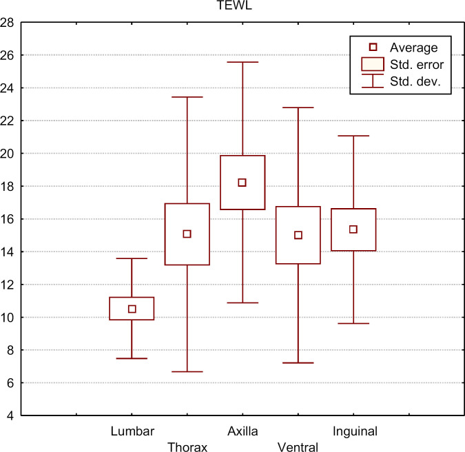
For TEWL, the lowest values were observed in the lumbar region (15.53g/h/m2), while the highest values were observed in the axillary fossa (18.22g/h/m2). TEWL was statistically significantly lower in the lumbar region as compared to the left side of the thorax region (P=0.016), the axillary fossa (P=0.0004), the ventral region (P=0.005), and the inguinal region (P=0.009) (Table 1 Fig 1).
Table 1.
TEWL in different regions in cats.
| Mean g/h/m2 | Median | SD | |
|---|---|---|---|
| Lumber region | 10.53 | 10.70 | 3.05 |
| Thorax | 15.06 | 13.3 | 8.38 |
| Axillary fossa | 18.22 | 16.30 | 7.34 |
| Ventral region | 15 | 12.25 | 7.79 |
| Inguinal region | 15.34 | 13.60 | 5.73 |
Fig 1.
TEWL in different regions in cats.
TEWL was higher in males (14.57 g/h/m2) than females (15.00 g/h/m2), but the differences were not statistically significant (P=0.89). No statistically significant differences were observed between males and females for TEWL in different body regions (lumbar region P=0.97, thorax region P=0.98, axilla P=0.91, ventral region P=0.06, inguinal region P=0.57) (Table 2, Figs 2 and 3).
Table 2.
TEWL in male and female cats.
| Mean g/h/m2 | Median | Variance | |
|---|---|---|---|
| Males | 14.57650 | 12.80000 | 49.50281 |
| Females | 15.00100 | 12.70000 | 50.60481 |
Fig 2.
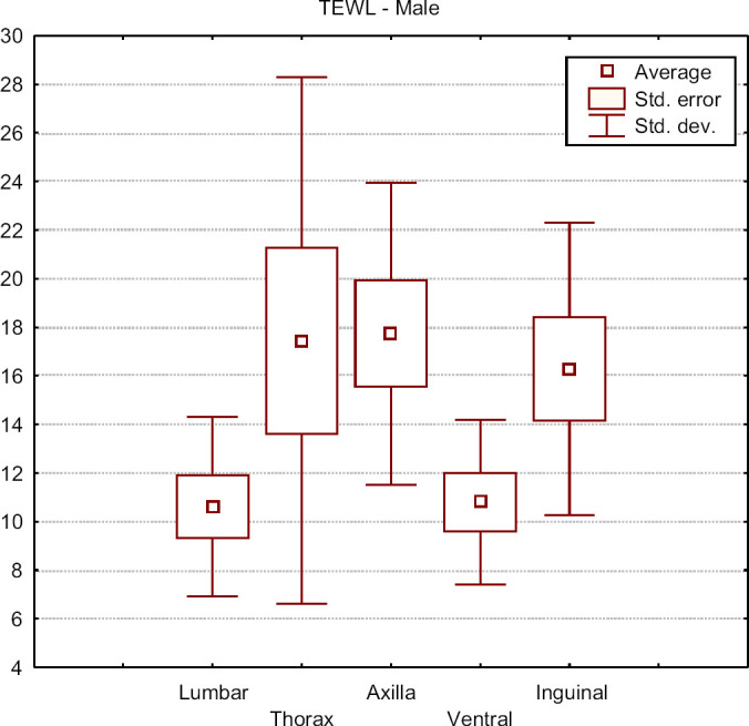
TEWL in different regions in male cats.
Fig 3.
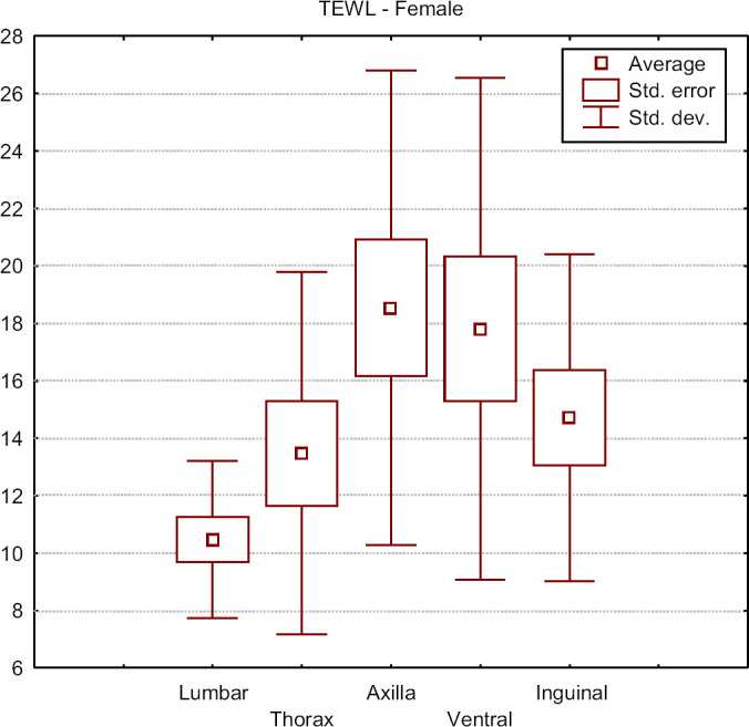
TEWL in different regions in female cats.
For skin hydration, the lowest values were observed in the lumbar region (4.62 CU) and the highest values in the inguinal region (18.29 CU). The value of this parameter was statistically significantly lower in the lumbar region than in the left thoracic region (P=0.000003), the axillary fossa (P=0.002), the ventral abdomen (P=0.03), and the inguinal region (P=0.0003). There were also statistically significant differences between the results for the left thoracic region and the ventral abdomen (P=0.005) (Table 3, Fig 4).
Table 3.
Skin hydration in different regions in cats.
| Mean (corneometer units) | Median | SD | |
|---|---|---|---|
| Lumber region | 4.62421 | 3.87000 | 4.35559 |
| Thorax | 14.18632 | 9.62000 | 15.02929 |
| Axillary fossa | 15.44421 | 13.08000 | 9.57414 |
| Ventral region | 13.38263 | 13.10000 | 7.51884 |
| Inguinal region | 18.29350 | 14.11500 | 10.75044 |
Fig 4.
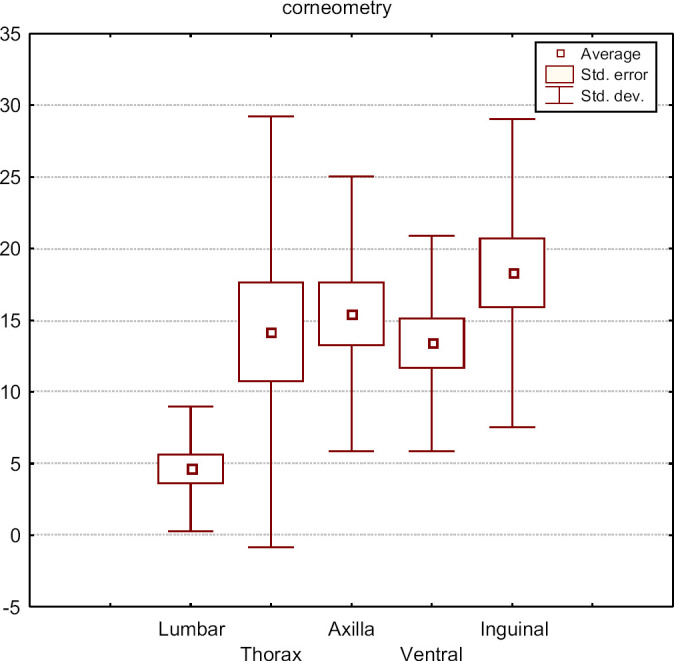
Skin hydration in different regions in cats.
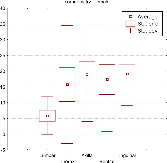
No statistically significant differences were observed for the results between males and females for skin hydration in corresponding body regions (P=0.81) (Table 4, Figs 5 and 6). In females, the values were higher (13.89 CU) than in males (12.28 CU).
Table 4.
Skin hydration in male and female cats.
| Mean | Median | SD | |
|---|---|---|---|
| Males | 12.28436 | 9.930000 | 9.04522 |
| Females | 13.89281 | 9.720000 | 12.05099 |
Fig 5.
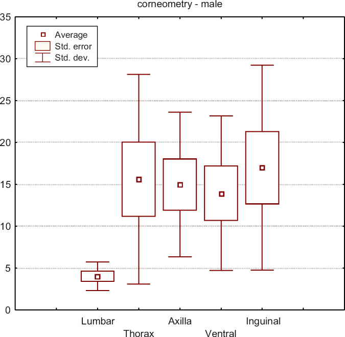
Skin hydration in different regions in male cats.
Fig 6.
Skin hydration in different regions in female cats.
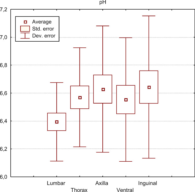
Skin pH ranged from 6.39 (the lumbar region) to 6.64 (the inguinal region). Statistically significant differences in skin pH were observed between the lumbar region and the axillary fossa (P=0.02), and between the lumbar region and the inguinal region (P=0.01) (Table 5 and Fig 7).
Table 5.
Skin pH in different regions in cats.
| Mean (pH) | Median | SD | |
|---|---|---|---|
| Lumber region | 6.394000 | 6.340000 | 0.281395 |
| Thorax | 6.570000 | 6.480000 | 0.354933 |
| Axillary fossa | 6.628500 | 6.625000 | 0.452842 |
| Ventral region | 6.553684 | 6.460000 | 0.443123 |
| Inguinal region | 6.643158 | 6.600000 | 0.509826 |
Fig 7.
Skin pH in different regions in cats.
The mean skin pH value for males (pH 6.94) was more basic than the mean female skin pH (pH 6.54) (P=0.004). A statistically significant difference between males and females was found for skin pH from the left thoracic region (P=0.004) but not for the ventral abdomen (P=0.09), the lumbar region (P=0.39), the axilla (P=0.18) or the inguinal region (P=0.49) (Figs 8 and 9).
Fig 8.
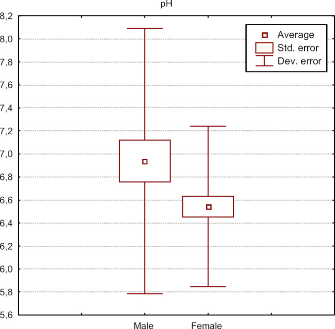
Skin pH in male and female cats.
Fig 9.
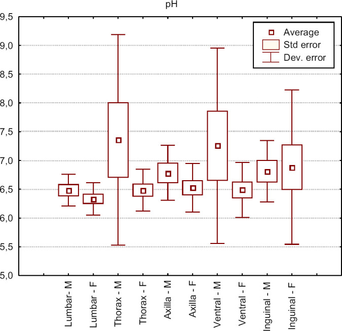
The distribution of skin pH in different regions for male and female cats.
The mean skin pH value for males (pH 6.94) was more basic than the mean female skin pH (pH 6.54) (P=0.004). A statistically significant difference between males and females was found for skin pH from the left thoracic region (P=0.004) but not for the ventral abdomen (P=0.09), the lumbar region (P=0.39), the axilla (P=0.18) or the inguinal region (P=0.49) (Figs 8 and 9).
Discussion
TEWL, skin hydration and skin pH measurements are considered to be useful techniques to assess the damage of skin in humans and are widely used to evaluate skin barrier function in patients with atopic dermatitis as well as to evaluate the therapeutic efficacy of locally administered treatments. 1–5,8,9
The integrity of skin barrier function is important in the aetiopathogenesis of atopic dermatitis. In dogs with atopic dermatitis, there are numerous defects in skin barrier function. Marsella et al found ultrastructural changes in the epidermis in such animals. 24 Atopic dogs have abnormalities in lipid lamellae organisation and wider intracellular species containing abnormal lipid material. 24 Lamellar lipids are reduced in number and highly disorganised. 29 Lipid lamella are also markedly heterogenous compared to normal dogs. 30 It is hypothesised that these defects are responsible for enhanced penetration of environmental allergens and increased risk of sensitisation in predisposed patients. 24 Reiter et al and Pin et al also observed a decrease in the amount of ceramides in the skin in atopic dogs. 31,32 Ceramides are the largest group of stratum corneum lipids. A ceramide deficiency is associated with an increase in TEWL and may be involved in impaired skin barrier function. 31
In veterinary medicine, information regarding these biophysical parameters in different diseases is limited. 21,22,26 There is also little information concerning the baseline values of these parameters in different animal species with most information obtained from canine studies. With the exception of pH, TEWL and skin hydration have not been investigated in cats.
Research conducted by other authors has pointed to statistically significant differences in the case of TEWL between different dog breeds. Hestler et al determined that TEWL values differ significantly between Beagles and Basset Hounds. 16 Differences in TEWL between breeds have also been described by Young et al. 15
TEWL may vary in different body regions in people 27 and dogs. 11,17 Oh and Oh found that TEWL in Beagles is the lowest for ear pinnae and for the lumbar region, as compared to other body regions, 11 with the highest values found on the head and the tail. Yoshihara et al, who also took measurements with Tewameter TM 300, showed that the lowest values of TEWL are found in the lumbar region. 18 A similar relationship was found in the present study, in that TEWL was lowest in the lumbar region, which was statistically significantly different from values obtained for other regions. In contrast, Watson et al, determined that the lowest TEWL values were found in the ventrum in dogs. 17 Previous studies regarding values of this parameter have not been conducted in cats, therefore, a direct comparison of results in the present investigation and other studies cannot be made.
The influence of body region on skin pH in animals was investigated by Meyer et al 28 In this investigation, various animal species (cattle, horses, goat, sheep), including dogs of different breeds and cats were studied. They concluded that there were no statistically significant differences in skin pH of different body regions. This is in contrast with the results of the present study. Similarly, research in humans has shown that pH values vary according to site. 25 Having compared the values of skin pH, Mayer et al concluded that skin pH obtained from most sites in cats was slightly acidic at 5.94–6.81, as compared to the results in the present study of 6.39–6.64.
Young et al assessed the influence of sex on TEWL, skin hydration, and skin pH 15 in Beagles, Fox Terriers, Labrador Retrievers and Manchester Terriers. In this study, the sex did not significantly influence the parameters, although males exhibited a larger range in skin pH than females. 15 These results correlate with the results obtained in the present study in regards to TEWL and the hydration of the epidermis. However, in the present study a significant difference in skin pH between males and females was observed (6.94 in males, 6.54 in females). The influence of sex on skin pH was also examined by Mayer et al and Bourdeau et al 13,28 These authors also failed to observe any influence of sex on skin pH in cats, but in cattle, the pH values of males were more basic than in females. 28 Matousek and Campbell also found that male dogs had a more basic pH than females in dogs. 14
We anticipate that the assessment of biophysical parameters of skin, especially TEWL, will be useful in understanding atopic dermatitis in cats. Much remains to be known about atopic dermatitis in cats, and the diagnosis of this disease can be challenging. The diagnosis is made on the basis of historical and clinical features and immunological tests (positive skin tests or increase of specific IgE) and the exclusion of other skin diseases which exhibit similar clinical signs. 33 In dogs, it is known that there are differences in TEWL in non-lesional skin between healthy dogs and dogs with atopy, with an increase in TEWL in atopic dogs. It is possible that such a relationship in cats exists, and disturbances in biophysical parameters of skin may be useful tools in the early diagnosis of atopy in this species.
The knowledge of skin pH in cats may also be useful in topical therapy. It is known that in dogs a increase of pH is found in pyoderma. 26 In dogs the use of topical products containing ethyl lactate, benzoyl peroxide, chlorhexidine, calicic acid, and sulfur causes a normalisation in skin pH, and it is possible to shorten the duration of topical therapy by using agents with pH similar to dogs skin. 26
In cats, similar to dogs and humans, there are differences between body regions in biophysical parameters of skin. Further research is necessary in order to specify the range of values of the biophysical parameters in healthy cats, and to assess them in various cutaneous diseases.
Acknowledgements
The authors thank Wiesław Sitkowski for statistical review.
References
- 1.Fluhr J.W., Feingold K.R., Elias P.M. Transepidermal water loss reflects permeability barier status: validation in human and rodent in vivo and ex vivo models, Exp Dermatol 15, 2006, 483–492. [DOI] [PubMed] [Google Scholar]
- 2.Dirschka T., Tronnier H., Fo R., Holst L. Clinical and laboratory investigations of epithelial barrier function and atopic diathesis in rosacea and perioral dermatitis, Br J Dermatol 150, 2004, 1136–1141. [DOI] [PubMed] [Google Scholar]
- 3.Gupta J., Grube E., Ericksen M.B., et al. Intrinsically defective skin barrier function in children with atopic dermatitis correlates with disease severity, J Allergy Clin Immunol 121, 2008, 725–730. [DOI] [PubMed] [Google Scholar]
- 4.Eberlein-König B., Schäfer T., Huss-Marp J., et al. Skin surface pH, stratum corneum hydration, trans-epidermal water loss and skin roughness related to atopic eczema and skin dryness in a population of primary school children: clinical report, Acta Derm Venereol, 2000, 188–191. [DOI] [PubMed]
- 5.Rudolph R., Kownatzki E. Corneometric, sebumetric and TEWL measurements following the cleaning of atopic skin with a urea emulsion versus a detergent cleanser, Contact Derm 50, 2004, 354–358. [DOI] [PubMed] [Google Scholar]
- 6.Suk-Jin Choi, Min-Gyu Song, Whan-Tae Sung, et al. Comparison of transepidermal water loss, capacitance and pH values in the skin between intrinsic and extrinsic atopic dermatitis patients, J Korean Med Sci 18, 2003, 93–96. [DOI] [PMC free article] [PubMed] [Google Scholar]
- 7.Loffler H., Steffes A., Happle R., Effendy I. Allergy and irritation: an adverse association in patients with atopic eczema, Acta Derm Venereol 83, 2003, 328–331. [DOI] [PubMed] [Google Scholar]
- 8.Biro K., Thaçi D., Ochsendorf F.R., Kaufmann R., Boehncke W.H. Efficacy of dexpanthenol in skin protection against irritation: a double-blind, placebo-controlled study, Contact Derm 49, 2003, 80–84. [DOI] [PubMed] [Google Scholar]
- 9.Aschoff R., Schwanebeck U., Bräutigam M., Meurer M. Skin physiological parameters confirm the therapeutic efficacy of pimecrolimus cream 1% in patients with mild-to-moderate atopic dermatitis, Exp Dermatol 18, 2009, 24–29. [DOI] [PubMed] [Google Scholar]
- 10.Laudańska H., Reduta T., Szmitkowska D. Evaluation of skin barrier function in allergic contact dermatitis and atopic dermatitis using method of the continuous TEWL measurement, Ann Acad Med Bialost 48, 2003, 124–127. [PubMed] [Google Scholar]
- 11.Oh W.-S., Oh T.-H. Measurement of transepidermal water loss from clipped and unclipped anatomical sites on the dog, Aust Vet J 87, 2009, 409–412. [DOI] [PubMed] [Google Scholar]
- 12.Shimada K., Yoshihara T., Yamamoto M., et al. Transepidermal water loss (TEWL) reflects skin barrier function of dogs, J Vet Med Sci 70, 2008, 841–843. [DOI] [PubMed] [Google Scholar]
- 13.Bourdeau P., Taylor K.W., Nguyen P., Biourge V. Evaluation of the influence of sex, diet and time on skin pH and surface lipids of cats, Vet Dermatol 15 (suppl 1), 2004, 41–69. [Google Scholar]
- 14.Matousek J., Campbell K.L.A. Comparative review of cutaneous pH, Vet Dermatol 13, 2002, 293–300. [DOI] [PubMed] [Google Scholar]
- 15.Young L.A., Dodge J.C., Guest K.J., Cline J.L., Kerr W.W. Age, breed, sex and period effects on skin biophysical parameters for dogs fed canned dog food, J Nutr 132, 2002, 1695S–1697S. [DOI] [PubMed] [Google Scholar]
- 16.Hester S.L., Rees C.A., Kennis R.A., et al. Evaluation of corneometry (skin hydration) and transepidermal water-loss measurements in two canine breeds, J Nutr 134, 2004, 2110S–2113S. [DOI] [PubMed] [Google Scholar]
- 17.Watson A., Fray T., Clarke S., Yates D., Markwell P. Reliable use of the ServoMed Evaporimeter EP-2 to assess transepidermal water loss in the canine, J Nutr 132, 2002, 1661S–1664S. [DOI] [PubMed] [Google Scholar]
- 18.Yoshihara T., Shimada K., Momoi Y., Konno K., Iwasaki T. A new method of measuring transepidermal water loss (TEWL) of dogs skin, J Vet Med Sci 69, 2007, 289–292. [DOI] [PubMed] [Google Scholar]
- 19.Yoshihara T., Endo K., Konno K., Iwasaki T. A new method for measuring canine transepidermal water loss, Vet Dermatol 15, 2004, 39. [Google Scholar]
- 20.Beco L., Fontaine J. Corneometry and transepidermal water loss measurements in the canine species: validation of these techniques in normal beagle dogs [Corneometrie et perte d'eau transepidermique: validation des technique chez des chiens sains], Ann Med Vet 144, 2000, 329–333. [Google Scholar]
- 21.Shimada K., Yoshihara T., Konno K., Nishifuji K. Increase in transepidermal water loss and decrease in ceramide content in the lesional and non-lesional skin of canine atopic dermatitis, Vet Dermatol 20, 2009, 541–546. [DOI] [PubMed] [Google Scholar]
- 22.Hightower K., Marsella R., Flynn-Lurie A. Effects of age and allergen exposure on transepidermal water loss in house dust mite-sensitized beagle model of atopic dermatitis, Vet Dermatol 21, 2010, 89–96. [DOI] [PubMed] [Google Scholar]
- 23.Shah J.H., Zhai H., Maibach H.I. Comparative evaporimetry in man, Skin Res Technol 11, 2005, 205–208. [DOI] [PubMed] [Google Scholar]
- 24.Marsella R., Samuelson D., Doerr K. Transmission electron microscopy studies in an experimental model of canine atopic dermatitis, Vet Dermatol 21, 2010, 81–88. [DOI] [PubMed] [Google Scholar]
- 25.Schmid-Wendtner M.H., Korting H.C. The pH of the skin surface and its impact on the barrier function, Skin Pharmacol Physiol 19, 2006, 296–302. [DOI] [PubMed] [Google Scholar]
- 26.Popiel J., Nicpoń J. Relacje pomiędzy ph skóry w przebiegu pyoderm u psów przed i po zastosowaniu preparatów działających zewnętrznie, Acta Sci Pol Medicina Veterinaria 3, 2004, 53–60. [Google Scholar]
- 27.Marrakchi S., Maibach H.I. Biophysical parameters of skin: map of human face, regional, and age-related differences, Contact Derm 57, 2007, 28–34. [DOI] [PubMed] [Google Scholar]
- 28.Mayer W., Neurad K. Comparison of skin pH in domesticated and laboratory mammals, Arch Dermatol Res 283, 1991, 16–18. [DOI] [PubMed] [Google Scholar]
- 29.Piekutowska A., Pin D., Rème C.A., Gatto H., Haftek M. Effects of a topically applied preparation epidermal lipids on the stratum corneum barrier of atopic dogs, J Comp Path 138, 2008, 197–203. [DOI] [PubMed] [Google Scholar]
- 30.Inman A.O., Olivry T., Dunston S.M., Monteiro-Riviere N.A., Gatto H. Electron microscopic observations of stratum corneum intercellular lipids in normal and atopic dogs, Vet Pathol 38, 2001, 720–723. [DOI] [PubMed] [Google Scholar]
- 31.Reiter L.V., Torres S.M.F., Wertz P.W. Characterization and quantification of ceramides in non-lesional skin of canine patient with atopic dermatitis compare with controls, Vet Dermatol 20, 2009, 260–266. [DOI] [PubMed] [Google Scholar]
- 32.Pin D., Popa I., Piekutowska A., et al. Biochemical analysis of epidermal lipids in normal and atopic dogs, before and after administration of an oral omega-6/omega-3 fatty acid supplement (Megaderm), Vet Dermatol 19 (suppl 1), 2008, 68, [abstract] [Google Scholar]
- 33.Scott D.W., Miler H.W., Griffin C.E. Small animal dermatology, 6th edn, 2001, WB Saunders: Philadelphia. [Google Scholar]


