Abstract
The interosseous part of the distal tibia is one of the regions in which osteochondroma can occur. Osteochondroma typically occurs among growing children and causes gradual ankle deformity by its pressure effect on the fibula. We presented six patients (Five boys and one girl with median age of 13 years old) with distal tibial interosseous osteochondroma. They were treated by a 180̊ fibular osteotomy around its longitudinal axis just proximal and distal to the lesion. All patients were treated without any complication except for one who developed non-union of the site of the fibular osteotomy. In the last follow-up, all the patients were pain-free, and no recurrence was reported. Various methods have been described for resecting interosseous osteochondroma of the distal tibia, with or without fibular osteotomy and with or without acute correction of ankle deformity during resection surgery. Still, there is no consensus over the best method for resecting such lesions.
Key Words: Excision, Fibula, Osteochondroma, Osteotomy, Tibia
Introduction
Osteochondroma is a benign lesion of the bone that usually occurs during the second decades of life. Although most osteochondroma lesions are solitary, approximately 10% are associated with hereditary multiple exostoses (HME) syndrome.1 Osteochondromas can develop in various upper or lower extremities regions, such as the elbow, knee, and ankle. The clinical significance of osteochondroma is its effect on its adjacent structures, gross deformity of the limb, and risk of transforming into a malignant lesion.2 the interosseous part of the distal tibia is one of the regions where osteochondromas can grow. Such osteochondromas affect ambulation by causing pain and limitation of ankle motion. It can cause valgus deformity of the ankle by its pressure effect on the fibula during growth and even its stress fracture.3,4 Additionally, it can compress the adjacent neurovascular structures.5 Since interosseous osteochondroma of the distal tibia usually occurs in growing children, its delayed management can lead to more deformity of the ankle.6 Several studies report the interosseous osteochondroma of the distal tibia and describe their technique for mass resection. Some surgeons prefer to resect the lesion from the tibia and fibula junction with an anterolateral3,4,7-10 or posterolateral6,11 approach by keeping the fibula intact. However, other surgeons perform the resection after fibular osteotomy to increase the exposure and reduce the risk of incomplete resection and, consequently, recurrence.12-15 Some others have corrected the ankle deformity concomitantly during mass resection surgery.16,17 In the present study, we aimed to present six patients with interosseous osteochondroma of the distal tibia treated by mass resection and ankle deformity correction. Furthermore, we reviewed and summarized the current literature on the surgical management of interosseous osteochondroma of the distal tibia.
Case Presentation
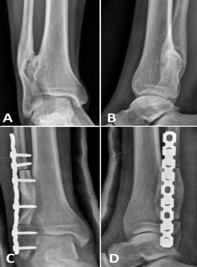
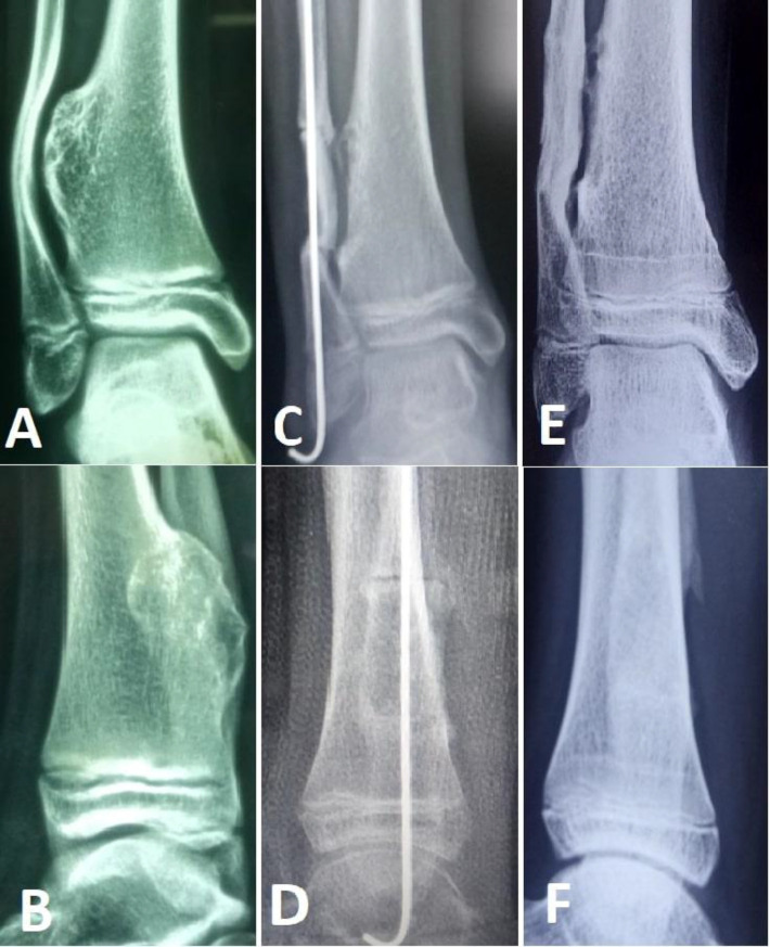
We retrospectively reviewed six consecutive patients with interosseous osteochondroma of the distal tibia. They were treated in our center during 2010-2020. Five patients were male, and one was female. The median age of the patients was 13 years old (with a range of 12-18 years old). Four lesions were in the right ankle and two in the left ankle. All the lesions were solitary osteochondroma (no HME). They were all presented with complaints of gross ankle deformity. Examinations of adjacent neurovascular structures were intact in all the patients. Standard anteroposterior, lateral, and mortise radiograph was taken. All the patients were operated on with the same technique. The operation was performed under general anesthesia in a lateral position. The thigh tourniquet was inflated for bleeding control. One gram of cephazolin was administered for infection prophylaxis. After sterile prep and draping through lateral approach, skin and subcutaneous incised at the apex of deformity, and fibular bone was exposed. Transverse osteotomy of the fibula was performed using a sagittal saw just proximal and just distal to the lesion. The isolated fibular bone was detached from the surrounding tissues to expose the mass. En-block excision of osteochondroma was performed using a saw and bone osteotome. Bone wax was placed at the site of the resected tumor to avoid synostosis. Finally, the isolated fibular bone was replaced after it was rotated 180̊ around its longitudinal axis from its original position to correct the valgus ankle deformity. The osteotomized fibular bones were fixed with reconstruction plates in four patients [Figure 1] and intramedullary Kirschner wires in two patients [Figure 2]. We chose reconstruction plates for those patients with larger body sizes. One of the patients was operated on for the second time because of recurrence. He underwent mass resection in another center with an anterolateral approach without fibular osteotomy. After the operation, a non-weight-bearing short-leg splint was applied. They were followed for at least two years with physical examination and radiography.
Figure 1.
Anteroposterior and lateral views of interosseous osteochondroma of the distal tibia before (A, B) and after (C, D) mass resection and deformity correction using 180̊ rotational osteotomy of the fibula. The isolated fibular bone was fixed with a reconstruction plate
Figure 2.
Anteroposterior and lateral views of interosseous osteochondroma of the distal tibia before (A, B), just after (C, D) mass resection, and fibular osteotomy. The intra-medullary pin was removed after the union of the sites of the fibular osteotomy (E, F)
The apparent deformity of the ankles was corrected after surgery. All the patients were treated without complications except one whose fibula was fixed with a plate. He developed with non-union of the proximal side of fibular osteotomy, so a second surgery was performed for him. The mean follow-up period for the patients was five years (range of 3-7 years). In the last follow-up, the patients did not have any pain, and they had a normal gait and normal ankle range of motion. No recurrence happened. No limb length discrepancy or gross deformity was found.
Discussion
Osteochondroma is a common benign bone tumor usually found at the metaphysis of the distal femur and proximal part of the tibia. Osteochondroma is not common near the ankle joint, and it is more prevalent in cases of HME.18,19 However, all our patients were cases of solitary exostosis. Osteochondroma of the distal tibia causes valgus deformity of the ankle. The valgus deformity of the ankle is characterized by tapering of distal tibia epiphysis from medial to lateral and shortening of the fibular bone.20 Valgus deformity of the ankle has to be treated to avoid osteoarthritis of the ankle in the future.
Table 1 summarizes the studies that reported distal tibial interosseous osteochondroma [Table 1]. Different methods have been applied to resect the lesion. Some surgeons resected the lesions through anterolateral or posterolateral approaches without fibula osteotomy. The problem with osteochondroma resection without osteotomy of the fibula is its limited access to the lesion; thus, identifying the borders of osteochondroma is hard, and its complete resection is difficult. Danielsson et al.7 reported recurrence after resection through a posterior approach without fibula osteotomy. We saw similar events in one of our patients that underwent mass resection through an anterolateral approach without fibula osteotomy in another center because of mass recurrence. Although previous investigations that applied anterolateral or posterolateral approaches for mass resection did not report any complications, almost all had short follow-ups to judge their recurrence rates. Some other surgeons used fibular osteotomy to have a better access to osteochondroma. Mahajan et al.12 resected the osteochondroma with a segment of the fibular bone adhered to it. Gupte et al.15 performed fibular osteotomy in two sites (just proximal to the lesion and just distal to the distal tibiofibular joint). Then, the isolated fibular segment was hinged anteriorly on the anterior distal tibiofibular ligament. Appy-Fedida et al.14 osteotomized the fibula in a similar pattern, but they excised the fibular fragment and used it after mass resection as a free autograft. Yang et al.13 applied a concomitant Volkmann tuberosity osteotomy beside fibular osteotomy to avoid injury to the posterior tibiofibular ligament.
Table 1.
Summary of previous studies that reported distal tibial interosseous osteochondroma
| Author (year) | Number of cases | Age, mean, years | Sex (M:F) | Side (R:L) | Method of mass resection | Method of fibula fixation | Mean F/U length, months | Complication |
|---|---|---|---|---|---|---|---|---|
| Danielsson LG et al. (1990) 7 | 3 | 13.3 | 2:1 | 2:1 | Anterior approach without fibula osteotomy | - | 7.5 | Recurrence after resection through the posterior approach |
| Gupte CM, et al. (2003) 15 | 2 | 13 | 2:0 | 1:1 | Fibula osteotomy in two sites and reflected anteriorly on the anterior distal tibiofibular ligament | Semi-tubular plates | 2.75 | none |
| Gil-Albarova J, et al. (2007) 16 | 5 | 11 | 2:3 | 3:2 | Fibula rotational osteotomy | IM pin | 4.4 | Synostosis in one patient |
| Ismail BE, et al. (2008) 8 | 1 | 14 | 0:1 | 1:0 | Anterior approach without fibula osteotomy | - | 2 | none |
| Wani IH, et al. (2009) 4 | 1 | 16 | 1:0 | 0:1 | Anterior approach without fibula osteotomy | - | 1 | none |
| Thakur GB, et al. (2012) 17 | 1 | 16 | 0:1 | 1:0 | Fibula sofield osteotomy | IM pin | 1 | none |
| Herrera-Perez M, et al. (2013) 20 | 1 | 13 | 0:1 | 1:0 | Anterior approach without fibula osteotomy | - | 0.5 | none |
| Genc B, et al. (2014) 5 | 1 | 17 | 1:0 | - | - | - | - | none |
| Tayara B, et al. (2016) 6 | 1 | 16 | 1:0 | 1:0 | Posterolateral approach without fibular osteotomy | - | 0.25 | none |
| Appy-Fedida B, et al. (2017) 14 | 8 (10 ankles) | 10.6 | 7:1 | 4:6 | Fibula osteotomy | IM pin | 5.9 | none |
| Mehraj M, et al. (2018) 9 | 3 | 16-18 | 2:1 | - | Anterior approach without fibula osteotomy | - | 0.25 | none |
| Mahajan NP, et al. (2020) 12 | 1 | 10 | 0:1 | 1:0 | Posterolateral approach with excision of overlying fibula | - | 1 | none |
| Yang H, et al. (2020) 13 | 11 | 27.5 | 7:4 | 5:6 | Fibula and Volkmann tuberosity osteotomy | Fibular LCP | 1 | superficial infection in one and nonunion of the site of fibular osteotomy in one patient |
| Panta S, et al. (2021) 11 | 1 | 18 | 1:0 | 1:0 | Posterolateral approach without fibular osteotomy | - | 0.5 | none |
| Singh CM, et al. (2021) 3 | 1 | 14 | 1:0 | 1:0 | Anterolateral approach without fibula osteotomy | - | 0.5 | none |
| John AM, et al. (2022) 10 | 1 | 24 | 1:0 | 1:0 | Anterolateral approach without fibula osteotomy | - | - | none |
| Present study | 6 | 14.3 | 5:1 | 4:2 | Transverse Fibula rotational osteotomy | Recon Plates or IM pins | 5 | One recurrence after resection through the anterolateral approach without fibular osteotomy and one non-union of fibula osteotomy site after plate fixation |
M: F, male to female ratio; R: L, right side to left side ratio; F/U, follow-up; IM pins, intramedullary pins (kirschner wires); Recon plates, reconstruction plates
The mentioned studies fixed the fibular bone fragment to its original place after resection of osteochondroma, so they did not correct the probable valgus deformity of the ankle, which was caused by the fibular bone deformity. Thakur et al.17 applied the sofield technique after fibula osteotomy to correct the fibula deformity. In addition, Gil-Albarova et al.16 used a so-called fibula rotational osteotomy technique for five patients by rotating the isolated fibular bone fragment around its axis for 180̊ to correct the ankle deformity. We used a similar technique for six patients unaware of the mentioned study. Our method had some differences. We performed transverse fibular osteotomy while they performed chevron osteotomy. We used a plate for fibula fixation for four patients and a pin for two patients, but they fixed all the fibular bones with pins. We used plates for older patients or those with larger body sizes. Moreover, to avoid further tibiofibular synostosis, we placed bone wax at the tibiofibular junction after osteochondroma resection and before fibula fixation. Gil-Albarova et al.16 placed free subcutaneous fat between the tibia and fibula for their patients except one, who developed tibiofibular synostosis, although he was asymptomatic. One of our patients, whose ankle was fixed with a plate, experienced non-union.
Conclusion
Various methods have been described for resecting interosseous osteochondroma of the distal tibia, with or without fibular osteotomy and with or without acute correction of ankle deformity during resection surgery. However, there is no consensus over the best method for resection of such lesions. More controlled investigations with larger sample sizes are required to identify the best surgical method.
Acknowledgment
The authors would like to thank Shiraz University of Medical Sciences, Shiraz, Iran, and also the Center for Development of Clinical Research of Nemazee Hospital and Dr. Nasrin Shokrpour for editorial assistance.
Conflict of interest:
None
Funding:
None
References
- 1.Tepelenis K, Papathanakos G, Kitsouli A, et al. Osteochondromas: An updated review of epidemiology, pathogenesis, clinical presentation, radiological features and treatment options. in vivo. 2021;35(2):681–691. doi: 10.21873/invivo.12308. [DOI] [PMC free article] [PubMed] [Google Scholar]
- 2.Bailescu I, Popescu M, Sarafoleanu LR, et al. Diagnosis and evolution of the benign tumor osteochondroma. Exp Ther Med. 2022;23(1):1–6. doi: 10.3892/etm.2021.11026. [DOI] [PMC free article] [PubMed] [Google Scholar]
- 3.Singh CM, Magar MT, Sud AD. Osteochondroma of the Distal Tibia Leading to Deformity and Stress Fracture of the Fibula-A Case Report. Medical Journal of Shree Birendra Hospital. 2021;20(2):173–176. [Google Scholar]
- 4.Wani IH, Sharma S, Malik FH, Singh M, Shiekh I, Salaria AQ. Distal tibial interosseous osteochondroma with impending fracture of fibula–a case report and review of literature. Cases J. 2009;2(1):1–4. doi: 10.1186/1757-1626-2-115. [DOI] [PMC free article] [PubMed] [Google Scholar]
- 5.Genc B, Solak A, Kalaycioğlu SK, Sahin N. Distal tibial osteochondroma causing fibular deformity and deep peroneal nerve entrapment neuropathy: a case report. Acta Orthop Traumatol Turc. 2014;48(4):463–466. doi: 10.3944/AOTT.2014.2741. [DOI] [PubMed] [Google Scholar]
- 6.Tayara B, Uddin F, Al-Khateeb H. Distal tibial osteochondroma causing fibular deformation resected through a posterolateral approach: a case report and literature review. Current Orthopaedic Practice. 2016;27(2):E12–E14. [Google Scholar]
- 7.Danielsson LG, Ei-Haddad I, Quadros O. Distal tibial osteochondroma deforming the fibula. Acta Orthop Scand. 1990;61(5):469–470. doi: 10.3109/17453679008993566. [DOI] [PubMed] [Google Scholar]
- 8.Ismail BE, Kissel CG, Husain ZS, Entwistle T. Osteochondroma of the distal tibia in an adolescent: a case report. J Foot Ankle Surg. 2008;47(6):554–558. doi: 10.1053/j.jfas.2008.07.004. [DOI] [PubMed] [Google Scholar]
- 9.Mehraj M, Shah I. Osteochondroma of distal tibia: A case series. International Journal of Orthopaedics. 2018;4(1):665–666. [Google Scholar]
- 10.John AM, Thomas V, Pillai MG, Theckanal JG, Sebastian JP. Solitary osteochondroma from interosseous border of distal tibia. Journal of Orthopaedic Association of South Indian States. 2021;18(2):85. [Google Scholar]
- 11.Panta S, Thapa SK, Paudel KP. Distal tibia interosseous osteochondroma with fibula deformity. Journal of Chitwan Medical College. 2021;11(3):148–150. [Google Scholar]
- 12.Mahajan NP, Wadia F, GS PK, Yadav AK, Narvekar M, Kondewar P. Segmental Fibulectomy to Excise the Adherent Distal Tibia Osteochondroma in a Case of Hereditary Multiple Exostosis–A Rare Case Report. J Orthop Case Rep. 2020;10(4):1. doi: 10.13107/jocr.2020.v10.i04.1780. [DOI] [PMC free article] [PubMed] [Google Scholar]
- 13.Yang H, Shou K, Wei S, et al. A revised surgical strategy for the distal tibiofibular interosseous osteochondroma. Biomed Res Int. 2020: 6371456. doi: 10.1155/2020/6371456. [DOI] [PMC free article] [PubMed] [Google Scholar]
- 14.Appy-Fedida B, Krief E, Deroussen F, et al. Mitigating Risk of Ankle Valgus From Ankle Osteochondroma Resection Using a Transfibular Approach: A Retrospective Study With Six Years of Follow-Up. J Foot Ankle Surg. 2017;56(3):564–567. doi: 10.1053/j.jfas.2017.01.029. [DOI] [PubMed] [Google Scholar]
- 15.Gupte CM, DasGupta R, Beverly MC. The transfibular approach for distal tibial osteochondroma: an alternative technique for excision. J Foot Ankle Surg. 2003;42(2):95–98. doi: 10.1016/s1067-2516(03)70008-8. [DOI] [PubMed] [Google Scholar]
- 16.Gil-Albarova J, Gil-Albarova R, Bregante-Baquero J. Fibular rotational osteotomy for the treatment of distal tibial osteochondroma: a technical modification for deformity correction and improved outcomes. J Foot Ankle Surg. 2007;46(6):474–479. doi: 10.1053/j.jfas.2007.08.001. [DOI] [PubMed] [Google Scholar]
- 17.Thakur GB, Jain M, Bihari AJ, Sriramka B. Transfibular excision of distal tibial interosseous osteochondroma with reconstruction of fibula using Sofield's technique–A case report. J Clin Orthop Trauma. 2012;3(2):115–118. doi: 10.1016/j.jcot.2012.09.003. [DOI] [PMC free article] [PubMed] [Google Scholar]
- 18.Masoum S H F. Multiple rib exostoses in a boy: a rare case resulting in surgery secondary to cosmetic concerns. Arch Bone Jt Surg. 2014;2: 243. [PMC free article] [PubMed] [Google Scholar]
- 19.Suranigi S, Rengasamy K, Najimudeen S, Gnanadoss J. Extensive osteochondroma of talus presenting as tarsal tunnel syndrome: report of a case and literature review. Arch Bone Jt Surg. 2016;4(3):269. [PMC free article] [PubMed] [Google Scholar]
- 20.Takikawa K, Haga N, Tanaka H, Okada K. Characteristic factors of ankle valgus with multiple cartilaginous exostoses. J Pediatr Orthop. 2008;28(7):761–765. doi: 10.1097/BPO.0b013e3181847511. [DOI] [PubMed] [Google Scholar]
- 21.Noonan KJ, Feinberg JR, Levenda A, Snead J, Wurtz LD. Natural history of multiple hereditary osteochondromatosis of the lower extremity and ankle. J Pediatr Orthop. 2002;22(1):120–124. [PubMed] [Google Scholar]
- 22.Herrera-Perez M, De Mendoza MA, De Bergua-Domingo JM, Pais-Brito JL. Osteochondromas around the ankle: report of a case and literature review. Int J Surg Case Rep. 2013;4(11):1025–1027. doi: 10.1016/j.ijscr.2013.08.015. [DOI] [PMC free article] [PubMed] [Google Scholar]


