



Abstract
Introduction: Adjuvant immune checkpoint inhibitor trials in renal cell carcinoma (RCC) call for improved recurrence risk stratification. Due to limitations of circulating tumor DNA (ctDNA) use in RCC, the use of hypermethylated SHOX2 gene (mSHOX2) in circulating cell-free DNA is explored as a surrogate marker for identifying high-risk patients after RCC surgery. Methods: Liquid biopsies were collected post-surgery from 45 RCC patients (mean duration 4.3 days). Real-time polymerase chain reaction was used to analyze SHOX2 methylation in circulating cell-free DNA. Patients were categorized as mSHOX2 positive or negative by cut-off. Metastasis-free survival (MFS), cancer-specific survival (CSS), and overall survival (OS) were assessed using Cox regression and Log-rank analyses (median follow-up time: 60 months). Results: 17 patients were mSHOX2 positive, showing unfavorable OS/CSS (Log-rank P = 0.004 and 0.02) and nearly 6-fold higher recurrence risk (hazard ratio 5.89, 95% CI 1.46-23.8). Multivariable Cox analysis confirmed mSHOX2 as an independent recurrence risk factor, disregarding TNM-based stratification. Conclusions: mSHOX2 effectively identifies high-risk RCC patients post-surgery, indicating minimal residual disease. This easy to implement biomarker has potential for guiding of adjuvant therapy decisions.
Keywords: Renal cell carcinoma, recurrence, biomarker, SHOX2, adjuvant therapy
Introduction
Adjuvant immunotherapy has entered genitourinary oncology. The KEYNOTE-564 trial demonstrated a disease-free survival benefit for adjuvant therapy with the immune checkpoint inhibitor (ICI) pembrolizumab in patients with renal cell carcinoma (RCC) at high risk of recurrence [1].
While some patients treated with adjuvant ICI appear to have a survival benefit, for a yet uncharacterized proportion of patients this therapy regimen represents an overtreatment with potentially severe side effects [1-3]. This issue was emphasized in the 2022 updated European Association of Urology (EAU) guidelines [4]. The reasons may include insufficient patient selection through histopathological TNM staging alone [3]. This implies new challenges, but also, due to new molecular analysis technologies, opportunities to identify subgroups of patients who will particularly benefit from adjuvant therapy or who do not need adjuvant therapy [5].
Adjuvant therapy can only be effective in patients who harbor micrometastases after tumor surgery that are not yet radiologically detectable, a stage called minimal residual disease (MRD) [3]. The concept of identifying MRD to guide adjuvant therapy decisions in RCC was recently highlighted at The Society for Immunotherapy of Cancer’s annual meeting (SITC 2022). Among the possible approaches, the detection of circulating tumor DNA (ctDNA) following tumor surgery is an emerging and powerful therapeutic concept to identify MRD patients at highest risk of disease recurrence [6]. The ctDNA-guided adjuvant treatment for patients with stage II colon cancer reduces the use of adjuvant chemotherapy without compromising recurrence-free survival [7]. This concept can also be applied to patients with urothelial carcinoma, where only ctDNA-positive patients at high risk of recurrence had a survival benefit from adjuvant therapy with atezolizumab in the phase 3 IMvigor010 trial [8]. Imvigor011 (NCT04660344) will further explore this therapeutic concept by administering either atezolizumab or placebo to patients with ctDNA detection after cystectomy.
This promising new therapeutic concept to guide adjuvant treatment decision on the detection of MRD may also support adjuvant therapy decisions for patients with RCC.
However, detection of ctDNA is costly and not widely available, and for RCC in particular, further technical difficulties arise for this diagnostic tool: Detected levels of ctDNA in patients with RCC are low, and highly sensitive methods are required [9]. Also, the fraction of ctDNA in liquid biopsy was found to be comparatively low in RCC patients compared to other solid tumor entities [10]. In addition, clear cell RCC is the prime example of a heterogeneous tumor with substantial intrapatient heterogeneity between metastatic lesions throughout tumor evolution; thus, detection of relevant mutations for ctDNA monitoring is complicated [11]. Therefore, even for truncal mutations such as the Von Hippel-Lindau gene, DNA-based genomic profiling in peripheral blood for metastatic RCC resulted in low detection rates [12]. As an alternative approach, the measurement of tumor-specific methylation signatures seems to be a promising concept to detect ctDNA in patients with RCC [9].
Research has identified specific methylation patterns across various entities with the potential to serve as pan-cancer biomarkers [13]. Hypermethylated short stature homeobox gene 2 (SHOX2) within circulating cell-free DNA (ccfDNA) is a highly sensitive surrogate parameter for the presence of ctDNA in diverse entities including RCC, where it has high prognostic potential as a surrogate for tumor burden prior to nephrectomy [14]. An U.S. Food and Drug Administration (FDA)-approved test kit (Epi proLung) detecting hypermethylated SHOX2 (mSHOX2) for diagnostic purposes in lung cancer would allow a straight forward clinical implementation of this promising liquid biopsy biomarker for patients with RCC as well.
Therefore, in our study, we aimed to investigate whether the presence of mSHOX2 in ccfDNA after nephrectomy represents a surrogate for MRD to identify patients at risk for disease recurrence who might particularly benefit from adjuvant immunotherapy.
Methods
Patient cohort
From a prospectively collected biobank registry, a single-center observational cohort study comprising 45 patients with RCC was assembled between 2014 and 2015. Male and female patients undergoing renal tumor surgery for radiologically suspected RCC aged 18-99 years were included. Patients with no histological evidence of malignancy after surgery were excluded retrospectively.
Assessment of SHOX2-methylation
A plasma blood sample was collected at a mean of 4.3 days (interquartile range 2.3-6.7 days) after surgery. The timing was chosen to exclude false positive data obtained by high pre-surgical levels regarding the half-life of ctDNA (approximately 2 hours) [15]. The method of measuring SHOX2-methylation in ccfDNA has been described previously in detail [13,14,16,17]. In brief, blood samples were collected in ethylenediaminetetraacetic acid (EDTA)-containing tubes and processed to plasma within 6 hours. Next, ccfDNA was bisulfite converted and purified [16]. Afterwards, SHOX2-methylation was measured using a methylation-specific quantitative real time polymerase chain reaction amplifying a 112 base pair fragment within the body of the SHOX2 gene as described in this publicly available protocol [18]. Relative methylation levels were calculated using the ΔΔCq method [17]. Using the previously validated cutoff > 0.25%, samples were dichotomized as either SHOX2 hypermethylated (mSHOX2-positive) or not hypermethylated (mSHOX2-negative) [14].
Follow-up data and statistical analysis
The patient cohort was observed from enrollment until December 2021. Overall survival (OS), cancer-specific survival (CSS), and metastasis-free survival (MFS) after surgery were estimated using univariable Kaplan-Meier regressions and tested with log-rank tests. Starting with the day of surgery, OS was defined as the period until death of any cause, CSS as the period until death causally related to RCC and MFS as the period until tumor recurrence or metastasis. Patients with completely resected metastatic disease without radiological evidence of further metastases were included as stage M1 with no evidence of disease (M1 NED) as defined in KEYNOTE-564 [19]. Patients undergoing palliative cytoreductive nephrectomy in the metastatic disease stage were excluded from the MFS analyses. Uni- and multivariable Cox regression analyses were performed to compare the independent prognostic value of SHOX2 hypermethylation with baseline characteristics (age, sex, Eastern Cooperative Oncology Group (ECOG) performance status, and high risk of recurrence based on histopathological findings according to the KEYNOTE-564 trial: pT2, G4; pT3-4; pN+; pM+ [19]). Variables were only included in the multivariable Cox regression models if survival effects were significant in the univariable analyses. Statistical analyses were performed with RStudio (version 1.4.1106) using “base” and “survminer” package (version 0.4.9, https://CRAN.R-project.org/package=survminer). Categorical variables are reported as median, frequency, range, and interquartile range (IQR). The Kruskal-Wallis rank sum test and Wilcoxon-Mann-Whitney test were used for intergroup comparisons. All tests were two-sided, and P-values < 0.05 were considered significant.
Results
Characteristics of the study cohort
45 patients with RCC who underwent surgery between 2014 and 2015 met the inclusion criteria. 73% of patients were male, and the median age at nephrectomy was 67 years (interquartile range (IQR): 57-74 years). Only 1 patient (2.2%) had simultaneously undergone resection of a singular pancreatic distant metastasis (M1 NED), while in another 6 patients (13.3%), the surgical intention was a cytoreductive palliative nephrectomy with known distant metastases. The main histologic subtype was clear cell renal carcinoma in 30 patients (66.7%), as well as 9 patients (20%) with papillary and 5 patients (11%) with chromophobe RCC. Data are summarized in Table 1. The median follow-up period was 63 months (mean: 51.8; IQR: 20-73 months). 18 (40%) patients died during the follow-up period, and 9 patients (20%) experienced disease recurrence.
Table 1.
Comparison of baseline parameters between patients with detection or absence of hypermethylated short stature homeobox 2 (mSHOX2-positive vs. -negative) in circulating cell-free DNA (ccfDNA)
| Characteristic | Overall, N = 45 | mSHOX2-positive, N = 17 | mSHOX2-negative, N = 28 | p-value1 |
|---|---|---|---|---|
| Age | 0.223 | |||
| Median, IQR | 67 (56.5, 74) | 70 (61.5, 75) | 67 (47, 73.5) | |
| Range | 23, 89 | 56, 89 | 23, 86 | |
| Sex | 0.760 | |||
| Male | 33 (73.3%) | 12 (70.6%) | 21 (75%) | |
| Female | 12 (26.7%) | 5 (29.4%) | 7 (25%) | |
| ECOG | 0.795 | |||
| 0 | 30 (66.7%) | 11 (64.7%) | 19 (67.9%) | |
| 1 | 14 (31.1%) | 5 (29.4%) | 9 (32.1%) | |
| 2 | 1 (2.2%) | 1 (5.9%) | 0 (0%) | |
| Histological Subtype | 0.150 | |||
| Clear cell | 30 (66.7%) | 14 (82.2%) | 16 (57.1%) | 0.072 |
| Non clear-cell | 15 (33,3%) | 3 (17,8%) | 13 (42,9%) | |
| Papillary | 9 (20%) | 2 (11.8%) | 7 (25%) | 0.262 |
| Chromophobe | 5 (11.1%) | 0 (0%) | 5 (17.9%) | 0.099 |
| Tubulocysytic | 1 (2.2%) | 1 (5.9%) | 0 (0%) | 0.427 |
| AJCC Stage | 0.013* | |||
| I | 24 (53.3%) | 4 (23.5%) | 20 (71.4%) | |
| II | 3 (6.7%) | 2 (11.8%) | 1 (3.6%) | |
| III | 11 (24.4%) | 6 (35.3%) | 5 (17.9%) | |
| IV | 7 (15.6%) | 5 (29.4%) | 2 (7.1%) | |
| Risk classification2 | 0.005** | |||
| High-risk | 18 (40%) | 11 (64.7%) | 7 (25%) | |
| Low-risk | 27 (60%) | 6 (35.3%) | 21 (75%) |
mSHOX2 = hypermethylated short stature homeobox 2, IQR = interquartile range, ECOG = Eastern Cooperative Oncology Group performance status, AJCC = American Joint Committee on Cancer.
Wilcoxon-Mann-Whitney-Test; Kruskal-Wallis rank sum test.
According to KEYNOTE-564 [19]: High-risk = pT2, G4; pT3-4, pN+, pM+ (M1 NED No Evidence of Disease).
P < 0.05;
P < 0.01.
SHOX2 hypermethylation and survival outcom
17 patients (37.8%) were defined as mSHOX2-positive and 28 patients (62.2%) were mSHOX2-negative. There were significant differences in baseline clinical parameters (Table 1); mSHOX2-positive patients were of higher tumor stage and risk of recurrence according to stratification based on histopathological findings.
In univariable Cox regression, the detection of mSHOX2 was associated with shortened OS (hazard ratio (HR) = 3.65, 95% Confidence Interval [CI] 1.41-9.46, P = 0.004), CSS (HR = 3.59, 95% CI 1.17-11.0, P = 0.02), and MFS (HR = 5.89, 95% CI 1.46-23.8, P = 0.005). The other factor significantly associated with survival outcomes was high-risk constellation based on histopathological TNM staging, namely pT2, G4; pT3-4; pN+ or pM+ tumors (OS: HR = 2.99, 95% CI 1.18-7.64, P = 0.02; CSS: HR = 5.16 95% CI 1.58-16.9, P = 0.002; MFS: HR = 5.15, 95% CI 1.37-19.4, P = 0.007). Age, sex, and ECOG performance status showed no statistically significant association with survival outcomes. In the multivariable Cox analysis, mSHOX2 was found to be an independent risk factor for both reduced OS and MFS (P = 0.034 and 0.042, respectively), while high-risk tumors were independently associated with reduced CSS (P = 0.030). The Cox regression analyses are presented in Table 2.
Table 2.
Uni- and multivariable Cox regression analysis regarding overall survival, cancer-specific survival and metastasis-free survival after surgery
| Univariable | OS | CSS | MFS | ||||||
|
|
|
|
|||||||
| Characteristic | HR | 95% CI | p-value | HR | 95% CI | p-value | HR | 95% CI | p-value |
|
| |||||||||
| Sex: Female | 0.7 | 0.23-2.1 | 0.522 | 1.11 | 0.34-3.6 | 0.872 | 1.30 | 0.32-5.4 | 0.715 |
| Age | 1.04 | 0.99-1.1 | 0.086 | 1.04 | 0.99-1.1 | 0.107 | 1.01 | 0.97-1.1 | 0.462 |
| ECOG | 2.30 | 0.90-5.9 | 0.084 | 2.14 | 0.67-6.7 | 0.195 | 0.57 | 0.07-4.6 | 0.598 |
| mSHOX2-positive | 3.59 | 1.41-9.5 | 0.004** | 3.59 | 1.17-11 | 0.026* | 5.89 | 1.46-23.8 | 0.005** |
| High-risk-tumor1 | 2.99 | 1.18-7.6 | 0.022* | 5.16 | 1.58-16.9 | 0.002** | 5.15 | 1.37-19.4 | 0.007** |
|
| |||||||||
| Multivariable | OS | CSS | MFS | ||||||
|
|
|
|
|||||||
| Characteristic | HR | 95% CI | p-value | HR | 95% CI | p-value | HR | 95% CI | p-value |
|
| |||||||||
| High-risk-tumor | 2.22 | 0.83-5.9 | 0.11 | 3.95 | 1.14-13 | 0.030* | 3.63 | 0.92-14 | 0.066 |
| mSHOX2-positive | 2.94 | 1.09-7.9 | 0.034* | 2.36 | 0.72-7.7 | 0.153 | 4.51 | 1.05-19 | 0.042* |
OS = overall survival, CSS = cancer-specific survival, MFS = metastasis-free survival, HR = hazard ratio, 95% CI = 95% confidence interval, mSHOX2 = hypermethylated short stature homeobox 2, ECOG = Eastern Cooperative Oncology Group performance status.
According to KEYNOTE-564 [19]: High-risk = pT2, G4; pT3-4, pN+, pM+ (M1 NED No Evidence of Disease).
P < 0.05;
P < 0.01.
The Kaplan-Meier estimators (KM) are presented in Figure 1. Detection of mSHOX2 was associated with significantly reduced OS (Figure 1A; log-rank P = 0.004), CSS (Figure 1B; log-rank P = 0.017), and MFS (Figure 1C; log-rank P = 0.005). Additional KM estimators, excluding patients with metastatic disease stages, are depicted in Figure 2.
Figure 1.

Kaplan-Meier estimators of survival after renal cell carcinoma surgery: All 45 patients were divided by methylation status of short stature homeobox 2 (SHOX2) gene in circulating cell-free DNA into either hypermethylated (mSHOX2-positive) or non-hypermethylated (mSHOX2-negative). Significant survival benefit was shown for mSHOX2-negative patients in: (A) Overall survival (Log rank P = 0.004), (B) Cancer-specific-survival (Log rank P = 0.017) and (C) Metastasis-free-survival (MFS, Log rank P = 0.005). For MFS analysis primary metastatic cases were excluded. (D) By composing mSHOX2 with high-risk criteria according to KEYNOTE-564 trial (pT2, G4; pT3-4; pN+, pM+ [19]) one event of recurrence is missed out (Log rank P < 0.001).
Figure 2.
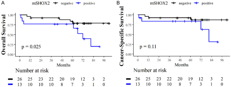
Kaplan-Meier estimators of survival after renal cell carcinoma surgery. After exclusion of primary metastatic cases, 39 patients are divided by methylation status of short stature homeobox 2 (SHOX2) gene in circulating cell-free DNA into either hypermethylated (mSHOX2-positive) or non-hypermethylated (mSHOX2-negative). A significant survival benefit was shown for mSHOX2-negative patients in (A) overall survival (Log rank P = 0.025) with a non-significant trend for (B) cancer-specific-survival (Log rank P = 0.11).
Of 17 mSHOX2-positive patients, 6 met the low-risk criteria according to TNM staging, excluding them from eventual adjuvant treatment according to current approval, while out of these patients 3 (50%) suffered from disease recurrence. The MFS was significantly shortened in low-risk patients with SHOX2 hypermethylation (P = 0.01). In high-risk patients, no significant difference was found in survival outcomes depending on SHOX2 methylation status (P = 0.73). The KM plots are shown in Figure 3.
Figure 3.
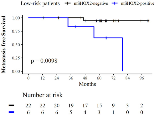
Kaplan-Meier estimator of metastasis-free survival in the low-risk subgroup after renal cell carcinoma surgery. Additional analysis including only patients with low risk of recurrence according to KEYNOTE-564 trial (pT1-2, G1-3, N0, M0) [19]. Hypermethylation of short stature homeobox 2 (mSHOX2) in circulating cell-free DNA was associated with impaired MFS (Log rank P < 0.01).
In an additional MFS analysis, we included all 16 patients (41.0%) with either mSHOX2 hypermethylation or a high-risk status as a composite score. By combining these two predictive indicators, only 1 event of recurrence was missed (Figure 1D).
Discussion
Recurrence of renal cell carcinoma is common, and to date, we have not progressed beyond predicting the risk of recurrence based on histopathology alone. Since the approval of pembrolizumab as an adjuvant treatment, accurate risk assessment has become increasingly important. As the IMmotion010 (ClinicalTrials.gov Identifier: NCT03024996) and CheckMate914 (NCT03138512) trials failed to show a benefit of adjuvant treatment with atezolizumab or nivolumab + ipilumumab, this further underscores the paramount clinical importance of accurate patient selection [20,21]. In the era of precision oncology, molecular biomarkers that identify patients who will benefit from adjuvant ICB would be desirable to guide adjuvant treatment decisions [22].
Postoperative detection of ctDNA as a surrogate for MRD has strong potential to guide adjuvant treatment decisions. This concept has been proven in large phase 3 clinical trials, such as the DYNAMIC and IMvigor010 [7,8]. Adjuvant atezolizumab only prolonged disease-free survival in the ctDNA-positive subgroup of the negative IMvigor010 trial [8], which now led to the initiation of the IMvigor011 trial, which tested atezolizumab versus placebo in patients with high-risk muscle-invasive bladder cancer who are ctDNA-positive following cystectomy (NCT04660344).
However, there are limitations in the determination of ctDNA, particularly in RCC, owing to difficult detection and intratumor heterogeneity as recently highlighted by Geertsen et al. [9]. Even in advanced RCC, detection of ctDNA is achieved within approximately half of the patients at most [23-26]. Therefore, despite its proven relevance throughout other solid tumor entities and indications for potential in MRD detection even in RCC, ctDNA is not gaining widespread use among RCC patients [26]. In order to avoid these technical difficulties, DNA methylation markers such as mSHOX2 might provide a promising approach.
Numerous studies have suggested the diagnostic value of methylation markers in RCC [27]. However, to date there are very limited data on the prognostic role of liquid biopsy in RCC. Nevertheless, our results demonstrating a significant survival difference according to postoperative SHOX2 hypermethylation status, as well as the identification of mSHOX2 as an independent risk factor in addition to TNM staging, are in line with previously discovered survival signals obtained from liquid biopsy studies: Jung et al. found that SHOX2 hypermethylation in preoperative blood samples of RCC patients is a prognostic factor for overall survival and a molecular staging parameter [14]. Yamamoto et al. found that increased fragmentation of ccfDNA after surgery was negatively correlated with progression-free survival (PFS) [28]. Bacon et al. observed shorter OS and PFS with fist-line therapy in ctDNA-positive patients, but only included metastatic RCC [29].
From our results we conclude that liquid biopsy for SHOX2 methylation assessment may have an additive value in recurrence risk stratification, a field in particular need of improvement [22]. Within days after surgery, we were able to identify patients with an approximately 6-fold increased risk of recurrence, including several “low-risk” patients with late-onset metastases. These findings indicate that mSHOX2-positive patients might derive increased benefits from adjuvant ICI therapy, especially when forming a composite score with TNM staging.
The eventual utility of these findings remains to be assessed; however, it seems likely that advising our patients regarding adjuvant ICI therapy after surgery will sometimes not be an easy and straightforward decision [5]. Given the high number needed to treat and adverse events following overtreatment without additional clinical benefit, adjuvant therapies must be used rationally. While in KEYNOTE-564, patients with stage M1 NED or tumors with sarcomatoid features had the strongest disease-free survival benefit on adjuvant pembrolizumab; for example, in non-sarcomatoid pT3a, G3, N0, and R0 tumors, additional tools may play a helpful role in evaluating the risk of recurrence. Therefore, under the objective of precision oncology, we envision mSHOX2 as a supportive biomarker.
However, a biomarker should not only be useful; it should also have potential for broad use in clinical practice. As an FDA-approved test kit already exists for mSHOX2, we believe that there is potential for simple implementation in clinical workflow. Pre-analytics should be considered, as the isolation of cell-free DNA should be completed within 6 h after blood plasma sample collection to avoid dilution of methylation patterns by background ccfDNA from disintegrating blood cells.
Our results have limitations. The study was monocentric; likewise, the cohort size was not large, accounting for 45 patients. The design deliberately focused on a single measurement point and dichotomy between mSHOX2-positive and -negative patients, refraining from quantitative analyses or longitudinal observations.
Conclusion
The findings of the study indicate that the evaluation of mSHOX2 methylation through liquid biopsy holds promise as a valuable biomarker in predicting the likelihood of recurrence and aiding in the selection of appropriate adjuvant treatments for patients with renal cell carcinoma.
Acknowledgements
We thank the BioBank Bonn of the Bonn University Medical Faculty and the University Hospital Bonn for the support of this study.
Written informed consent was obtained from all the participants.
Disclosure of conflict of interest
DD has been an employee of Epigenomics AG, Berlin, Germany. This company aims to commercialize DNA methylation biomarker SHOX2. DD is co-inventor and owns patents on methylation biomarkers and related technologies. DD receives inventor’s compensation from Epigenomics AG.
Abbreviations
- RCC
Renal Cell Carcinoma
- ICI
Immune Checkpoint Inhibitor
- MRD
Minimal Residual Disease
- ctDNA
Circulating Tumor DNA
- SHOX2
Short Stature Homeobox 2
- mSHOX2
Hypermethylated Short Stature Homeobox 2
- ccfDNA
Circulating Cell-Free DNA
- NED
No Evidence of Disease
References
- 1.Powles T, Tomczak P, Park SH, Venugopal B, Ferguson T, Symeonides SN, Hajek J, Gurney H, Chang YH, Lee JL, Sarwar N, Thiery-Vuillemin A, Gross-Goupil M, Mahave M, Haas NB, Sawrycki P, Burgents JE, Xu L, Imai K, Quinn DI, Choueiri TK, Tomczak P, Choueiri T, Park SH, Venugopal B, Ferguson TR, Hajek J, Lin TP, Symeonides SN, Lee JL, Sawrycki P, Haas NB, Gurney HP, Mahave M, Sarwar N, Thiery-Vuillemin A, Gross-Goupil M, Chevreau C, Burke JM, Doshi G, Melichar B, Topart D, Oudard S, Kopyltsov E, Hammers HJ, Quinn DI, Alva A, Menezes JdJ, Silva AGe, Winquist EW, Hamzaj A, Procopio G, Karaszewska B, Nowakowska-Zajdel EM, Alekseev BY, Gafanov RA, Izmailov A, Semenov A, Afanasyev SG, Lipatov ON, Powles TB, Srinivas S, McDermott D, Kochuparambil ST, Davis ID, Peltola K, Sabbatini R, Chung J, Shkolnik MI, Matveev VB, Gajate Borau P, McCune S, Hutson TE, Dri A, Sales SC, Yeung C, Alcala Castro CM, Bostrom P, Laguerre B, Buttigliero C, de Giorgi U, Fomin EA, Zakharia Y, Hwang C, Singer EA, Yorio JT, Waterhouse D, Kowalyszyn RD, Alfie MS, Yanez Ruiz E, Buchler T, Kankaanranta K, Ferretti G, Kimura G, Nishimura K, Masumori N, Tamada S, Kato H, Kitamura H, Danielewicz I, Wojcik-Tomaszewska J, Sala Gonzalez N, Chiu KY, Atkins MB, Heath E, Rojas-Uribe GA, Gonzalez Fernandez ME, Feyerabend S, Pignata S, Numakura K, Cybulska Stopa B, Zukov R, Climent Duran MA, Maroto Rey PJ, Montesa Pino A, Chang CH, Vengalil S, Waddell TS, Cobb PW, Hauke R, Anderson DM, Sarantopoulos J, Gourdin T, Zhang T, Jayram G, Fein LE, Harris C, Beato PMM, Flores F, Estay A, Rubiano JA, Bedke J, Hauser S, Neisius A, Busch J, Anai S, Tsunemori H, Sawka D, Sikora-Kupis B, Arranz JA, Delgado I, Chen CH, Gunderson E, Tykodi S, Koletsky A, Chen K, Agrawal M, Kaen DL, Sade JP, Tatangelo MD, Parnis F, Barbosa FM, Faucher G, Iqbal N, Marceau D, Paradis JB, Hanna N, Acevedo A, Ibanez C, Villanueva L, Galaz PP, Durango IC, Manneh R, Kral Z, Holeckova P, Hakkarainen H, Ronkainen H, Abadie-Lacourtoisie S, Tartas S, Goebell PJ, Grimm MO, Hoefner T, Wirth M, Panic A, Schultze-Seemann W, Yokomizo A, Mizuno R, Uemura H, Eto M, Tsujihata M, Matsukawa Y, Murakami Y, Kim M, Hamberg P, Marczewska-Skrodzka M, Szczylik C, Humphreys AC, Jiang P, Kumar B, Lu G, Desai A, Karam JA, Keogh G, Fleming M, Zarba JJ, Leiva VE, Mendez GA, Harris SJ, Brown SJ, Antonio Junior JN, Costamilan RdC, Rocha RO, Muniz D, Brust L, Lalani AK, Graham J, Levesque M, Orlandi F, Kotasek R, Deville JL, Borchiellini D, Merseburger A, Rink M, Roos F, McDermott R, Oyama M, Yamamoto Y, Tomita Y, Miura Y, Ioritani N, Westgeest H, Kubiatowski T, Bal W, Girones Sarrio R, Rowe J, Prow DM, Senecal F, Hashemi-Sadraei N, Cole SW, Kendall SD, Richards DA, Schnadig ID, Gupta M. Pembrolizumab versus placebo as post-nephrectomy adjuvant therapy for clear cell renal cell carcinoma (KEYNOTE-564): 30-month follow-up analysis of a multicentre, randomised, double-blind, placebo-controlled, phase 3 trial. Lancet Oncol. 2022;23:1133–1144. doi: 10.1016/S1470-2045(22)00487-9. [DOI] [PubMed] [Google Scholar]
- 2.Cosso F, Roviello G, Nesi G, Shabani S, Spatafora P, Villari D, Catalano M. Adjuvant therapy for renal cell carcinoma: hype or hope? Int J Mol Sci. 2023;24:4243. doi: 10.3390/ijms24044243. [DOI] [PMC free article] [PubMed] [Google Scholar]
- 3.Dibajnia P, Cardenas LM, Lalani AA. The emerging landscape of neo/adjuvant immunotherapy in renal cell carcinoma. Hum Vaccin Immunother. 2023;19:2178217. doi: 10.1080/21645515.2023.2178217. [DOI] [PMC free article] [PubMed] [Google Scholar]
- 4.Bedke J, Albiges L, Capitanio U, Giles RH, Hora M, Ljungberg B, Marconi L, Klatte T, Volpe A, Abu-Ghanem Y, Dabestani S, Fernández-Pello S, Hofmann F, Kuusk T, Tahbaz R, Powles T, Bex A. The 2022 updated european association of urology guidelines on the use of adjuvant immune checkpoint inhibitor therapy for renal cell carcinoma. Eur Urol. 2023;83:10–14. doi: 10.1016/j.eururo.2022.10.010. [DOI] [PubMed] [Google Scholar]
- 5.Marchioni M, Amparore D, Marandino L, Bertolo R, Erdem S, Ingels A, Muselaers S, Kara O, Pavan N, Roussel E, Carbonara U, Pecoraro A, Diana P, Pecoraro A, Campi R European Association of Urology Young Academic Urologists Renal Cancer Working Group. Is adjuvant immunotherapy worth for all patients with clear-cell renal cell carcinoma at high risk of recurrence? Eur Urol Open Sci. 2022;46:39–42. doi: 10.1016/j.euros.2022.10.002. [DOI] [PMC free article] [PubMed] [Google Scholar]
- 6.Dasari A, Grothey A, Kopetz S. Circulating tumor DNA-defined minimal residual disease in solid tumors: opportunities to accelerate the development of adjuvant therapies. J. Clin. Oncol. 2018;36:JCO2018789032. doi: 10.1200/JCO.2018.78.9032. [DOI] [PMC free article] [PubMed] [Google Scholar]
- 7.Tie J, Cohen JD, Lahouel K, Lo SN, Wang Y, Kosmider S, Wong R, Shapiro J, Lee M, Harris S, Khattak A, Burge M, Harris M, Lynam J, Nott L, Day F, Hayes T, McLachlan SA, Lee B, Ptak J, Silliman N, Dobbyn L, Popoli M, Hruban R, Lennon AM, Papadopoulos N, Kinzler KW, Vogelstein B, Tomasetti C, Gibbs P DYNAMIC Investigators. Circulating tumor DNA analysis guiding adjuvant therapy in stage II colon cancer. N Engl J Med. 2022;386:2261–2272. doi: 10.1056/NEJMoa2200075. [DOI] [PMC free article] [PubMed] [Google Scholar]
- 8.Powles T, Assaf ZJ, Davarpanah N, Banchereau R, Szabados BE, Yuen KC, Grivas P, Hussain M, Oudard S, Gschwend JE, Albers P, Castellano D, Nishiyama H, Daneshmand S, Sharma S, Zimmermann BG, Sethi H, Aleshin A, Perdicchio M, Zhang J, Shames DS, Degaonkar V, Shen X, Carter C, Bais C, Bellmunt J, Mariathasan S. ctDNA guiding adjuvant immunotherapy in urothelial carcinoma. Nature. 2021;595:432–437. doi: 10.1038/s41586-021-03642-9. [DOI] [PubMed] [Google Scholar]
- 9.Geertsen L, Koldby KM, Thomassen M, Kruse T, Lund L. Circulating tumor DNA in patients with renal cell carcinoma. A systematic review of the literature. Eur Urol Open Sci. 2022;37:27–35. doi: 10.1016/j.euros.2021.12.006. [DOI] [PMC free article] [PubMed] [Google Scholar]
- 10.Husain H, Pavlick DC, Fendler BJ, Madison RW, Decker B, Gjoerup O, Parachoniak CA, McLaughlin-Drubin M, Erlich RL, Schrock AB, Frampton GM, Das Thakur M, Oxnard GR, Tukachinsky H. Tumor fraction correlates with detection of actionable variants across > 23,000 circulating tumor DNA samples. JCO Precis Oncol. 2022;6:e2200261. doi: 10.1200/PO.22.00261. [DOI] [PMC free article] [PubMed] [Google Scholar]
- 11.Gerlinger M, Rowan AJ, Horswell S, Math M, Larkin J, Endesfelder D, Gronroos E, Martinez P, Matthews N, Stewart A, Tarpey P, Varela I, Phillimore B, Begum S, McDonald NQ, Butler A, Jones D, Raine K, Latimer C, Santos CR, Nohadani M, Eklund AC, Spencer-Dene B, Clark G, Pickering L, Stamp G, Gore M, Szallasi Z, Downward J, Futreal PA, Swanton C. Intratumor heterogeneity and branched evolution revealed by multiregion sequencing. N Engl J Med. 2012;366:883–892. doi: 10.1056/NEJMoa1113205. [DOI] [PMC free article] [PubMed] [Google Scholar]
- 12.Kotecha RR, Gedvilaite E, Ptashkin R, Knezevic A, Murray S, Johnson I, Shapnik N, Feldman DR, Carlo MI, Shah NJ, Dunigan M, Huberman K, Benayed R, Zehir A, Berger MF, Ladanyi M, Tsui DWY, Motzer RJ, Lee CH, Voss MH. Matched molecular profiling of cell-free DNA and tumor tissue in patients with advanced clear cell renal cell carcinoma. JCO Precis Oncol. 2022;6:e2200012. doi: 10.1200/PO.22.00012. [DOI] [PMC free article] [PubMed] [Google Scholar]
- 13.de Vos L, Jung M, Koerber RM, Bawden EG, Holderried TAW, Dietrich J, Bootz F, Brossart P, Kristiansen G, Dietrich D. Treatment response monitoring in patients with advanced malignancies using cell-free SHOX2 and SEPT9 DNA methylation in blood: an Observational Prospective Study. J Mol Diagn. 2020;22:920–933. doi: 10.1016/j.jmoldx.2020.04.205. [DOI] [PubMed] [Google Scholar]
- 14.Jung M, Ellinger J, Gevensleben H, Syring I, Lüders C, de Vos L, Pützer S, Bootz F, Landsberg J, Kristiansen G, Dietrich D. Cell-free SHOX2 DNA methylation in blood as a molecular staging parameter for risk stratification in renal cell carcinoma patients: a Prospective Observational Cohort Study. Clin Chem. 2019;65:559–568. doi: 10.1373/clinchem.2018.297549. [DOI] [PubMed] [Google Scholar]
- 15.Diehl F, Schmidt K, Choti MA, Romans K, Goodman S, Li M, Thornton K, Agrawal N, Sokoll L, Szabo SA, Kinzler KW, Vogelstein B, Diaz LA Jr. Circulating mutant DNA to assess tumor dynamics. Nat Med. 2008;14:985–990. doi: 10.1038/nm.1789. [DOI] [PMC free article] [PubMed] [Google Scholar]
- 16.Schröck A, Leisse A, de Vos L, Gevensleben H, Dröge F, Franzen A, Wachendörfer M, Schröck F, Ellinger J, Teschke M, Wilhelm-Buchstab T, Landsberg J, Holdenrieder S, Hartmann G, Field JK, Bootz F, Kristiansen G, Dietrich D. Free-circulating methylated DNA in blood for diagnosis, staging, prognosis, and monitoring of head and neck squamous cell carcinoma patients: an Observational Prospective Cohort Study. Clin Chem. 2017;63:1288–1296. doi: 10.1373/clinchem.2016.270207. [DOI] [PubMed] [Google Scholar]
- 17.Dietrich D, Jung M, Puetzer S, Leisse A, Holmes EE, Meller S, Uhl B, Schatz P, Ivascu C, Kristiansen G. Diagnostic and prognostic value of SHOX2 and SEPT9 DNA methylation and cytology in benign, paramalignant and malignant pleural effusions. PLoS One. 2013;8:e84225. doi: 10.1371/journal.pone.0084225. [DOI] [PMC free article] [PubMed] [Google Scholar]
- 18.Jung M, Kristiansen G, Dietrich D. DNA methylation analysis of free-circulating DNA in body fluids. In: Tost J, editor. DNA Methylation Protocols. New York, NY: Springer New York; 2018. pp. 621–641. [DOI] [PubMed] [Google Scholar]
- 19.Choueiri TK, Tomczak P, Park SH, Venugopal B, Ferguson T, Chang YH, Hajek J, Symeonides SN, Lee JL, Sarwar N, Thiery-Vuillemin A, Gross-Goupil M, Mahave M, Haas NB, Sawrycki P, Gurney H, Chevreau C, Melichar B, Kopyltsov E, Alva A, Burke JM, Doshi G, Topart D, Oudard S, Hammers H, Kitamura H, Bedke J, Perini RF, Zhang P, Imai K, Willemann-Rogerio J, Quinn DI, Powles T KEYNOTE-564 Investigators. Adjuvant pembrolizumab after nephrectomy in renal-cell carcinoma. N Engl J Med. 2021;385:683–694. doi: 10.1056/NEJMoa2106391. [DOI] [PubMed] [Google Scholar]
- 20.Pal SK, Uzzo R, Karam JA, Master VA, Donskov F, Suarez C, Albiges L, Rini B, Tomita Y, Kann AG, Procopio G, Massari F, Zibelman M, Antonyan I, Huseni M, Basu D, Ci B, Leung W, Khan O, Dubey S, Bex A. Adjuvant atezolizumab versus placebo for patients with renal cell carcinoma at increased risk of recurrence following resection (IMmotion010): a multicentre, randomised, double-blind, phase 3 trial. Lancet. 2022;400:1103–1116. doi: 10.1016/S0140-6736(22)01658-0. [DOI] [PubMed] [Google Scholar]
- 21.Motzer RJ, Russo P, Gruenwald V, Tomita Y, Zurawski B, Parikh OA, Buti S, Barthelemy P, Goh JCH, Ye D, Lingua A, Lattouf JB, Escudier B, George S, Shuch B, Simsek B, Spiridigliozzi J, Chudnovsky A, Bex A. LBA4 adjuvant nivolumab plus ipilimumab (NIVO+IPI) vs placebo (PBO) for localized renal cell carcinoma (RCC) at high risk of relapse after nephrectomy: results from the randomized, phase III checkmate 914 trial. Ann Oncol. 2022;33:S1430. [Google Scholar]
- 22.Ciccarese C, Strusi A, Arduini D, Russo P, Palermo G, Foschi N, Racioppi M, Tortora G, Iacovelli R. Post nephrectomy management of localized renal cell carcinoma. From risk stratification to therapeutic evidence in an evolving clinical scenario. Cancer Treat Rev. 2023;115:102528. doi: 10.1016/j.ctrv.2023.102528. [DOI] [PubMed] [Google Scholar]
- 23.Ball MW, Gorin MA, Guner G, Pierorazio PM, Netto G, Paller CJ, Hammers HJ, Diaz LA, Allaf ME. Circulating tumor DNA as a marker of therapeutic response in patients with renal cell carcinoma: a Pilot Study. Clin Genitourin Cancer. 2016;14:e515–e520. doi: 10.1016/j.clgc.2016.03.019. [DOI] [PMC free article] [PubMed] [Google Scholar]
- 24.Maia MC, Bergerot PG, Dizman N, Hsu J, Jones J, Lanman RB, Banks KC, Pal SK. Association of circulating tumor DNA (ctDNA) detection in metastatic renal cell carcinoma (mRCC) with tumor burden. Kidney Cancer. 2017;1:65–70. doi: 10.3233/KCA-170007. [DOI] [PMC free article] [PubMed] [Google Scholar]
- 25.Kim YJ, Kang Y, Kim JS, Sung HH, Jeon HG, Jeong BC, Seo SI, Jeon SS, Lee HM, Park D, Park WY, Kang M. Potential of circulating tumor DNA as a predictor of therapeutic responses to immune checkpoint blockades in metastatic renal cell carcinoma. Sci Rep. 2021;11:5600. doi: 10.1038/s41598-021-85099-4. [DOI] [PMC free article] [PubMed] [Google Scholar]
- 26.Smith CG, Moser T, Mouliere F, Field-Rayner J, Eldridge M, Riediger AL, Chandrananda D, Heider K, Wan JCM, Warren AY, Morris J, Hudecova I, Cooper WN, Mitchell TJ, Gale D, Ruiz-Valdepenas A, Klatte T, Ursprung S, Sala E, Riddick ACP, Aho TF, Armitage JN, Perakis S, Pichler M, Seles M, Wcislo G, Welsh SJ, Matakidou A, Eisen T, Massie CE, Rosenfeld N, Heitzer E, Stewart GD. Comprehensive characterization of cell-free tumor DNA in plasma and urine of patients with renal tumors. Genome Med. 2020;12:23. doi: 10.1186/s13073-020-00723-8. [DOI] [PMC free article] [PubMed] [Google Scholar]
- 27.Francini E, Fanelli GN, Pederzoli F, Spisak S, Minonne E, Raffo M, Pakula H, Tisza V, Scatena C, Naccarato AG, Loda M, Nuzzo PV. Circulating cell-free DNA in renal cell carcinoma: the new era of precision medicine. Cancers (Basel) 2022;14:4359. doi: 10.3390/cancers14184359. [DOI] [PMC free article] [PubMed] [Google Scholar]
- 28.Yamamoto Y, Uemura M, Nakano K, Hayashi Y, Wang C, Ishizuya Y, Kinouchi T, Hayashi T, Matsuzaki K, Jingushi K, Kato T, Kawashima A, Ujike T, Nagahara A, Fujita K, Imamura R, Nonomura N. Increased level and fragmentation of plasma circulating cell-free DNA are diagnostic and prognostic markers for renal cell carcinoma. Oncotarget. 2018;9:20467–20475. doi: 10.18632/oncotarget.24943. [DOI] [PMC free article] [PubMed] [Google Scholar]
- 29.Bacon JVW, Annala M, Soleimani M, Lavoie JM, So A, Gleave ME, Fazli L, Wang G, Chi KN, Kollmannsberger CK, Wyatt AW, Nappi L. Plasma circulating tumor DNA and clonal hematopoiesis in metastatic renal cell carcinoma. Clin Genitourin Cancer. 2020;18:322–331. e322. doi: 10.1016/j.clgc.2019.12.018. [DOI] [PubMed] [Google Scholar]