

Abstract
Objective
The purpose of this review was to provide dental professionals with information regarding the various types of zirconia restorations, their mechanical and optical properties, tooth preparation design, and bonding protocol in an effort to enhance the longevity and durability of zirconia restorations.
Overview
The yttria content of zirconia ceramics determines their classification. The mechanical and optical properties of each type are discussed, with an emphasis on the effect of yttria concentration on the properties of zirconia. The processing and sintering methods are also discussed as they have a direct impact on the properties of zirconia. The design of tooth preparation, specifically occlusal reduction, varies depending on the type of zirconia used in each case. Finally, a protocol for zirconia restoration bonding is described to ensure optimal bonding to the tooth structure.
Conclusion
Not all zirconia restorations are the same. The selection of zirconia type based on yttria concentration, processing and sintering methods, tooth preparation design, and adherence to the bonding protocol are all critical to the success and longevity of zirconia restorations.
Clinical Significance
Zirconia restorations are the most commonly used indirect restorative material. The selection of the most appropriate zirconia type based on its yttria content, which determines its strength and translucency, is critical to the success and the longevity of the restoration. Tooth preparation design also influences the strength and translucency of the zirconia. Air-borne particle abrasion, followed by a ceramic primer and resin cement, can ensure a durable bond to the tooth structure.
Keywords: Zirconia, Ceramics, Properties, Prosthodontics, Operative Dentistry, Restorative Dentistry
1. INTRODUCTION
When a clinician writes a laboratory prescription to his or her dental technologist requesting a “monolithic zirconia restoration,” the statement is incomplete unless the type of zirconia necessary for a particular case is specified. This review will describe the many types of zirconia materials depending on their yttria concentration and structure design. In addition, the mechanical and optical properties of each zirconia type, as well as how to optimize the bonding of the zirconia restoration to the tooth structure, will be discussed, along with clinical recommendations for selecting the most appropriate type of zirconia restoration based on tooth locations.
2. ZIRCONIA CERAMICS AND YTTRIA CONCENTRATION
Yttria-stabilized tetragonal zirconia polycrystal (Y-TZP) is the most robust, biocompatible, and corrosion resistant of all restorative ceramics. It exists in three phases: monoclinic (at room temperature), tetragonal (above ~1170°C), and cubic (above ~2370°C) with tetragonal being the strongest and most durable. Yttria is added to the zirconia powder to stabilize tetragonal zirconia at room temperature.1 There are traces of other oxides in zirconia ceramics and with no glass, therefore, zirconia cannot be etched with conventional hydrofluoric acid used for etching glass ceramics. The zirconia ceramics’ strength is a major factor in its widespread use in dentistry.
The first generation of zirconia ceramics contained 3 mol% of yttria and 0.25 wt% alumina and is referred to as 3Y-zirconia. This generation was characterized by its high strength and very low translucency. However, due to its affordable cost, improvements had to be made to the restoration’s translucency for it to be accepted. To achieve that, the alumina content was reduced from 0.25 to 0.05 wt%. This effectively reduced the concentration of alumina particles at the boundaries of the tetragonal grains, allowing more light to transmit through than the first generation.2–4 This second generation contains >70% tetragonal and <30% cubic, depending on the sintering temperature, and is also referred to as 3Y-zirconia.5 It possesses high strength due to a phenomenon called transformation toughening. This indicates that when a crack starts to propagate it triggers the surrounding tetragonal particles to partially transform to the monoclinic phase. The monolithic crystals are larger in size and volume than the tetragonal crystals, creating a compressive stress around the crack that prevents its propagation.6, 7 As the use of monolithic zirconia as a full contour restoration increased, manufacturers investigated the possibility of further increasing zirconia’s translucency so that it can be used in the anterior dentition for more esthetically demanding cases. By increasing the concentration of yttria from 3 to 5 even 6 mol%, researchers were able to improve the translucency of zirconia, and this “third (5Y)” generation zirconia restoration was advertised as cubic (containing) or translucent zirconia. The push for 5Y-zirconia as a translucent and long-lasting and esthetic indirect restorative material aimed to compete with glass ceramics. What was overlooked about 5Y-zirconia was that the increase in its translucency came with the sacrifice of its strength, yet it was nonetheless classified as monolithic zirconia. The explanation of this to the clinician and technician was not made clear. When the concentration of yttria increases from 3 to 5 mol%, the proportion of cubic content to tetragonal content increases to an estimated ratio ranging from 50:50 to 70:30.5 It is not possible for cubic zirconia to undergo phase transformation. Therefore, the zirconia’s resistance to crack propagation reduces, and its fracture strength decreases significantly. As a rule, as the percentage of yttria increases, the translucency increases and the strength decreases (Table 1).
TABLE 1.
Mechanical properties of different zirconia types based on yttria concentration.
| Zirconia type | Yttria concentration (mol%, wt%) | Strength (MPa) | Fracture toughness (MPa m1/2) | Elastic modulus (GPa) |
|---|---|---|---|---|
| 3Y-zirconia | 3 mol%, 5–6 wt% | 900–1300 | 3.5–4.5 | 200–210 |
| 4Y-zirconia | 4 mol%, 7–8 wt% | 600–800 | 2.5–3.5 | 200–210 |
| 5Y- zirconia | 5 mol%, 8–9 wt% | 300–600 | 2.2–2.7 | 200–210 |
| 5Y/3Y-zirconia | 3–5 mol%, 5–9 wt% | 300–1200 | 2.2–4.5 | 200–210 |
| 5Y/4Y-zirconia | 4–5 mol%, 7–9 wt% | 300–600 | 2.2–3.5 | 200–210 |
Unfortunately, there are no specific clinical studies that support the use of 5Y-zirconia. Long-term follow up of any clinical trial involving monolithic zirconia restorations is minimal to conclude any clinical effectiveness. Communications between clinicians, technicians, and researchers have risen due to the increased observation in monolithic zirconia fractures, and there was much confusion as to why such a robust ceramic material might fracture. It is now acknowledged that increasing the yttria concentration weakens the material and is the primary cause of zirconia restorations failing prematurely. This issue was recognized by manufacturers and a 4Y-zirconia was introduced to increase the strength while keeping an acceptable level of translucency in comparison to 3Y zirconia. With 60%–75% tetragonal or 25%–40% cubic contents, 4Y-zirconia is promoted as a zirconia type that can combines strength and translucency.5 Chairside-zirconia milling has been a favor for this type of zirconia such as Chairside Zirconia (3M, USA) and Katana STML (Kuraray Noritake, Japan).
Recently, a multi-yttria type of zirconia (5 and 3Y) and (5 and 4Y) has been introduced. This type of zirconia is intended for the lower yttria concentration to be designed in the cervical-middle third of the crown (for strength) and higher yttria concentration in the middle-occlusal third (for translucency). With the weakest part of zirconia located on the functional portion of the crown, there is a risk that under load, cracks that originate in the occlusal third may progress, resulting in chip fracture or, in the best-case scenario, ceasing in the middle-cervical third.8 A recent chewing simulation study9 compared the fracture resistance and survival rate of multi-yttria layered zirconia (5/3Y, ZirCAD Prime and 5/4Y ZirCAD MT Multi; Ivoclar Vivadent: Schaan, Liechtenstein) to 4 and 3Y-zirconia (ZirCAD LT and MT; Ivoclar Vivadent: Schaan, Liechtenstein). The yttria content significantly affected the fracture resistance of the crowns. The mean fracture resistance, from highest to lowest was 3Y-PSZ, 4Y-PSZ, followed by the 5/3Y and 5/4Y zirconia. The fracture resistance of multilayer zirconia crowns is dictated by the amount of the weaker zirconia phase in the occlusal portion of the restoration, and not by the stronger zirconia in the cervical portion.
Certificates issued by the Division of Identalloy Council describing the manufacturer of the zirconia crown and the composition should now be mandatory to present with every case returned by the laboratory. Importantly, the percentage of yttria has always been presented to clinicians by unit mol%. It is presented on the certificate by unit wt%. This means 5–6 wt% equals 3 mol%, 7–8 wt% equals 4 mol%, and 8–9 wt% equals 5 mol%.
A clinician is strongly encouraged to collaborate with a dental technician who is not only talented but also knowledgeable. It is equally important for the technician to understand the differences between the various types of zirconia and to follow the request made by the clinician on the lab form. The clinician could request that the laboratory to check the IFU for the zirconia to determine the published Y wt%. Alternatively, if the technician believes that the clinician’s request is not appropriate for the case, he or she can provide their opinion regarding the most appropriate option. In a recent study, zirconia specimens were ordered from 9 dental laboratories for posterior (high strength) and anterior (high translucency) clinical indications. The specimens were then tested and evaluated for their mechanical, physical, and optical properties.10 The outcome was quite interesting and confirming that there is a large discrepancy in strength and translucency based on yttria content. Moreover, dental laboratories DID NOT always adhere to the details requested in a prescription for zirconia.
3. ZIRCONIA PROCESSING AND SINTERING
Regardless of the type of zirconia, pre-sintered zirconia pucks are milled and then sintered. Zirconia’s mechanical, physical, and optical properties are significantly influenced by the sintering process.11 The traditional sintering process can take up to 12 h. As the processing and sintering processes are carried out in the laboratory and require attention to detail, it is essential to work with a technician who possesses the necessary expertise. Clinician only sees the end result and has no idea how the zirconia was processed and sintered, as most zirconia restorations appear identical; however, their properties can be severely compromised.
Focus has been placed on reducing sintering time to allow zirconia to be processed in 1 day. Currently, furnaces are equipped with sintering programs that allow zirconia to be sintered in as little as 10 min. Due to the function of sintering, the primary concern with changing the sintering program was the potential impact on the zirconia’s properties. In vitro studies have confirmed that speed sintering has no effect on zirconia’s properties,12, 13 which is great news for same-day dentistry as zirconia restorations can be delivered on the same day. It is essential to note, however, that not all zirconias can be speed sintered. When speed sintered, certain zirconia types may result in the formation of voids between particles14; certain restoration geometrical parameters, notably thickness, may cause nonuniform densification and even introduce cracks.15, 16 It is crucial to calibrate the type of zirconia that can be speed sintered by contacting the manufacturer.
4. ZIRCONIA RESTORATION PROPERTIES
Zirconia can be manufactured as a core material that is then veneered with porcelain, or as a monolithic restoration. Monolithic zirconia strength can range from 400 to 1300 MPa depending on yttria content. It also has a fracture toughness that can range from 2.4 to 6 MPa m1/2. Due to the relatively high mechanical properties, the hardness (around 14 GPa) and elastic modulus (around 210 GPa) of zirconia are high as well (Table 1).17 These properties are significantly higher as compared to those of a natural tooth. Concerns regarding such a strong, hard, and stiff material should not be disregarded. What long-term effect will this material have on the periodontal ligament or an osteointegrated implant? Only long-term clinical follow-up will accurately answer this question.
Optical characteristics of zirconia has been a focus of researchers and manufacturers alike. Translucency parameter values at 1 mm thickness can range from 12 (3Y-zirconia) to 25 (5Y-zirconia), the smaller the value the lower the translucency.3–5 In addition, contrast ratio value of 1 is a complete opaque state. It is reported that contrast ratio value at 1 mm for 3Y-zirconia has been reported to be around 0.90, and for 5Y-zirconia around 0.70.3–5 Understanding how the eyes perceive translucency is crucial from a clinical standpoint. These reported values exceed the translucency threshold, indicating that you can perceive the difference in translucency between 3Y- and 5Y-zirconia, with the latter being more translucent (Table 2). Clinically, it is important to understand these properties when deciding which type of zirconia to use to mask a dark underlying structure, as the more translucent 5Y-zirconia will have difficulty masking it effectively. Alternately, and because of its low translucency, 3Y-zirconia should be considered when there is a dark underlying structure, such as an amalgam or cast core, or a titanium abutment. Cast post and core were planned to be restored with a zirconia fixed dental prosthesis, as shown in Figure 1A. For the purpose of clinically demonstrating the significance of selecting the proper type of zirconia when masking dark underlying structure, a 5Y zirconia restoration (Figure 1B) was utilized to demonstrate how a more translucent zirconia has a difficult time masking the cast post and core. The more appropriate selection in this case would be a 3Y zirconia restoration (Figure 1C), with a thickness more than 1 mm, combined with using a white opaque-shade luting resin-based cement, ultimately has enough opacity to mask the cast post and core. For enhanced characterization, layering the facial with porcelain would be ideal to result in the most esthetic outcome. Another study18 has also confirmed that layered zirconia and the white-opaque resin-based luting agent presented a significant effect on the substrate masking ability.
TABLE 2.
Optical properties of different zirconia types based on yttria concentration.
| Zirconia type | Yttria concentration (mol%, wt%) | Translucency parameter | Contrast ratio |
|---|---|---|---|
| 3Y-zirconia | 3 mol%, 5–6 wt% | 12–14 | 0.90–0.93 |
| 4Y-zirconia | 4 mol%, 7–8 wt% | 15–18 | 0.80–0.85 |
| 5Y-zirconia | 5 mol%, 8–9 wt% | 18–25 | 0.72–0.78 |
| 5Y/3Y-zirconia | 3–5 mol%, 5–9 wt% | 12–25 | 0.72–0.93 |
| 5Y/4Y-zirconia | 4–5 mol%, 7–9 wt% | 15–25 | 0.72–0.85 |
FIGURE 1.
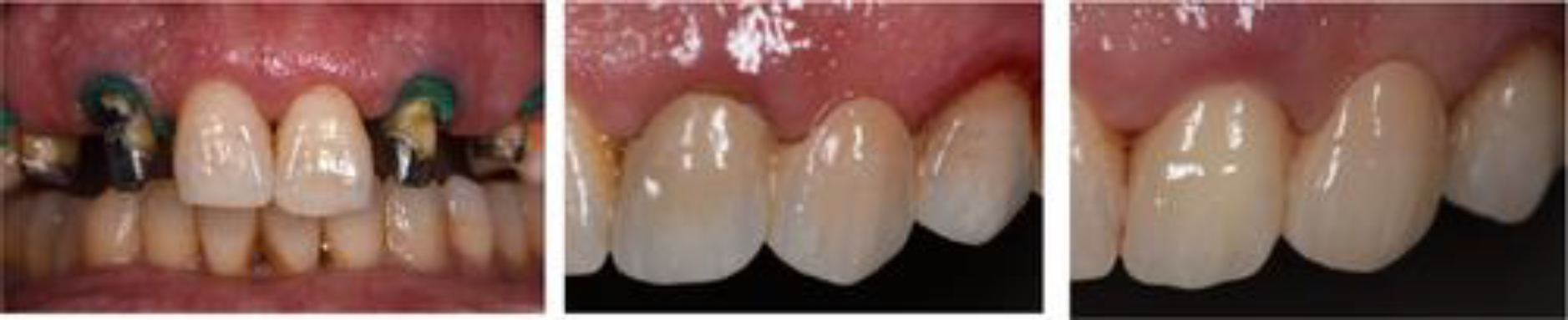
(A) Cast post and core (teeth #s 5–7, and 10–12) that was restored for demonstration purposes with a 5Y zirconia fixed dental prosthesis (10–12) (B) to show lack of ability to mask dark underlying structure. (C) 3Y zirconia fixed dental prosthesis (10–12) was used as final restoration, which shows its capability of masking dark underlying structures.
Most anterior monolithic zirconia restorations are externally stained and glazed to try and emulate the appearance of natural teeth. This is concerning because the stability of stains and glazes is questionable. A 5-year toothbrush simulation study evaluated the color stability of ceramics externally stained and glazed according to manufacturer instructions.19 Glass ceramics retained color better than 3- and 5Y zirconias, while all ceramics loss 40%–50% of its gloss. Interestingly, a more abrasive (charcoal) toothpaste resulted in less color stability and greater loss of gloss than a regular and less abrasive toothpaste. Clinically, when selecting monolithic zirconia for esthetic cases, the facial aspect should be cut back and layered with porcelain to achieve the best esthetic outcome instead of relying on stains/glazes.
5. TOOTH PREPARATION DESIGN
Understanding the tooth preparation requirements for any type of indirect restoration is fundamental for its longevity and outcome of the restoration. For gold and porcelain-fused-to-metal restorations, classic prosthodontic preparation principles have been elucidated. Many classical principles were recommended for contemporary ceramics, such as zirconia, but this is not necessarily the optimal design to maximize the longevity of this restoration. Based on many manufacturers’ recommendation for a layered zirconia restoration is to reduce the tooth structure sufficiently to make room for the bilayered restoration comparable to porcelain fused to metal. A 1 mm reduction of axial wall is minimal with a 1.5–2 mm reduction of incisal and occlusal surfaces. A skilled technician will design the core to conform to the tooth’s anatomy while also providing adequate support for the veneering porcelain. A flat core design will create occlusal anatomy with unsupported porcelain, which has been one of the primary causes of porcelain chipping. There are no defined tooth reduction design principles for monolithic zirconia preparation design. Some manufacturers have recommended that a 0.5 mm reduction is sufficient, while a 1 mm reduction is optimal. Others recommended reducing the tooth structure like a gold restoration. Important to consider when preparing for a monolithic zirconia restoration: Are tooth preparation guidelines the same for all types of zirconia?
A fatigue study evaluated the fracture and fatigue resistance of 3-, 4-, and 5Y zirconias with 0.7- and 1.2-mm thicknesses, simulating a 5-year clinical outcome.20 Fatiguing had minimal effect on the fracture strength of zirconia. However, the most clinically relevant outcome is survivability. 3Y zirconia specimens had zero failure post-fatiguing for both thicknesses. This can be explained simply by the transformation toughening phenomenon, as previously explained, because 3Y zirconia is predominantly tetragonal and can transform to monoclinic and resist crack propagation. The 4Y zirconia at 0.7 mm thickness had 50% fracturing before completing the 5-year clinical simulation. When the thickness was increased to 1.2 mm thickness, no fracture occurred. For the 5Y zirconia specimens, 75% fractured at 0.7 mm thickness, while 35% fractured at 1.2 mm thickness. The lack of fracture resistance in 5Y zirconia can also be explained by the transformation toughening phenomenon, in which a lower proportion of tetragonal particles and a higher proportion of cubic particles reduces the ability to resist crack propagation.
The contention that ceramics are strengthened when a resin cement is used to bond the restoration to the structure is controversial. A recent review asked the question: Does adhesive luting reinforce the mechanical properties of dental ceramics used as restorative materials? Twenty studies were included in the meta-analysis, and the conclusion was that adhesive luting strengthens the mechanical properties of glass–ceramic restorations but not zirconia restorations.21 Regardless if you agree or not with this conclusion, when strength is of the essence, it is important to select a zirconia type with optimal strength that can resist fracture in heavy-load-bearing areas, if adhesive luting enhances the strength in any way, that would be a “cherry on top.”
In response to the question: are tooth preparation guidelines the same for all zirconia types? The obvious response is NO. Based on the outcome of the fatigue study,20 the occlusal reduction guideline for zirconia restorations should be 1 mm when a 3Y zirconia is selected, 1.2 mm minimal reduction for a 4Y zirconia, and 1.5 minimal reduction if a 5Y zirconia ceramic is used. Axial wall reduction must be a minimum of 1 mm with a light chamfer margin design. Some clinicians have advocated a feather edge design for simplicity and tooth conservation; however, when communicating with technicians, this will present difficulties in finishing the margin and preventing chipping. Once the crown with a feather-edge margin has been bonded to the tooth, the concern is alleviated. Finally, respect and follow an anatomical preparation, particularly occlusally in the central groove area, where a flat-cut preparation will challenge the technician to design occlusal anatomy, resulting in reduced thickness in the area where clinical fractures are most likely initiate.
6. CEMENTATION/BONDING OF ZIRCONIA RESTORATIONS
When proper resistance and retention form are present in the abutment, any conventional cement may be utilized. However, when there is insufficient retention form, the zirconia restoration must be bonded using a resin cement. If a diligent bonding protocol is followed, a zirconia restoration can be bonded to the tooth structure.22 The bonding protocol is a combination mechanical and chemical pre-treatment. The mechanical pre-treatment involves air-borne particle abrasion with 50 μm Al2O3 particles, 2.5 bar pressure for 10 s at a 10 mm standoff distance. While the chemical pre-treatment involves using a ceramic primer that contains silane and a 10-MDP monomer. An adhesive or self-adhesive resin cement is used to bond the zirconia to the tooth structure. These steps were simplified by Blatz et al.23 using the acronym APC, which stands for Air-borne particle abrasion, Primer, and Resin Cement.
Debonding of zirconia has been an issue in recent years, causing many clinicians to lose confidence in bonding zirconia restorations. Due to zirconia’s high affinity for salivary, blood lipids, and proteins, contamination during the try-in phase was a leading cause of debonding. Decontamination cannot be achieved with conventional methods used for glass ceramics for example phosphoric acid, steaming, ultrasonic bathing, and so forth. There have been numerous cleaning solutions that have been introduced to address this issue. A study examined cleaning methods and solutions of contaminated zirconia material.24 Rinsing the contaminated surface with water, applying phosphoric acid, and applying a ceramic primer prior to contamination were all ineffective at decontaminating the zirconia surface and significantly decreased the bond strength. Designated zirconia cleaning solutions (Katana Cleaner; Kuraray, Ivoclean; Ivoclar, ZirClean; Bisco) performed similarly and as expected by chemically decontaminating the zirconia surface. Airborne-particle abrasion was the most effective method for removing salivary contamination from zirconia, as it mechanically decontaminates the surface.
After cleaning the zirconia surface, a ceramic primer is applied. It is recommended to scrub the primer with agitation and allow it to soak into the dense particle surface of zirconia rather than quickly blasting the primed surface with air.23, 24 After applying the primer for 2–3 min, a gentle stream of air is used to evaporate any excess solvents. An adhesive or self-adhesive resin cement is used to bond the zirconia to the tooth structure.
It is recommended that the clinician takes responsibility of air-borne particle abrasion themselves after try in, due to the critical parameters that must be meticulously followed. Moreover, it has been researched that air-borne particle abrasion instantly after try in, followed by primer application within this brief period, creates a positively charged surface whose surface energy allows a better priming capability.25 On the other hand, if a technician were to abrade the zirconia 2–3 days before sending the restoration to the clinician, the surface energy is lost and priming capabilities are not as efficient resulting in significantly lower bond strength. The recommended sequence for optimally bonding zirconia is as follows: ask the technician not to abrade the surface, try in the restoration, followed by air-particle abrasion according to the previously defined parameters, steam clean, or air-blast the surface, then primer application followed by the resin cement.
Proper polymerization of the resin requires sufficient light curing. This is frequently overlooked by clinicians and can be a significant cause of zirconia debonding. Zirconia attenuates light due to its opacity, preventing sufficient light energy from penetrating to the resin. Thickness of the restoration is also a confounding factor.3 To overcome and prevent under-curing the resin cement, sufficient energy can be delivery by increasing the curing time.3 To achieve this, it is recommended to cure each surface for at least 40 s. Be cautious of curing lights with high irradiance that claim to shorten curing times; they do not provide sufficient energy to the resin cement.
7. AFTER ZIRCONIA CEMENTATION
Occlusal adjustment is a common clinical practice to ensure proper occlusion. When zirconia is the restoration required to adjust, it is important to understand that this ceramic restoration behaves differently than other indirect restorations. Generally, chairside ceramic adjustment introduces microcracks, weakens the ceramic, and can result in ceramic fracture. Due to the ceramic nature of zirconia, its adjustment is significantly more challenging. Any adjustment of the zirconia could result in phase transformation. For 3Y and, to a lesser extent, 4Y zirconias, phase transformation can initially increase strength; however, as more adjustments are made, more cracks are introduced, and phase transformation can no longer prevent crack propagation, resulting in premature fracture.6, 26, 27 This is more challenging with 5Y zirconia because it does not undergo phase transformation to the same extent as 3- and 4Y zirconia due to its lower tetragonal content. It has been proved that chairside adjustment reduces zirconia strength regardless of yttria content.28, 29 If zirconia must be adjusted, it is advised to use a fine-grit diamond rotary instrument rather than a coarse-grit instrument, followed by a meticulous polishing method.30 When polishing is inadequate, a rough zirconia surface can cause the opposing tooth to wear. However, a highly polished unglazed zirconia is the friendliest to an opposing natural tooth compared to other ceramics and even enamel to enamel.31 Communicate with your technician when completing the zirconia restoration to first polish the surface of the zirconia and then, if desired, apply the glaze. After the glaze has worn away, a highly polished zirconia surface will be revealed.
Clinical survivability of layered zirconia restorations has acceptable level of evidence. A small number of studies concluded a 5-year cumulative survival rate of 96% for tooth-supported layered zirconia restorations and 97% survival rate for implant-supported restorations.32 Well-designed controlled clinical trials are extremely lacking, especially for monolithic zirconia. A retrospective multicentric study in private practices aimed to evaluate the outcomes of 619 3- and 4Y monolithic zirconia crowns followed up between 18 and 84 months.33 The survival rate was 99% with 1 crown fracturing and 9 debonded. Laboratory survey studies have been helpful in providing an early sense of indication of a newly introduced ceramic system, especially given the paucity of clinical evidence for the majority of these systems.34 Laboratories surveyed provide a 5-year warranty for ceramics they fabricated, which can enforce the numbers, allowing them to evaluate a large number of ceramic restorations in relatively short period of time. Zirconia restorations were classified into four categories: single crowns, fixed dental prosthesis, monolithic and layered restorations. Over 77,000 monolithic single unit 3Y zirconia restorations were reviewed for remake reasons due to fracture only, resulting in a fracture rate of approximately 0.5%, and 33,036 layered zirconia restorations had a fracture rate of approximately 2.8%. For fixed dental prosthesis, approximately 16,500 monolithic 3Y zirconia were reviewed, resulting in a fracture rate of 1.30%. While approximately 13,000 layered 3Y fixed dental prosthesis resulted in a fracture rate of 1.50%.34 Studies of this nature are not a replacement for clinical trials, but they can provide early insight and information regarding a recently introduced ceramic material that is being provided to patients with little to no clinical evidence.
8. CONCLUDING REMARKS
Yttria concentration in zirconia composition is integral to the optical and mechanical properties of zirconias. In general, as the concentration of yttria increases, the translucency increases while the strength decreases.
The most recent type of zirconia is composed of multi-layering of yttria (5- and 3Y) or (5- and 4Y) intended to mimic the translucency of the tooth structure from cervical to incisal or occlusal. Due to the fact that the weakest zirconia is on the functional surfaces of the crown, where the majority of cracks originate, caution is required when selecting this type of zirconia.
Fast sintering of zirconia enables the delivery of zirconia crowns in a single visit. More research is required to understand and confirm any possible effect such sintering process may have on the properties of zirconia.
The bonding of zirconia restorations to tooth structure can be long-lasting if a strict and meticulous protocol is followed; air-particle abrasion, ceramic primer containing the MDP monomer, and a resin cement. Light cure efficiently (40 s/surface) to ensure proper light energy passing through zirconia for optimal resin cement curing.
Regardless of yttria content, chairside zirconia adjustment weakens the zirconia. It is best to prevent zirconia adjustments whenever possible. If a zirconia adjustment is necessary, it is advised to use a fine diamond bur rather than a coarse bur.
Monolithic tooth- and implant-supported zirconia restorations are arguably the most prescribed indirect ceramic material, despite the severe lack of clinical evidence to support their use. This ceramic material possesses high hardness, is resistant to wear, and does not wear when polished. These characteristics are not similar to the behavior of the tooth structure, which makes the long-term success of these restorations questionable, especially concerning biological aspects of the tooth.
ACKNOWLEDGEMENTS
Yu Zhang would like to acknowledge funding from the U.S. National Institutes of Health/National Institute of Dental and Craniofacial Research (grant numbers: R01DE033545, R01DE026279, and R01DE026772).
Footnotes
CONFLICT OF INTEREST STATEMENT
The authors declare that they do not have any financial interest in the companies whose materials are included in this article.
REFERENCES
- 1.Kelly JR, Denry I. Stabilized zirconia as a structural ceramic: an overview. Dent Mater, 2008;24:289–298. [DOI] [PubMed] [Google Scholar]
- 2.Tong H, Tanaka CB, Kaiser MR, Zhang Y. Characterization of three commercial Y-TZP ceramics produced for their high translucency. Ceram Int, 2016;42(1 Pt B):1077–1085. [DOI] [PMC free article] [PubMed] [Google Scholar]
- 3.Sulaiman TA, Abdulmajeed AA, Donovan TE, Ritter AV, Vallittu PK, Närhi TO, Lassila LV. Optical properties and light irradiance of monolithic zirconia at variable thicknesses. Dent Mater, 2015;31(10):1180–1187. [DOI] [PubMed] [Google Scholar]
- 4.Zhang Y Making Yttria-Stabilized Tetragonal Zirconia Translucent. Dent Mater, 2014;30:1195–1203. [DOI] [PMC free article] [PubMed] [Google Scholar]
- 5.Lim CH, Vardhaman S, Reddy N, Zhang Y. Composition, processing, and properties of biphasic zirconia bioceramics: Relationship to competing strength and optical properties. Ceram Inter., 2022;48(12):17095–17103. [DOI] [PMC free article] [PubMed] [Google Scholar]
- 6.Garvie RC, Hannink RH, Pascoe RT. Ceramic steel? Nature, 1975;258:703–704. [Google Scholar]
- 7.Hannink RH, Kelly PM, Muddle BC. Transformation toughening in zirconia-containing ceramics. J Amer Ceram Soc, 2000;83(3):461–87. [Google Scholar]
- 8.Zhang Y, Sailer I, Lawn BR. Fatigue of Dental Ceramics. J Dent., 2013;41:1135–1147. [DOI] [PMC free article] [PubMed] [Google Scholar]
- 9.Badr Z, Culp L, Duqum I, Lim CH, Zhang Y, A. Sulaiman T. Survivability and fracture resistance of monolithic and multi-yttria-layered zirconia crowns as a function of yttria content: A mastication simulation study. J Esthet Rest Dent., 2022;34:633–640. [DOI] [PMC free article] [PubMed] [Google Scholar]
- 10.Liao Y, Gruber M, Lukic H, McLees J, Chen S, Boghosian A, Megremis S. Survey of the mechanical and physical behaviors of yttria-stabilized zirconia from multiple dental laboratories. J Amer Dent Assoc, 2023(1);2:100018. [Google Scholar]
- 11.Sulaiman TA, Abdulmajeed AA, Donovan TE, Vallittu PK, Närhi TO, Lassila LV. The effect of staining and vacuum sintering on optical and mechanical properties of partially and fully stabilized monolithic zirconia. Dent Mater J, 2015;34:605–610. [DOI] [PubMed] [Google Scholar]
- 12.Kaizer MR, Gierthmuehlen PC, Dos Santos MB, Cava SS, Zhang Y. Speed sintering translucent zirconia for chairside one-visit dental restorations: Optical, mechanical, and wear characteristics. Ceram Intern, 2017;43:10999–1005. [DOI] [PMC free article] [PubMed] [Google Scholar]
- 13.Jansen JU, Lümkemann N, Letz I, Pfefferle R, Sener B, Stawarczyk B. Impact of high-speed sintering on translucency, phase content, grain sizes, and flexural strength of 3Y-TZP and 4Y-TZP zirconia materials. J Prosthet Dent., 2019;122:396–403. [DOI] [PubMed] [Google Scholar]
- 14.Lawson NC, Maharishi A. Strength and translucency of zirconia after high-speed sintering. J Esthet Restor Dent, 2020;32:219–225. [DOI] [PubMed] [Google Scholar]
- 15.Nonaka K, Teramae M, Pezzotti G. Evaluation of the effect of high-speed sintering and specimen thickness on the properties of 5 mol% yttria-stabilized dental zirconia sintered bodies. Mater., 2022;15(16):5685. [DOI] [PMC free article] [PubMed] [Google Scholar]
- 16.Alshahrani AM, Lim CH, Kim J, Zhang Y. Transient thermal stresses developed during speed sintering of 3 mol% yttria-stabilized tetragonal zirconia polycrystals. Dent Mater, 2023;39(5):522–528. [DOI] [PMC free article] [PubMed] [Google Scholar]
- 17.Zhang Y, Lawn BR. Novel zirconia materials in dentistry. J Dent Res, 2018;97:140–147. [DOI] [PMC free article] [PubMed] [Google Scholar]
- 18.Bacchi A, Boccardi S, Alessandretti R, Pereira GK. Substrate masking ability of bilayer and monolithic ceramics used for complete crowns and the effect of association with an opaque resin-based luting agent. J Prosthodont Res. 2019; 63(3): 321–326 [DOI] [PubMed] [Google Scholar]
- 19.Sulaiman TA, Camino RN, Cook R, Delgado AJ, Roulet JF, Clark WA. Time-lasting ceramic stains and glaze: a toothbrush simulation study. J Esthet Restor Dent, 2020;32:581–585. [DOI] [PubMed] [Google Scholar]
- 20.Abdulmajeed A, Sulaiman T, Abdulmajeed A, Bencharit S, Närhi T. Fracture load of different zirconia types: A mastication simulation study. J Prosthodont., 2020;29:787–791. [DOI] [PubMed] [Google Scholar]
- 21.da Rosa LS, Dapieve KS, Dalla-Nora F, Rippe MP, Valandro LF, Sarkis-Onofre R, Pereira GK. Does Adhesive Luting Reinforce the Mechanical Properties of Dental Ceramics Used as Restorative Materials? A Systematic Review and Meta-Analysis. J Adhes Dent, 2022;24:209–222. [DOI] [PMC free article] [PubMed] [Google Scholar]
- 22.Inokoshi M, De Munck J, Minakuchi S, Van Meerbeek B. Meta-analysis of bonding effectiveness to zirconia ceramics. J Dent Res, 2014;93:329–334. [DOI] [PubMed] [Google Scholar]
- 23.Blatz MB, Alvarez M, Sawyer K, Brindis M. How to bond zirconia: the APC concept. Compend Contin Educ Dent, 2016;37:611–618. [PubMed] [Google Scholar]
- 24.Sulaiman TA, Altak A, Abdulmajeed A, Rodgers B, Lawson N. Cleaning zirconia surface prior to bonding: A comparative study of different methods and solutions. J Prosthodont, 2022;31:239–244. [DOI] [PubMed] [Google Scholar]
- 25.Al-Akhali M, Al-Dobaei E, Wille S, Mourshed B, Kern M. Influence of elapsed time between airborne-particle abrasion and bonding to zirconia bond strength. Dent Mater, 2021;37:516–522. [DOI] [PubMed] [Google Scholar]
- 26.Zhang Y, Lawn BR, Malament KA, Thompson VP, Rekow ED. Damage accumulation and fatigue life of particle-abraded ceramics. Interl J Prosthodont., 2006;19(5). [PubMed] [Google Scholar]
- 27.Srikanth R, Kosmac T, Della Bona A, Yin L, Zhang Y. Effects of cementation surface modifications on fracture resistance of zirconia. Dent Mater, 2015;31(4):435–442. [DOI] [PMC free article] [PubMed] [Google Scholar]
- 28.Khayat W, Chebib N, Finkelman M, Khayat S, Ali A. Effect of grinding and polishing on roughness and strength of zirconia. J Prosthet Dent, 2018;119:626–631. [DOI] [PubMed] [Google Scholar]
- 29.Wongkamhaeng K, Dawson DV, Holloway JA, Denry I. Effect of surface modification on in-depth transformations and flexural strength of zirconia ceramics. J Prosthodont., 2019;28:e364–75. [DOI] [PMC free article] [PubMed] [Google Scholar]
- 30.Abdulmajeed A, Sulaiman TA, Abdulmajeed AA, Närhi TO. Strength and phase transformation of different zirconia types after chairside adjustment. J Prosthet Dent., 2022. [DOI] [PubMed] [Google Scholar]
- 31.Aljomard YR, Altunok EÇ, Kara HB. Enamel wear against monolithic zirconia restorations: A meta-analysis and systematic review of in vitro studies. J Esthet Restor Dent, 2022;34:473–489. [DOI] [PubMed] [Google Scholar]
- 32.Larsson C, Wennerberg A. The clinical success of zirconia-based crowns: a systematic review. Int J Prosthodont., 2014;27:33–43. [DOI] [PubMed] [Google Scholar]
- 33.Valenti M, Valenti A, Schmitz JH, Cortellini D, Canale A. Survival analysis up to 7 years of 621 zirconia monolithic single crowns with feather-edge margins fabricated with a cast-free workflow starting from intraoral scans: A multicentric retrospective study. J Prosthet Dent, 2023;129:76–82. [DOI] [PubMed] [Google Scholar]
- 34.Sulaiman TA, Abdulmajeed AA, Delgado A, Donovan TE. Fracture rate of 188695 lithium disilicate and zirconia ceramic restorations after up to 7.5 years of clinical service: A dental laboratory survey. J Prosthet Dent., 2020;123:807–810. [DOI] [PubMed] [Google Scholar]