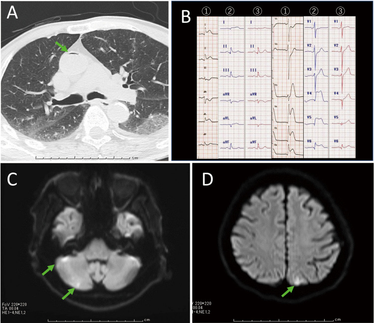
A 76-year-old man lost consciousness while attempting to transfer to a wheelchair following CT-guided needle biopsy for suspected lung cancer. CT images showed air moving in the pulmonary veins (Supplementary Movie 1) and air in the aorta (Figure A). ECG showed ST-segment elevation in leads I, II, aVL, and V4–V6 (Figure B ①). Transthoracic echocardiography (TTE) showed marked hypokinesis of the anterior and anterolateral wall from mid to apex and increased brightness of the posterior wall and both papillary muscles (Supplementary Movie 2 ①). ST-segment elevation showed changes over time (Figure B). TTE performed on the following day showed disappearance of the increased myocardial brightness and improvement of LV wall motion (Supplementary Movie 2 ②). Creatine kinase enzyme increased to 739 U/L. Magnetic resonance imaging of his brain revealed multiple cerebral infarctions (Figure C,D). Air in the coronary arteries became microbubbles, creating large differences in acoustic impedance between the myocardium and the microbubbles, enhancing tissue brightness. Multidetector CT showed no stenosis in the coronary arteries.
Figure.
(A) Air in the aorta (arrow). (B) Time course of ECG ① after loss of consciousness ② 44 min and ③ 118 min after (C,D) multiple cerebral infarctions (arrows).
The incidence of air embolism during CT-guided lung biopsy is rare, reported to be 0.061%.1 In this case, air was introduced from the puncture site into the pulmonary vein due to atmospheric pressure during removal of the puncture needle that had penetrated the pulmonary vein. If air enters the left heart system, there is a method of aspirating air through the catheter;2 it is important to prevent cerebral embolism by lowering the head. Increased myocardial brightness may be an early and important signal of air embolism.
Disclosures
None.
Supplementary Files
Supplementary Movie 1.
Supplementary Movie 2.
References
- 1. Tomiyama N, Yasuhara Y, Nakajima Y, Adachi S, Arai Y, Kusumoto M, et al.. CT-guided needle biopsy of lung lesions: A survey of severe complication based on 9,783 biopsies in Japan. Eur J Radiol 2006; 59: 60–64. [DOI] [PubMed] [Google Scholar]
- 2. Tamura H, Ogura R, Izumi T, Hosokawa S.. Successful air bubble aspiration in the left atrium using three-dimensional transesophageal echocardiography. Circ J 2023; 87: 581. [DOI] [PubMed] [Google Scholar]
Associated Data
This section collects any data citations, data availability statements, or supplementary materials included in this article.
Supplementary Materials
Supplementary Movie 1.
Supplementary Movie 2.


