ABSTRACT
ER+ breast cancers (BC) are characterized by the elevated expression and signaling of estrogen receptor alpha (ESR1), which renders them sensitive to anti-endocrine therapy. While these therapies are clinically effective, prolonged treatment inevitably results in therapeutic resistance, which can occur through the emergence of gain-of-function mutations in ESR1. The central importance of ESR1 and development of mutated forms of ESR1 suggest that vaccines targeting these proteins could potentially be effective in preventing or treating endocrine resistance. To explore the potential of this approach, we developed several recombinant vaccines encoding different mutant forms of ESR1 (ESR1mut) and validated their ability to elicit ESR1-specific T cell responses. We then developed novel ESR1mut-expressing murine mammary cancer models to test the anti-tumor potential of ESR1mut vaccines. We found that these vaccines could suppress tumor growth, ESR1mut expression and estrogen signaling in vivo. To illustrate the applicability of these findings, we utilize HPLC to demonstrate the presentation of ESR1 and ESR1mut peptides on human ER+ BC cell MHC complexes. We then show the presence of human T cells reactive to ESR1mut epitopes in an ER+ BC patient. These findings support the development of ESR1mut vaccines, which we are testing in a Phase I clinical trial.
KEYWORDS: Breast cancer, estrogen receptor, neoepitopes, resistance mutations, cancer vaccines, preventative vaccination, immunotherapy
Introduction
Approximately 75% of breast cancers (BC) are estrogen receptor α+ (ER+) and treated with anti-estrogen (endocrine) therapies, such as tamoxifen and aromatase inhibitors. While these therapies are often highly effective, ~25% of patients with localized cancers (Stage I–III) and nearly all with metastatic cancers (Stage IV) develop resistance to these therapies.1–7 A common resistance mechanism in these heavily treated patients occurs through gain-of-function mutations in ESR1, found in 20–40% of patients with metastatic ER+ BC who received endocrine therapies, but rarely in primary tumors.5,8,9 These gain-of-function mutations are clustered within the ligand-binding domain (LBD) of ESR1 at one of three neighboring amino acids, all of which lead to ligand-independent ER activity and neomorphic activation of various pathways as a major mechanism of acquired endocrine resistance.1,8,10–12
ESR1 is highly expressed in ER+ breast cancer and its mutations cause amino acid alterations, which generate potential neoantigens that can be recognized and targeted immunologically. Neoantigens are formed by mutations to self-proteins that render them immunologically non-self, which many studies have suggested to be a central factor in generating anti-tumor immunity.13,14 While most neoantigens are formed by randomly occurring mutations,14 emergence of ESR1 activating mutations in ER+ BC after prolonged endocrine therapy occurs in a predictable manner as a mechanism of therapeutic resistance. In addition to their ligand-independent estrogen signaling, studies have also documented that mutated ESR1 confers neomorphic oncogenic properties, making it a driver of tumor progression and escape.10–12 We have previously demonstrated the critical importance of targeting oncogenic pathways, in comparison to non-essential tumor associated antigens, for the efficacy of anti-tumor vaccines.15–17 Thus, given the critical importance of ESR1 mutations in sustaining ER+ BC oncogenic signaling, their predictable emergence as neoantigens, and the elevated expression of ESR1 in ER+ BC, we hypothesized that a vaccination strategy targeting mutant forms of ESR1 (ESR1mut) could be an effective therapeutic approach to immunologically prevent or treat endocrine resistant cancers.
In our study, we developed vaccines to several mutant forms of human ESR1 and determined that these vaccines could all elicit robust ESR1-specific T cell responses. We then developed immune competent models of human ESR1mut expressing murine mammary cancer and demonstrated that different human ESR1mut vaccines could elicit cross-protective anti-tumor responses that suppressed ESR1mut expression and estrogen signaling. We then validated the presentation of ESR1 peptides on human ER+ BC MHC complexes. These results led to the initiation of a clinical trial targeting ESR1mut using peptide vaccines. Analysis of one of the trial participants documents the existence of ESR1mut-specific responses even prior to vaccination. This result suggests the potential to augment these responses with ESR1mut vaccination to prevent the clinical emergence of ESR1mut+ BC and prevent the development of ESR1mut-mediated endocrine resistance.
Results
Development of an adenoviral vaccine targeting ESR1 mutants in vivo
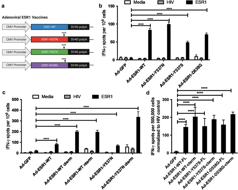
To test the potential of vaccination against mutant forms of ESR1, we developed a series of 1st generation [E1,E3-] adenoviral vectors expressing wild-type ESR1 or one of the three dominant ESR1 mutant genes (comprising >75% of ESR1 mutations) to immunologically target wild-type or endocrine resistance-associated mutant ER18,19 (Figure 1a). While ESR1 is highly homologous between mice and human (~90%),20 we found that immunizing mice with any of these vaccines was sufficient to break immune tolerance and induce human ESR1-specific T cell immunity that was comparable between ESR1-WT and various ESR1mut vaccines in C57BL/6 mice, without signs of obvious autoimmunity (Figure 1b). Given the parity of responses, we next wanted to determine if ESR1-specific responses in mice were concentrated against particular regions of ESR1, so we constructed vaccines encoding different subunits of ESR1 to ascertain if certain regions of ESR1 harbored more immunodominant epitopes.21,22 We constructed three roughly equal (~200 amino acid) ESR1 subunits, comprised of an N-terminal, a middle, and C-terminal domain (containing the neoepitope site), which were all incapable of stimulating estrogen signaling (Figure S1). To determine if these vaccines could induce presentation on human HLA, we vaccinated human HLA-A2+ transgenic mice. These studies revealed that N-terminal and C-terminal vaccines yielded potent ESR1-specific T cell responses, but that the Y537N mutant form of the C-terminal vaccine had the greatest response (Figure 1c). We then expanded our vaccination of HLA-A2+ transgenic mice to include additional ESR1 C-terminal mutant vaccines (ESR1-Y537S and D538G). In these studies, we again observed that C-terminal vaccines elicited stronger T cell responses compared to full-length versions, but no significant differences were observed between different ESR1-WT and ESR1mut vaccines (Figure 1d). Given the large amount of overlap between the sequences encoded by ESR1-WT and ESR1mut vaccines, splenocytes from mice vaccinated with the c-terminal ESR1 vectors were assessed by IFN-gamma ELISPOT after stimulation with peptides from the region that forms the neoepitope. This included the vaccine matched neoepitope peptides (peptides specific for the mutation, for instance a peptide containing Y537N for an Ad-ESR1-Y537N vaccine) and stimulation using peptides from non-matched mutants (Figure S2A). These assays revealed weak induction of neoepitope specific peptide responses in individual ESR1mut vaccinated mice in comparison to control Ad-GFP or Ad-ESR1-WT-cterm mice.
Figure 1.

Development and testing of human ESR1 targeting vaccines. (a) Diagram of E1 region for [E1-,E3-] adenoviral ESR1 vaccines. (b,c) C57BL/6J mice (b) and HLA-A2-Tg (c) were vaccinated with indicated viruses and assessed for ESR1-specific T cell responses at 2 weeks post vaccination through IFNγ ELISPOT (n = 5 mice/group). (d) HLA-A2 tg mice were vaccinated with the indicated viruses and assessed for ESR1-specific T cell responses at 2 weeks post vaccination through IFNγ ELISPOT (n = 3–4 mice/group). Error bars represent SEM. Select p values shown. Significance determined via T test with Bonferroni correction for multiple comparisons. ****p < .0001.
As peptide specific responses are highly sensitive to MHC haplotypes for presentation and binding, we also utilized Ad-ESR1Y537N to vaccinate a cohort of diversity outbred mice (Figure S2B). While we found strong responses against adenoviral epitopes, we found far more variable responses against ESR1-specific and ESR1mut neoepitope peptides, with some mice having almost no response and other having a robust response. However, we did observe a positive correlation between ESR1-specific and ESR1 neoepitope responses and found evidence for cross-reactivity between ESR1-Y537N and Y537S neoepitopes. These results suggest that in mice, systemic ESR1 neoepitope-specific T cell responses can occur but are a minor population in the spleen, with the response dominated by the large number of shared epitopes between ESR1-WT and ESR1mut.
Oncogenicity of ESR1 mutants and development of immune competent ESR1mut cell line model
As no immune competent mouse cell line models of ERmut+ BC exist, we sought to develop one to test the impact of ESR1-WT and ESR1mut vaccination on tumor growth. As an initial step, we validated the ability of our ESR1mut genes to confer ligand-independent estrogen signaling (Fig S3A) and transduced an ER+ BC line (MCF-7) with ESR1-WT or ESR1-Y537N to assess their oncogenic functional capacity. In the absence of estrogen, RNAseq analysis of these tumors revealed that ESR1-Y537N cells have activation of hormonal signaling pathways, dysregulation of apoptotic and cytokine pathways, with upregulation of proliferative pathways (Figure S3B). More critically, we found that in mice without exogenous estrogen supplementation in vivo, MCF7/ESR1-Y573N permitted robust estrogen-independent growth, in comparison to MCF7/ESR1-WT cells (Figure S3C). Subsequent studies confirmed this for an ESR1-Y537S mutant (Figure S3D) and validated the oncogenic nature of different ESR1mut expression in ER+ BC, supporting that activating ESR1 mutant expression may enhance the growth of murine mammary cells.
To determine if these mutants also confer estrogen-independent activation to facilitate the growth of mouse mammary cells, we first expressed human ESR1 and various human ESR1 mutants in a mouse mammary line (MM3MG) and confirmed the robust stimulation of estrogen independent signaling pathways by ESR1mut expression with comparable levels of ESR1 and ESR1mut expression (Figure 2a,b). Next, we tested the in vivo growth of stably transduced MM3MG-ESR1-Y537N cells in comparison to previously generated stably transduced MM3MG-HERΔ16 (strong HER2 oncogene), which we found capable to transforming MM3MG cells.16,23 These studies revealed that an ESR1 mutant expression conferred a comparable growth advantage in the mammary fat pad (Figure 2c). We then compared MM3MG-ESR1-Y537N cells with MM3MG cells expressing either ESR1-WT or ESR1-Y537S and found that both ESR1 mutants elicited more robust growth in non-estrogen supplemented mice in comparison to ESR1-WT mice (Figure 2d). Collectively, these results suggested that ESR1mut expressing MM3MG cells represented a potential model of ESR1mut expressing cancers that could be interrogated by vaccination.
Figure 2.

Development of ERmut+ mouse mammary tumor model. (a) MM3MG cell lines expressing Y537N or Y537S doxycycline inducible ESR1 mutants were assessed at 24 hours post-dox/estrogen stimulation for the indicated pathway (n = 3/group). (b) QRT-PCR assessment of ESR1 expression in MM3MG lines. (c) Indicated MM3MG cells were implanted into BALB/c mice and allowed to grow, measured bi-weekly (n = 5 mice/group). (d) Indicated MM3MG cells were implanted into BALB/c mice and allowed to grow, measured bi-weekly (n = 5 mice/group).
Vaccination to prevent and treat ESR1mut+ BC
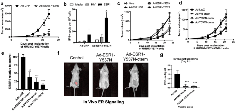
Given the ability of different Ad-ESR1mut vaccines to elicit ESR1-specific responses, we next wanted to determine if these vaccines could prime the immune system to prevent the outgrowth of tumors expressing an ESR1 mutant gene using this new model. To test this concept, we vaccinated mice with Ad-ESR1-Y537N and 2 weeks later implanted murine mammary tumor cells expressing ESR1-Y537N (Figure 3a). In this setting, preventative vaccination had a significant anti-tumor effect and elicited ESR1-specific T cell responses, suggesting the potential of vaccination to prevent the outgrowth of ESR1mut cancers (Figure 3b). Notably, when mice were vaccinated and implanted with ESR1-WT expressing cells, we observed no impact from vaccination, suggesting strong on-target anti-ESR1mut effects from vaccination (Fig S4A). Given that many patients have existing ER+ BC containing ESR1 mutations and that multiple types of mutations can occur, we next tested the impact of different ESR1mut and ESR1-WT therapeutic vaccinations in tumor-bearing mice. In this study we implanted ESR1-Y537N tumors and subsequently vaccinated tumor bearing mice with Ad-ESR1-wild-type, Y537N, and Y537S vaccines (Figure 3c). We found that both Ad-ESR1-Y537N and Ad-ESR1-Y537S were able to slow the growth of ESR1-Y537N expressing tumors, whereas we surprisingly observed that Ad-ESR1-WT had no effect. These data could suggest an anti-tumor role for ESR1mut reactive T cells in the tumor microenvironment, possible neoantigen immunodominance in the context of a developing tumor, antigen spreading following vaccination for ESR1mut, or cross-protection between different ESR1 mutants. To validate that this effect was specific for C-terminal subunit vaccines, we repeated this experiment using C-terminal truncated vaccines (for ESR1-WT, Y537N and Y537S), as well as with the inclusion of an Ad-LacZ control (Figure 3d). Here we used an MM3MG line expressing ESR1-Y537N linked to CD90.1 by a T2A self-cleaving peptide tag. This allows for assessment of the levels of ESR1 expression by staining for surface expression of CD90.1. This experiment again demonstrated that enhanced anti-tumor responses were afforded by both mutant vaccines, in contrast to control or ESR1-WT c-terminal vaccination. However, analysis of tumor outgrowths demonstrated that all ESR1 vaccines significantly suppressed ESR1-Y537N expression. Loss of the target antigen by tumors from vaccinated mice confirms the potency of all ESR1 vaccines to elicit a suppression of ESR1 expression, but with stronger anti-tumor responses by ESR1mut vaccines (Figure 3e). To determine if selection against ESR1mut expression suppressed ER signaling, we transduced MM3MG-ESR1-Y537N cells with a stable Estrogen signaling pathway reporter lentivirus (ERE-Luc) and repeated a vaccination with Ad-ESR1-Y537N, Ad-ESR1-Y537-cterm, or an Ad-GFP control. This experiment revealed a significant suppression of ER signaling in tumors in vivo from both ESR1mut vaccines (Figure 3f,g), which was again associated with reduced growth of Ad-ESR1-Y537N vaccinated mice in comparison to control (Figure S4B). Collectively, these experiments demonstrate that ESR1mut vaccines are capable of eliciting anti-tumor immune responses that suppress ESR1mut expression and signaling, thus negating the impact of ESR1mut expression and depriving cancer cells of this endocrine resistance mechanism.
Figure 3.

Anti-tumor impact of ESR1mut vaccines in a ERmut+ mouse mammary tumor model. (a) BALB/c mice were vaccinated with indicated virus and MM3MG-ESR1-Y537N cells were implanted two weeks post-vaccination and tumor growth measured biweekly. (b) spleens from mice in (a) were assessed by IFNγ ELISPOT assay (n = 5 mice/group). (c,d) BALB/c mice were implanted with MM3MG-ESR1-Y537N (c) or MM3MG-ESR1-Y537N-CD90.1 (d) expressing cells and vaccinated the following day with tumor growth measured bi-weekly (n = 5 mice/group). (e) Tumors from (d) were removed and CD90.1 expression was measured ex vivo as a surrogate of ESR1 expression. Shown as % of expression seen in control treated cells. Error bars represent SEM. *p < .05; **p < .05; ***p < .001. (f,g) Representative images and analysis (g) of MM3MG-ESR1-Y537N-CD90.1/ERE-Luc tumors vaccinated with ad-GFP control or ad-ESR1-Y537N as in (c,d).
Presentation and immunogenicity of ESR1 epitopes against human T cells
Having demonstrated some efficacy in ESR1mut vaccination in mouse models, we wanted to further explore its potential as a clinical immunotherapeutic target in humans. To first validate that peptides encoded by ESR1 were presented by major histocompatibility complexes (MHC), we utilized proteomic analysis of ESR1mut+ MCF7 cells through HLA class I immune-precipitation and peptide HPLC. These analyses identified multiple ESR1 peptides as being potentially presented by HLA-A2 complexes, including an Y537N mutant neoepitope (Table S1), thus supporting the potential ESR1-specific T cell recognition of ER+ BC.
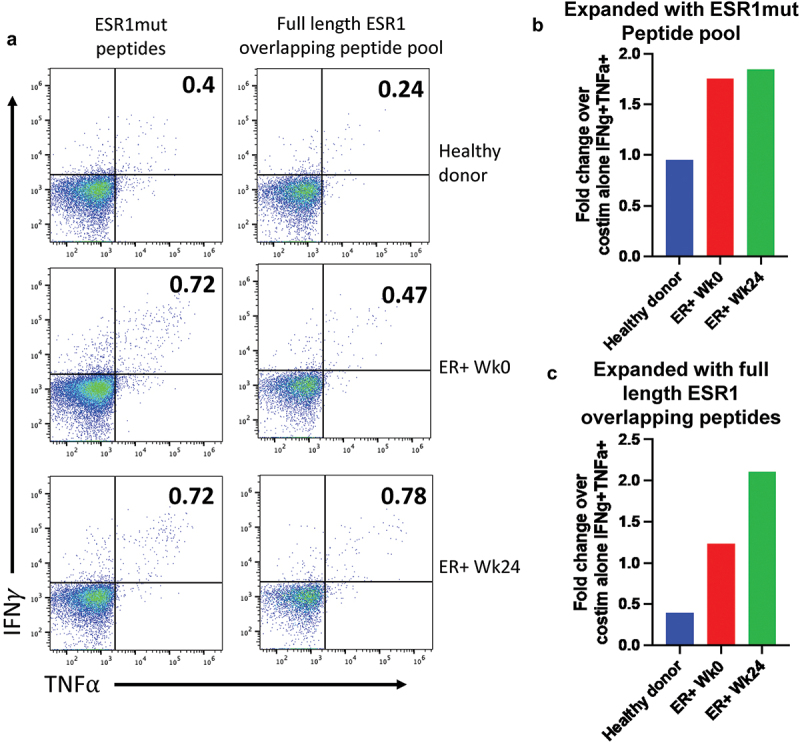
Given our ability to elicit ESR1 and ESR1mut-specific T cell responses, we subsequently initiated a Phase I clinical trial to determine if we could elicit or enhance T cell responses to ESR1 and ESR1mut through peptide vaccination, using a mix of four 10-mer ESR1mut and one 10-mer ESR1-WT peptide in Montanide with GM-CSF (NCT04270149). While ongoing, our preliminary studies document more ESR1-reactive T cells in PBMCs from ER+ BC patients (prior to vaccination, Wk 0), in comparison to levels observed from normal donors following ESR1-peptide stimulation (Figure 4a–c). Expansion and re-stimulation with a pool of the five vaccine containing peptides showed similar levels of IFNg+TNFa+ double producing CD8 T cells pre- and post-vaccine (Figure 4b). Notably, we observe greater responses against an overlapping peptide pool spanning the entire ESR1 protein in ER+ patient PBMCs after the completion of vaccination, suggesting some ability to enhance ESR1-specific immunity beyond just the vaccination epitopes (Figure 4c). While incomplete, these responses suggest the presence of ESR1-reactive T cells in ER+ BC patients, which may be augmented through vaccination. Our ongoing trial and future trials will determine if the vaccine strategies utilized will be sufficient to expand these populations to elicit a therapeutic effect and potentially prevent or delay the development of endocrine resistance.
Figure 4.

Expansion of ESR1-reactive human T cells. (a) PBMCs from a healthy donor and a patient enrolled in NCT04270149 at week 0 and week 24 after completing 6 vaccinations. Cells were expanded in vitro for 9 days with the vaccine containing ESR1mut peptides (left) or an overlapping peptide pool that encompassed the entire ESR1 protein (right). Cells were then restimulated, stained, and analyzed by spectral flow cytometry for IFNg and TNFa. Cells shown are pregated on live, singlets, CD3+, CD4–, CD8+, CD45RA–, CD45RO+. (b,c) the fold change in expression percent of ESR1mut peptides (b) or full length ESR1 (c) compared to a negative control that received costimulation but no peptide is shown for each sample.
Discussion
The predicable resistance to endocrine therapy in metastatic ER+ breast cancer creates an opportunity to target the outgrowth of predictably arising ER-LBD-mutant cells with a preventative vaccine to block progression and treatment failure for 20–40% of patients. The ability of vaccines to prevent diseases is well-established, and is based on their ability to generate potent immunity against pathogen xenoantigens capable of eradicating cells expressing the antigen target, through the induction of T cell-mediated immunity to limit expression of these genes and minimize downstream signaling, or reduce the number of cells highly expressing these genes. Based on this premise, we demonstrated the ability of multiple ESR1mut vaccines to elicit human ESR1-specific T cell responses and stimulate anti-tumor responses against a novel immune competent model of ESR1mut+ mammary cancer.
In our development of this novel ESR1mut+ model, we independently validated work from multiple groups that ESR1 LBD mutants induce estrogen-independent ER signaling and neomorphic signaling changes2,8,10–12 by demonstrating the capacity of ESR1 mutants to elicit enhanced estrogen oncogenic signaling in human and mouse cells, which promotes growth in vivo and suppression of interferon genes (such as IFI27 and SOCS2, Figure S3, Figure 2). These findings are consistent with other groups that have documented the capacity of estrogen signaling to suppress immune and inflammatory responses,24–26 as well as the suppression of Stat1 downstream of ESR1 targets, similar to the progesterone receptor.27
Using this ESR1mut+ model, we found that ESR1-WT and ESR1mut vaccination can elicit anti-tumor responses, critically reducing ESR1mut expression and suppressing estrogen signaling in tumor outgrowths (Figures 1 and 3). As mutated ESR1 confers intrinsic endocrine resistance and to neighboring cells through paracrine mechanisms,10 the reduction of ESR1mut expression through vaccination may be a critical means to eliminate the development of this type of resistance. In our studies, we found that prophylactic vaccination against ESR1mut could be highly effective in slowing the development of ESR1mut+ cancers (Figure 3a), as well as reducing ER expression and signaling in ESR1mut+ cancers (Figure 3e–g), thus suggesting the potential of vaccinating patients prior to the development of ESR1 mutations. It is important to note that while estrogen signaling was seen in our ESR1mut expressing cell lines, these tumors do not appear addicted to ER expression in the same manner as human ER+ tumors (Fig S3). This could partially explain the moderate anti-tumor responses observed despite the induction of a robust anti-ESR1 immune response. While the loss of antigen represented a viable escape mechanism for our tumor model, it may be less likely to occur in patients with ER+ breast cancer suggesting that these vaccines could be more impactful clinically. However, our data also suggest that local immune suppressive mechanisms are likely to play a key role in mitigating anti-tumor responses in established cancers, which has been repeatedly observed in preclinical and clinical studies.16–28–31 Strategies to overcome these limitations will be critical in extending the therapeutic efficacy of these types of cancer vaccines. As we observed anti-tumor cross-protection between vaccines targeting different ESR1 mutations (Figures 1 and 3c,d), our study also suggests the potential of ESR1mut vaccines to induce T cells with TCR capable of cross-reacting to multiple different mutations. This is likely to be critically important in a prophylactic setting where several different potential mutations might arise in ER+ BC cells.
The ESR1 vaccines described here encoded the human ESR1 gene and immunogenicity was measured in mice, making human ESR1 a potential foreign antigen. While this does represent a lower threshold for breaking immune tolerance with a vaccine than would typically be seen in patients, the human and mouse ESR1 genes share ~90% sequence homology.32 This, together with the evidence that tumor cells expressing human ESR1 were able to reliably grow without rejection in mice, suggests a level of tolerance to human ESR1. Further testing in a transgenic animals expressing human ESR1 is planned to better assess our vaccines ability to break immune tolerance, as we have performed with other human oncogenes.16,17
In our examination of human ER+ BC models, we found evidence for the presentation of different ER peptides, including mutated peptides (Table S1). Given this evidence of antigen presentation of ER peptides, we predicted that there would be a higher prevalence of T cells capable of recognizing ESR1-WT and ESR1mut peptides in patients, which we confirmed in the initial assessments of a single ER+ BC patient in our ongoing Phase I clinical trial (Figure 4). However, the potential clinical impact of ESR1-specific T cell responses remains unknown, as ER+ BC has been reported to have lower levels of HLA expression that may limit T cell responsiveness.33–35 This may be interpreted as evidence for immune surveillance, as TILs have been found to positively associate with outcome with survival in ER+ BC, with Foxp3+ T regulatory cells having an inverse association with survival.34–36 These studies suggest that ER+ BC may yet be responsive to anti-tumor T cells, which may explain clinical responses observed in trials testing PD-1 immune checkpoint inhibitors in ER+ BC.20 In this context, the existence of ESR1-specific responses in an ER+ BC patient at higher levels than those seen in non-tumor patients may suggest that vaccination could augment these responses. As such, tolerance to ESR1 may be ‘broken’ during the development of ER+ tumors to induce a small subdominant population of ESR1-reactive T cells, which may play a role in early immune surveillance.37 Supporting this notion, ESR1-specific IgG autoantibodies have been detected in ~ 50% of Systemic Lupus Erythematosus patients and in ~ 40% of patients with Systemic Sclerosis.38–40 However, this may also suggest that responses to ER readily occur but are effectively suppressed, which may set a high threshold for achieving ER-specific anti-tumor vaccine efficacy. While ER signaling plays a key role in multiple tissues for different physiologic processes, the systemic use of ER antagonists, inhibitors and degraders demonstrates that systemic inhibition of ER function is not overly toxic, which is corroborated by case reports of men and women with genetic loss of function mutations in ESR1 .41,42
The importance in ESR1 in ER+ BC coupled with its non-essential nature and the ability to break immune tolerance to ESR1 in patients collectively make it an ideal immunotherapeutic target to combat resistance in 20–40% of ER+ BC patients. Moreover, a focus on the mutated, neoepitope region of ESR1mut may allow for a more potent induction of T cell immunity due to a lower level of immune tolerance and local immunodominance that might translate into a more impactful prevention of endocrine resistance for a subsets of patients. Despite this, we would note that the clinically efficacy of vaccine-induced ER-specific T cell responses may be offset by a reduction of HLA expression or local immune suppression, as is the case in many types of tumors. These insights and preclinical data support our ongoing clinical trial testing the safety and immunologic efficacy of a mutant ESR1 multi-peptide vaccine in patients with metastatic ER+, HER2- BC (NCT04270149). This general approach of vaccines targeting oncogenic neoantigens may also find utility in other cancers as a therapy or preventative strategy to subvert resistance.
Methods
Cell line and adenoviral vector construction
Tumor cell lines 293T, MCF7, and MM3MG were obtained from and maintained as recommended by the American Tissue Culture Collection (ATCC). Cells were modified by stable lentivirus transduction43 and selection for expression of indicated genes. LeGO vectors (Addgene) were used to track MCF7-ESR1-WT and MCF7-ESR1-Y537N cells. Adenoviral vectors were generated using standard cloning techniques as previously described.44 ESR1 mutants were generated through site-directed mutagenesis and cloned into various lentiviral constructs (previously established in our laboratory) using Gateway cloning techniques (Invitrogen). Lentiviral vectors encoding ESR1-Y537N-T2A-CD90.1 were generated using Geneblocks (IDT) and assembled using Gibson Isothermal Assembly reactions (NEB) into lentiviral vectors. A Greenfire ER signaling reporter was purchased from SBI and utilized to infect MM3MG-ESR1-Y537N-CD90.1 cells to generate ER-Luc signaling reporter cells. Adenoviral vectors were generated using standard techniques utilized in our previous studies.16,17,45 All cloning details, plasmid maps, and sequences are available upon request.
Quantitative rt-PCR
Real-time PCR was performed using an ABI 7300 system using standard methods and intron spanning primers for ESR1, and control housekeeping genes. TaqMan probes for ESR1 specific mutations were generated (PrimeTime 5’ 6-FAM/ZEN/3’ IBFQ 18 bases; IDT). Expression differences were assessed using the ddCT method against GAPDH control gene and a control treatment group
Next-generation RNA sequencing
MCF7 cells expressing ESR1-WT or ESR1-Y537N were cultured in media supplemented with charcoal-dextran treated FBS (Omega Scientific) with or without [20 nM] estrogen. Total RNA was extracted using a RNeasy Kit (Qiagen) and sequencing libraries prepared using the Illumina TruSeq Stranded mRNA Library Prep Kit according to the manufacturer’s protocol. Sequencing was performed on the Illumina HiSeq 4000 (50bp/SR/~300 M reads), then aligned to the hg19 reference genome (STAR aligner) and annotated using Partek® Genomics Suite® software (version 9.0.20, Copyright©2018 Partek Inc).The data was normalized using the TMM method.46 Significance was defined as genes with a differential gene expression of <0.05 FDR and >|2| fold change.
Mice, vaccination and tumor cell implantation
SCID-beige (C.B-Igh-1b/GbmsTac-Prkdcscid-Lystbg N7; Taconic Biosciences, CBSCBG), BALB/c and DO (Jackson Labs, stock 000651), DO (009376) and C57BL/6-Mcph1Tg(HLA-A2.1)1Enge/J (Jackson Labs, stock 003475) were purchased and bred at Duke University. Mice were used between 6 and 12 weeks of age. MCF7 and MM3MG cells were injected subcutaneously into the mammary fat pad or flank of mice (1 × 10⁵–1 ×10⁶ cells per animal) and measured biweekly. Tumor measurements were made using calipers and volumes calculated using the formula (v = width × width ×(length/2)). Assessments of luciferase activity were performed by IP injection of 2.9 mg of D-luciferin (Gold Biotechnology) and imaging on a Pearl Imager (LiCOR) with analysis of Region of Interest (ROI) determined using ImageStudio v5.2. Mice were vaccinated with a single dose of Ad-ESR1 and Ad-ESR1-MUT constructs via footpad injection of 2 × 10⁹ particles/mouse (20 uL per footpad/40uL per mouse) while anesthetized with isoflurane. All mouse experiments were done in accordance with Duke Institutional Animal Care and Use Committee – approved protocol (A080–20-04). In vaccine experiments to assess immunogenicity, mice were vaccinated and immune responses assessed 2 weeks post-vaccination (Figures 1 and 2). In our preventative vaccine experimental strategies, mice were vaccinated 2 weeks prior to implantation of tumors (Figure 3a, S4A). Tumor treatment vaccine experiments were performed by implantation of tumor cells with vaccination 1 days post-implantation (Figure 3c,d, S4B).
Ethical use of lab animals
All mice were maintained, bred, and utilized experimentally in accordance with guidelines set out by the Animal Care and Use Program through Duke University’s Division of Laboratory Animal Resources (DLAR). All animal procedures, protocols and studies were approved by the Institutional Animal Care and Use Committee at Duke University (A115-17-05 and A043-23-02).
ESR1 signaling
In signaling assays, 293T cells stably expressing dox-inducible ESR1 mutants were transfected with dual luciferase reporter constructs (Cignal Reporter Assay Kit 336841, Qiagen) and harvested at 24–48 hrs for measurement of luciferase activity. Each condition was plated in quadruplicate and GFP control vectors were used as negative controls.
Flow cytometry of mouse samples
For flow cytometry, cells were isolated from cell lines or ex vivo tumors. Unless indicated, all flow cytometry was done on tumors from mice when tumors reached a terminal endpoint volume (~2000 mm3). Prior to staining, tumors were digested using a mix of collagenase (1 mg/mL), DNAse (20 U/mL), and hyaluronidase (100 μg/mL) for 90 minutes at 37°C. Digested tumors were mechanically dissociated by smashing through a 40-µm cell strainer (Greiner Bio-One). Red blood cells were lysed with RBC lysing buffer (Sigma). Fixable Aqua dye (Invitrogen) was added to assess cell viability. Cells were incubated with fluorochrome-conjugated antibodies and fixed with 1% formalin (Sigma). Antibodies used include: CD45 (30F11), CD8β (YTS156.7.7), CD4 (GK1.5), CD11b (M1/70), CD90.1 (OX-7) (all Biolegend). Data were collected using an LSR II flow cytometer (BD Bioscience) and analyzed with FlowJo software (Tree Star).
Peptide elution/mass spectrometry
109 MCF7 cells were washed with PBS to remove serum proteins and resuspended in lysis buffer (1% NP40, 150 mM NaCl, 10 mM Na2HPO4, 1 mM EDTA, protease inhibitors, Sigma-Aldrich). Cell lysates were subjected to two rounds of immunoprecipitation using 1 mg pan HLA class I-specific antibody and 1 mL of Protein A/G beads (Pierce Biotechnology). The sample solution was heated to 85°C and after cooling to room temperature, peptides were separated from the antibody and HLA molecules by size-exclusion centrifugation (Amicon Ultra-3 10 kDa molecular mass cutoff membrane filters, Millipore). The filtrate was concentrated using vacuum centrifugation and subjected to HPLC (high performance liquid chromatography) and MS (mass spectrometry) analyses. Lastly, synthetic peptides were obtained (New England Peptide) for the MHC class I-bound peptides that were identified by HPLC-MS/MS analyses, and the sequences was confirmed under identical conditions of collision used to identify the MHC class I bound peptides. The SYFPEITHI prediction algorithm (http://www.syfpeithi.de/bin/MHCServer.dll/EpitopePrediction.htm) was used to check binding affinity for HLA-A2 and all listed peptides were predicted to have medium-high binding affinity.
Expansion of human ESR1 peptide specific CTLs
PBMCs were thawed and expanded as previously described.47 Briefly, cells were plated (Day 0) with GM-CSF, IL-4, and Flt3-L overnight to mature antigen presenting cells. On Day 1, LPS, R848, and IL-1b were added with peptides (1 uM each). Starting on Day 2 and every 2–3 days after that IL-2, IL-7, and IL-15 were added. On Day 9 cells were washed, counted, and replated with anti-CD28, anti-CD49d, anti-CD107a and desired peptides for 8 hours prior to staining for flow cytometry.
Peptide sequences used:
ESR1WT535–544 PLYDLLLEML
ESR1Y535S PLSDLLLEML
ESR1Y573N PLNDLLLEML
ESR1D538G PLYGLLLEML
ESR1L544V PLYDLLLEMV
Spectral flow cytometry on human PBMCs
Restimulated cells were stained with ViaDye Red (Cytek Biosciences) for viability and then stained for surface markers indicated (Sup Table S2) in BrilliantViolet buffer (BD Biosciences). Cells were fixed, permeabilized, and stained for intracellular antibodies using the FoxP3 fix-perm kit (Tonbo Biosciences) according to manufacturer’s instructions. Samples were acquired using a Cytek Northern Lights spectral cytometer and analyzed using FlowJo V10 (Tree Star).
ELISPOT
Mouse (Mabtech Inc.) or human (Endogen Inc.) IFN-γ ELISPOT assays were performed according to according to manufacturer’s instructions. Briefly, splenocytes (500,000 cells/well) or CTLs from PBMCs (50,000 cells/well) were stimulated with ESR1 peptide pool (1 μg/ml/peptide, JPT) or irrelevant HIV-gag peptide mix (1 μg/ml/peptide, JPT) for 24 hours. PMA (50 ng/ml) and Ionomycin (1 μg/ml) (Sigma) were used as positive controls.
Statistical analysis
Data are presented as mean ± SEM. Data from experiments with 3 or more treatment groups were analyzed by 1-way ANOVA or 2-way ANOVA with Bonferroni’s multiple comparisons test. A 2-tailed, unpaired Student’s t test was used for experiments with only 2 groups. Tumor volumes were analyzed at the terminal endpoint only, unless otherwise indicated. Statistical analysis was performed using Prism (GraphPad). p values of .05 or less were considered statistically significant. Not all significant differences are shown in every graph. *p < .05; **p < .01; ***p < .001
Supplementary Material
Acknowledgments
The authors would like to thank and acknowledge all the members of the Applied Therapeutics Center for their helpful efforts, insights and discussion as well as Ramila Phillip (Immunotope, Inc.) for her assistance in proteomic analyses and discussions.
Funding Statement
This work was supported by the Department of Defense [BC170281 and BC230508 to ZCH and BC113107 to HKL], the National Cancer Institute [5K12-CA100639-09 to HKL and ZCH, 1R01-CA238217-01A1 to ZCH, T32-CA009111 to EJC and GPD, K22-CA262340-01 to EJC]. JD, CAR, GJL, JW, XYY, and TW declare no potential competing interests.
Disclosure statement
ZCH and HKL are both named inventors on patents for ESR1mut vaccination and both founders, equity holders, and on the scientific advisory board of Replicate Biosciences, which holds a license on these patents.
Data availability
All sequencing data that support the findings of this study have been deposited in the National Center for Biotechnology Information Gene Expression Omnibus (GEO) and are accessible through the GEO Series accession number GSE153033. All other relevant data are available from the corresponding author upon request.
Author contributions
Conception and study design: GPD, EJC, JD, AH, MM, HKL, ZCH; Data generation: GPD, EJC, JD, CAR, GJL, JW, XYY, AS, TW, ZCH; Analysis and interpretation of data: GPD, EJC, JD, ZCH; Drafting of manuscript: EJC, ZCH; Critical revision of manuscript: all authors; Statistical analysis: EJC, ZCH; Supervision and funding: HKL, EJC, ZCH. All authors reviewed and approved the final manuscript.
Supplementary material
Supplemental data for this article can be accessed on the publisher’s website at https://doi.org/10.1080/21645515.2024.2309693
References
- 1.Hanker AB, Sudhan DR, Arteaga CL.. Overcoming endocrine resistance in breast cancer. Cancer Cell. 2020;37(4):496–10. doi: 10.1016/j.ccell.2020.03.009. [DOI] [PMC free article] [PubMed] [Google Scholar]
- 2.Haque MM, Desai KV. Pathways to endocrine therapy resistance in breast cancer. Front Endocrinol (Lausanne). 2019;10:573. doi: 10.3389/fendo.2019.00573. [DOI] [PMC free article] [PubMed] [Google Scholar]
- 3.Gezer U, Bronkhorst AJ, Holdenrieder S. The clinical utility of droplet digital PCR for profiling circulating tumor DNA in breast cancer patients. Diagnostics (Basel). 2022;12(12):3042. doi: 10.3390/diagnostics12123042. [DOI] [PMC free article] [PubMed] [Google Scholar]
- 4.Stergiopoulou D, Markou A, Giannopoulou L, Buderath P, Balgkouranidou I, Xenidis N, Kakolyris S, Kasimir-Bauer S, Lianidou E. Detection of ESR1 mutations in primary tumors and plasma cell-free DNA in high-grade serous ovarian carcinoma patients. Cancers Basel. 2022;14(15):3790. doi: 10.3390/cancers14153790. [DOI] [PMC free article] [PubMed] [Google Scholar]
- 5.Brett JO, Spring LM, Bardia A, Wander SA. ESR1 mutation as an emerging clinical biomarker in metastatic hormone receptor-positive breast cancer. Breast Cancer Res. 2021;23(1):ARTN 85. doi: 10.1186/s13058-021-01462-3. [DOI] [PMC free article] [PubMed] [Google Scholar]
- 6.Sundaresan TK, Dubash TD, Zheng Z, Bardia A, Wittner BS, Aceto N, Silva EJ, Fox DB, Liebers M, Kapur R. et al. Evaluation of endocrine resistance using ESR1 genotyping of circulating tumor cells and plasma DNA. Breast Cancer Res Treat. 2021;188(1):43–52. doi: 10.1007/s10549-021-06270-z. [DOI] [PMC free article] [PubMed] [Google Scholar]
- 7.Muendlein A, Geiger K, Gaenger S, Dechow T, Nonnenbroich C, Leiherer A, Drexel H, Gaumann A, Jagla W, Winder T, et al. Significant impact of circulating tumour DNA mutations on survival in metastatic breast cancer patients. Sci Rep. 2021;11(1):6761. doi: 10.1038/s41598-021-86238-7. [DOI] [PMC free article] [PubMed] [Google Scholar]
- 8.Dustin D, Gu G, Fuqua SAW. ESR1 mutations in breast cancer. Cancer. 2019;125(21):3714–3728. doi: 10.1002/cncr.32345. [DOI] [PMC free article] [PubMed] [Google Scholar]
- 9.Ahn SG, Bae SJ, Kim Y, Ji JH, Chu C, Kim D, Lee J, Cha YJ, Lee K-A, Jeong J, et al. Primary endocrine resistance of ER+ breast cancer with ESR1 mutations interrogated by droplet digital PCR. NPJ Breast Cancer. 2022;8(1):58. doi: 10.1038/s41523-022-00424-y. [DOI] [PMC free article] [PubMed] [Google Scholar]
- 10.Jeselsohn R, Bergholz JS, Pun M, Cornwell M, Liu W, Nardone A, Xiao T, Li W, Qiu X, Buchwalter G, et al. Allele-specific chromatin recruitment and therapeutic vulnerabilities of ESR1 activating mutations. Cancer Cell. 2018;33(2):173–86 e175. doi: 10.1016/j.ccell.2018.01.004. [DOI] [PMC free article] [PubMed] [Google Scholar]
- 11.Gelsomino LPanza S, Giordano C, Barone I, Gu G, Spina E, Catalano S, Fuqua S, Andò S. Mutations in the estrogen receptor alpha hormone binding domain promote stem cell phenotype through notch activation in breast cancer cell lines. Cancer Lett. 2018;428:12–20. doi: 10.1016/j.canlet.2018.04.023. [DOI] [PubMed] [Google Scholar]
- 12.Gelsomino L, Gu G, Rechoum Y, Beyer AR, Pejerrey SM, Tsimelzon A, Wang T, Huffman K, Ludlow A, Andò S, et al. ESR1 mutations affect anti-proliferative responses to tamoxifen through enhanced cross-talk with IGF signaling. Breast Cancer Res Treat. 2016;157(2):253–265. doi: 10.1007/s10549-016-3829-5. [DOI] [PMC free article] [PubMed] [Google Scholar]
- 13.Yamamoto TN, Kishton RJ, Restifo NP. Developing neoantigen-targeted T cell–based treatments for solid tumors. Nat Med. 2019;25(10):1488–99. doi: 10.1038/s41591-019-0596-y. [DOI] [PubMed] [Google Scholar]
- 14.Hu Z, Ott PA, Wu CJ. Towards personalized, tumour-specific, therapeutic vaccines for cancer. Nat Rev Immunol. 2018;18(3):168–182. doi: 10.1038/nri.2017.131. [DOI] [PMC free article] [PubMed] [Google Scholar]
- 15.Crosby EJ, Acharya CR, Haddad A-F, Rabiola CA, Lei G, Wei J-P, Yang X-Y, Wang T, Liu C-X, Wagner KU, et al. Stimulation of Oncogene-specific tumor-infiltrating T cells through combined vaccine and αPD-1 enable sustained antitumor responses against established HER2 breast cancer. Clin Cancer Res. 2020;26(17):4670–81. doi: 10.1158/1078-0432.CCR-20-0389. [DOI] [PMC free article] [PubMed] [Google Scholar]
- 16.Crosby EJ, Gwin W, Blackwell K, Marcom PK, Chang S, Maecker HT, Broadwater G, Hyslop T, Kim S, Rogatko A, et al. Vaccine-induced memory CD8(+) T cells provide clinical benefit in HER2 expressing breast cancer: a mouse to human translational study. Clin Cancer Res. 2019;25(9):2725–36. doi: 10.1158/1078-0432.CCR-18-3102. [DOI] [PMC free article] [PubMed] [Google Scholar]
- 17.Hartman ZC, Wei J, Osada T, Glass O, Lei G, Yang X-Y, Peplinski S, Kim D-W, Xia W, Spector N, et al. An adenoviral vaccine encoding full-length inactivated human Her2 exhibits potent immunogenicity and enhanced therapeutic efficacy without oncogenicity. Clin Cancer Res. 2010;16(5):1466–77. doi: 10.1158/1078-0432.CCR-09-2549. [DOI] [PMC free article] [PubMed] [Google Scholar]
- 18.Kuang Y, Siddiqui B, Hu J, Pun M, Cornwell M, Buchwalter G, Hughes ME, Wagle N, Kirschmeier P, Jänne PA, et al. Unraveling the clinicopathological features driving the emergence of ESR1 mutations in metastatic breast cancer. NPJ Breast Cancer. 2018;4(1):22. doi: 10.1038/s41523-018-0075-5. [DOI] [PMC free article] [PubMed] [Google Scholar]
- 19.Fribbens C, Garcia Murillas I, Beaney M, Hrebien S, O’Leary B, Kilburn L, Howarth K, Epstein M, Green E, Rosenfeld N, et al. Tracking evolution of aromatase inhibitor resistance with circulating tumour DNA analysis in metastatic breast cancer. Ann Oncol. 2018;29(1):145–153. doi: 10.1093/annonc/mdx483. [DOI] [PMC free article] [PubMed] [Google Scholar]
- 20.Cardoso F, McArthur HL, Schmid P, Cortés J, Harbeck N, Telli ML, Cescon DW, O’Shaughnessy J, Fasching P, Shao Z, et al. LBA21 KEYNOTE-756: phase III study of neoadjuvant pembrolizumab (pembro) or placebo (pbo) + chemotherapy (chemo), followed by adjuvant pembro or pbo + endocrine therapy (ET) for early-stage high-risk ER+/HER2– breast cancer. Ann Oncol. 2023;34:S1260–S1261. doi: 10.1016/j.annonc.2023.10.011. [DOI] [Google Scholar]
- 21.Cunningham AL, Garçon N, Leo O, Friedland LR, Strugnell R, Laupèze B, Doherty M, Stern P. Vaccine development: from concept to early clinical testing. Vaccine. 2016;34(52):6655–64. doi: 10.1016/j.vaccine.2016.10.016. [DOI] [PubMed] [Google Scholar]
- 22.Schiller JT, Lowy DR. Raising expectations for subunit vaccine. J Infect Dis. 2015;211(9):1373–1375. doi: 10.1093/infdis/jiu648. [DOI] [PMC free article] [PubMed] [Google Scholar]
- 23.Tsao LC, Crosby EJ, Trotter TN, Agarwal P, Hwang B-J, Acharya C, Shuptrine CW, Wang T, Wei J, Yang X, et al. CD47 blockade augmentation of trastuzumab antitumor efficacy dependent on antibody-dependent cellular phagocytosis. JCI Insight. 2019;4(24). doi: 10.1172/jci.insight.131882. [DOI] [PMC free article] [PubMed] [Google Scholar]
- 24.Kurebayashi S, Miyashita Y, Hirose T, Kasayama S, Akira S, Kishimoto T. Characterization of mechanisms of interleukin-6 gene repression by estrogen receptor. J Steroid Biochem Mol Biol. 1997;60(1–2):11–17. doi: 10.1016/s0960-0760(96)00175-6. [DOI] [PubMed] [Google Scholar]
- 25.Ray A, Prefontaine KE, Ray P. Down-modulation of interleukin-6 gene expression by 17 beta-estradiol in the absence of high affinity DNA binding by the estrogen receptor. J Biol Chem. 1994;269(17):12940–12946. doi: 10.1016/S0021-9258(18)99966-7. [DOI] [PubMed] [Google Scholar]
- 26.Chan SR, Vermi W, Luo J, Lucini L, Rickert C, Fowler AM, Lonardi S, Arthur C, Young LJ, Levy DE, et al. STAT1-deficient mice spontaneously develop estrogen receptor α-positive luminal mammary carcinomas. Breast Cancer Res. 2012;14(1):R16. doi: 10.1186/bcr3100. [DOI] [PMC free article] [PubMed] [Google Scholar]
- 27.Goodman ML, Trinca GM, Walter KR, Papachristou EK, D’Santos CS, Li T, Liu Q, Lai Z, Chalise P, Madan R, et al. Progesterone receptor attenuates STAT1-mediated IFN signaling in breast cancer. J Immunol. 2019;202(10):3076–3086. doi: 10.4049/jimmunol.1801152. [DOI] [PMC free article] [PubMed] [Google Scholar]
- 28.Morse MA, Crosby EJ, Force J, Osada T, Hobeika AC, Hartman ZC, Berglund P, Smith J, Lyerly HK. Clinical trials of self-replicating RNA-based cancer vaccines. Cancer Gene Ther. 2023;30(6):803–11. doi: 10.1038/s41417-023-00587-1. [DOI] [PMC free article] [PubMed] [Google Scholar]
- 29.Dailey GP, Crosby EJ, Hartman ZC. Cancer vaccine strategies using self-replicating RNA viral platforms. Cancer Gene Ther. 2023;30(6):794–802. doi: 10.1038/s41417-022-00499-6. [DOI] [PMC free article] [PubMed] [Google Scholar]
- 30.Saxena M, van der Burg SH, Melief CJM, Bhardwaj N. Therapeutic cancer vaccines. Nat Rev Cancer. 2021;21(6):360–378. doi: 10.1038/s41568-021-00346-0. [DOI] [PubMed] [Google Scholar]
- 31.Hollingsworth RE, Jansen K. Turning the corner on therapeutic cancer vaccines. NPJ Vaccines. 2019;4(1):7. doi: 10.1038/s41541-019-0103-y. [DOI] [PMC free article] [PubMed] [Google Scholar]
- 32.Gonzalez TL, Rae JM, Colacino JA, Richardson RJ. Homology models of mouse and rat estrogen receptor-alpha ligand-binding domain created by in silico mutagenesis of a human template: molecular docking with 17ss-estradiol, diethylstilbestrol, and paraben analogs. Comput Toxicol. 2019;10:1–16. doi: 10.1016/j.comtox.2018.11.003. [DOI] [PMC free article] [PubMed] [Google Scholar]
- 33.Lee HJ, Song IH, Park IA, Heo S-H, Kim Y-A, Ahn J-H, Gong G. Differential expression of major histocompatibility complex class I in subtypes of breast cancer is associated with estrogen receptor and interferon signaling. Oncotarget. 2016;7(21):30119–32. doi: 10.18632/oncotarget.8798. [DOI] [PMC free article] [PubMed] [Google Scholar]
- 34.Sinn BV, Weber KE, Schmitt WD, Fasching PA, Symmans WF, Blohmer J-U, Karn T, Taube ET, Klauschen F, Marmé F, et al. Human leucocyte antigen class I in hormone receptor-positive, HER2-negative breast cancer: association with response and survival after neoadjuvant chemotherapy. Breast Cancer Res. 2019;21(1):142. doi: 10.1186/s13058-019-1231-z. [DOI] [PMC free article] [PubMed] [Google Scholar]
- 35.Chung YR, Kim HJ, Jang MH, Park SY. Prognostic value of tumor infiltrating lymphocyte subsets in breast cancer depends on hormone receptor status. Breast Cancer Res Treat. 2017;161(3):409–420. doi: 10.1007/s10549-016-4072-9. [DOI] [PubMed] [Google Scholar]
- 36.Liu F, Lang R, Zhao J, Zhang X, Pringle GA, Fan Y, Yin D, Gu F, Yao Z, Fu L, et al. CD8(+) cytotoxic T cell and FOXP3(+) regulatory T cell infiltration in relation to breast cancer survival and molecular subtypes. Breast Cancer Res Treat. 2011;130(2):645–55. doi: 10.1007/s10549-011-1647-3. [DOI] [PubMed] [Google Scholar]
- 37.Dunn GP, Old LJ, Schreiber RD. The three Es of cancer immunoediting. Annu Rev Immunol. 2004;22(1):329–360. doi: 10.1146/annurev.immunol.22.012703.104803. [DOI] [PubMed] [Google Scholar]
- 38.Giovannetti A, Maselli A, Colasanti T, Rosato E, Salsano F, Pisarri S, Mezzaroma I, Malorni W, Ortona E, Pierdominici M, et al. Autoantibodies to Estrogen receptor α in systemic sclerosis (SSc) as pathogenetic determinants and markers of progression. PloS ONE. 2013;8(9):e74332. doi: 10.1371/journal.pone.0074332. [DOI] [PMC free article] [PubMed] [Google Scholar]
- 39.Colasanti T, Maselli A, Conti F, Sanchez M, Alessandri C, Barbati C, Vacirca D, Tinari A, Chiarotti F, Giovannetti A, et al. Autoantibodies to estrogen receptor α interfere with T lymphocyte homeostasis and are associated with disease activity in systemic lupus erythematosus. Arthritis & Rheumatism. 2012;64(3):778–87. doi: 10.1002/art.33400. [DOI] [PubMed] [Google Scholar]
- 40.Maselli A, Conti F, Alessandri C, Colasanti T, Barbati C, Vomero M, Ciarlo L, Patrizio M, Spinelli FR, Ortona E, et al. Low expression of estrogen receptor β in T lymphocytes and high serum levels of anti-estrogen receptor α antibodies impact disease activity in female patients with systemic lupus erythematosus. Biol Sex Differ. 2016;7(1):3. doi: 10.1186/s13293-016-0057-y. [DOI] [PMC free article] [PubMed] [Google Scholar]
- 41.Quaynor SD, Stradtman EW, Kim H-G, Shen Y, Chorich LP, Schreihofer DA, Layman LC. Delayed puberty and Estrogen Resistance in a woman with Estrogen receptor α variant. N Engl J Med. 2013;369(2):164–71. doi: 10.1056/NEJMoa1303611. [DOI] [PMC free article] [PubMed] [Google Scholar]
- 42.Smith EP, Boyd J, Frank GR, Takahashi H, Cohen RM, Specker B, Williams TC, Lubahn DB, Korach KS. Estrogen resistance caused by a mutation in the estrogen-receptor gene in a man. N Engl J Med. 1994;331(16):1056–61. doi: 10.1056/NEJM199410203311604. [DOI] [PubMed] [Google Scholar]
- 43.Hartman ZC, Poage GM, den Hollander P, Tsimelzon A, Hill J, Panupinthu N, Zhang Y, Mazumdar A, Hilsenbeck SG, Mills GB, et al. Growth of triple-negative breast cancer cells relies upon coordinate autocrine expression of the proinflammatory cytokines IL-6 and IL-8. Cancer Res. 2013;73(11):3470–3480. doi: 10.1158/0008-5472.CAN-12-4524-T. [DOI] [PMC free article] [PubMed] [Google Scholar]
- 44.Hartman ZC, Black EP, Amalfitano A. Adenoviral infection induces a multi-faceted innate cellular immune response that is mediated by the toll-like receptor pathway in A549 cells. Virology. 2007;358(2):357–372. doi: 10.1016/j.virol.2006.08.041. [DOI] [PubMed] [Google Scholar]
- 45.Hartman ZC, Wei J, Glass OK, Guo H, Lei G, Yang X-Y, Osada T, Hobeika A, Delcayre A, Le Pecq J-B, et al. Increasing vaccine potency through exosome antigen targeting. Vaccine. 2011;29(50):9361–9367. doi: 10.1016/j.vaccine.2011.09.133. [DOI] [PMC free article] [PubMed] [Google Scholar]
- 46.Conesa A, Madrigal P, Tarazona S, Gomez-Cabrero D, Cervera A, McPherson A, Szcześniak MW, Gaffney DJ, Elo LL, Zhang X, et al. A survey of best practices for RNA-seq data analysis. Genome Biol. 2016;17(1):13. doi: 10.1186/s13059-016-0881-8. [DOI] [PMC free article] [PubMed] [Google Scholar]
- 47.Cimen Bozkus C, Blazquez AB, Enokida T, Bhardwaj N. A T-cell-based immunogenicity protocol for evaluating human antigen-specific responses. STAR Protoc. 2021;2(3):100758. doi: 10.1016/j.xpro.2021.100758. [DOI] [PMC free article] [PubMed] [Google Scholar]
Associated Data
This section collects any data citations, data availability statements, or supplementary materials included in this article.
Supplementary Materials
Data Availability Statement
All sequencing data that support the findings of this study have been deposited in the National Center for Biotechnology Information Gene Expression Omnibus (GEO) and are accessible through the GEO Series accession number GSE153033. All other relevant data are available from the corresponding author upon request.


