Abstract
Recombinant botulinum neurotoxin serotype A binding domain [BoNT/A(Hc)], expressed in Pichia pastoris, was developed as a vaccine candidate for preventing botulinum neurotoxin type A (BoNT/A) intoxication. After fermentation and cell disruption, BoNT/A(Hc) was purified by using a three-step chromatographic process consisting of expanded-bed chromatography, Mono S cation-exchange chromatography, and hydrophobic interaction chromatography. Two pools of immunogenic product were separated on the Mono S column and processed individually. Both products were more than 95% pure and indistinguishable by sodium dodecyl sulfate-polyacrylamide gel electrophoresis, Western blot analysis, and enzyme-linked immunosorbent assay (ELISA). Each protein was assayed for potency in mice at immunogen doses ranging from 2.4 ng to 10 μg, followed by challenge with 1,000 mouse intraperitoneal 50% lethal doses (i.p. LD50) of BoNT/A. The calculated 50% effective dose for both peaks was approximately 0.1 μg/mouse. Peak 1 was evaluated further in a mouse efficacy assay. Mice were injected either once, twice, or three times at five different doses and subsequently challenged with 100,000 mouse i.p. LD50 of BoNT/A. In general, multiple injections protected better than one, with complete or nearly complete protection realized at doses of ≥0.5 μg/mouse. Serum neutralization and ELISA titers were also determined. Tellingly, 82 of 83 mice with antibody titers of ≥1,600, as measured by ELISA, survived, but only 6 of 42 mice with titers of ≤100 survived. This work shows that the purified BoNT/A(Hc) produced was a highly effective immunogen, able to protect against a high challenge dose of neurotoxin.
The botulinum neurotoxins (BoNT) are the causative agents of botulism and represent a family of seven structurally similar but antigenically distinct serotypes (A to G). These toxins exert their action by blocking release of the neurotransmitter acetylcholine at the neuromuscular junction (15, 22, 30). BoNT are usually expressed in Clostridium botulinum as a single polypeptide chain and then posttranslationally nicked, forming a dichain consisting of a 100-kDa heavy chain and a 50-kDa light chain held together by a single disulfide bond (9, 10). Topologically, these neurotoxins are composed of three domains, a binding domain, a translocation domain, and a catalytic domain, each of which is believed to play a role in intoxication. The carboxy-terminal portion of the heavy chain is responsible for binding nerve cell receptor(s) (2, 18, 26). After toxin binding, it is thought to be internalized into an endosome through receptor-mediated endocytosis (27). It is believed that the 50-kDa amino-terminal domain of the heavy chain possesses channel-forming capabilities when in the acidic environment of the endosome, allowing internalization of the toxin (20, 30). The final step in the mechanism involves zinc-dependent proteolysis (22, 23) by the catalytic domain of key cytosolic substrates (19, 22, 24, 28) necessary for neurotransmitter release.
Inhibition of BoNT action at a key step of the process outlined above could abolish the onset of botulism. One approach to developing a vaccine against botulism would be to construct and express a gene encoding only the binding domain of BoNT [BoNT(Hc)] and purify the translated product. This material, when administered to an organism, would not cause botulism, because it lacks the enzyme and should not be able to enter the nerve cell without the translocation domain. Antibodies toward the product which neutralize BoNT serotype A (BoNT/A) toxicity when the host is directly challenged could be produced. This strategy was applied with fragments of tetanus toxin (12, 16) when researchers demonstrated that the binding domain protected mice against a challenge of 10 50% lethal doses (LD50) of tetanus toxin. Currently, a toxoid vaccine against BoNT serotypes A to E is used (1, 11, 14, 29). However, there are inherent problems with the toxoid. The product consists of a crude extract of clostridial proteins. The material is dangerous to produce, and thus there is a high cost associated with preparing the toxoid vaccine. The toxoid also contains formalin, which is very painful for the recipient. Finally, only five of the seven serotypes are represented in the formulation.
The aim of the work presented here was to develop a process for isolating a highly immunogenic recombinant BoNT(Hc) which could protect animals against a direct challenge of BoNT and be cheaper and less dangerous to produce. Ultimately, the developed process will be licensed as a vaccine. Previous work with BoNT/A demonstrated that the binding domain of serotype A [BoNT/A(Hc)] expressed in Escherichia coli partially protected mice challenged with up to 1,200 LD50 of BoNT/A (17). In this study, a synthetic gene encoding BoNT/A(Hc) (6) was modified, constructed, expressed in the yeast Pichia pastoris, and purified. The recombinant product was evaluated as a vaccine candidate for its ability to protect mice against a direct challenge of BoNT/A in potency and efficacy studies.
MATERIALS AND METHODS
Synthetic gene modification and vector construct.
A synthetic gene encoding BoNT/A(Hc) was inserted into the vector pHILD4 (gift from the Phillips Petroleum Company, Bartlesville, Okla.) and expressed intracellularly in P. pastoris GS115. The synthetic gene used for expression in yeast was initially constructed (6) with restriction enzyme sites NcoI at the 5′ end and SmaI at the 3′ end for insertion into the E. coli expression vector pTrc 99. PCR primers were designed to remove the NcoI site and a GCT (Ala) codon spacer at the 5′ end of the gene and the SmaI site at the 3′ end. The 5′ PCR primer provided an EcoRI site, an ACG codon (for translational regulation), and an initiation codon at the 5′ end, while a second EcoRI site was incorporated at the 3′ end. The modified gene was then inserted into the unique EcoRI site of pHILD4.
The vector harboring the BoNT/A(Hc) gene was linearized with SacI, and the cassette was integrated into the chromosomal AOX1 locus of P. pastoris (5). Yeast transformants expressing the selectable markers histidine dehydrogenase (7) and aminoglycoside phosphotransferase 3′ (I) (25) were isolated and shown to be capable of expressing recombinant BoNT/A(Hc). Stock seed cultures were prepared for protein expression.
Protein expression.
A stock seed culture of P. pastoris was grown at 30°C to an A600 of 20 in shake flasks containing 0.5 liter of YNB medium (13.4 g of yeast nitrogen base without amino acids, 20 g of glycerol, and 0.4 mg of biotin per liter, in 100 mM sodium phosphate [pH 6.0]). The shake flask culture was used to inoculate a 5-liter BioFlo 3000 fermentor (New Brunswick Scientific, Edison, N.J.) containing 5% glycerol in 2.5 liters of basal salt medium plus PTM4 trace mineral salts. Dissolved oxygen was maintained at 40%, and the pH was maintained at 5 with 30% ammonium hydroxide. After the initial glycerol was consumed, 50% (wt/vol) glycerol was added at a rate of 15 ml/liter/h for 3 h. Methanol feed was started at 2.5 ml/liter/h and gradually increased to a final feed rate of 11 ml/liter/h over 74.5 h. The methanol feed rate was adjusted by using the dissolved oxygen-spike method (4). A saturated solution of Casamino Acids (66.67% [wt/vol]) was added to the culture 1.5 h after the start of methanol feeding at a rate of 10 ml/h. After 74.5 h of methanol induction, the cells were harvested by centrifugation for 15 min at a relative centrifugal force of 12,000, using a Beckman JA-10 rotor (Beckman Instruments, Palo Alto, Calif.), and then stored frozen at −20°C.
Cell disruption.
For the purification, 222.2 g of frozen cell paste was dissolved in 2 liters of 20 mM Na-MES (morpholineethanesulfonic acid–5 mM EDTA–2 mM phenylmethylsulfonyl fluoride [pH 5.7]) at 4°C. Cells were disrupted with a Gaulin APV 30CD cell disrupter (Gaulin APV Inc., Everett, Mass.), in series with a 0.08-ft3 shell-and-tube heat exchanger (Allegheny Bradford Corp., Bradford, Pa.), equilibrated at 2°C. The homogenate was subjected to 10 passes through the disruption cell at 16,000 lb/in2, with alternate passes through the system at low pressure for cooling. The resulting cell lysate had a volume of 1,970 ml and a total protein concentration of 3.29 mg/ml.
Expanded-bed chromatography.
Expanded-bed chromatography was carried out with Streamline SP XL cation-exchange resin in a Streamline 25 column (Pharmacia, Uppsala, Sweden) with a 20-cm settled bed height (packed-column volume of 98 ml). The column was equilibrated with 20 mM Na-MES–10 mM NaCl–5 mM EDTA (pH 5.7) at 4°C in expanded-bed mode for 10 column volumes at a linear flow rate of 200 cm/h. At this flow rate, the height of the expanded volume was 42 cm. The cell lysate was loaded at 200 cm/h for a total of 1,970 ml or 6.5 g of total protein. The column was then washed with 5 column volumes of 20 mM Na-MES–10 mM NaCl–5 mM EDTA (pH 5.7) at 4°C in expanded-bed mode. Flow was then stopped, the bed was allowed to settle, and the column adapter was lowered to form a packed-bed column. The packed-bed column was connected to a BioCad 60 workstation (PerSeptive Biosystems, Framingham, Mass.) for programmed elution and monitoring of A280. The column was washed in downward flow for 3 column volumes and then eluted with 4 column volumes of 20 mM Na-MES–400 mM NaCl–5 mM EDTA (pH 5.7). A product peak fraction of 248 ml was collected based on the resulting UV absorbance chromatogram trace with a protein concentration of 0.923 mg/ml. This material was immediately frozen at −20°C.
FPLC purification.
All protein purification steps, except as described above for expanded-bed chromatography, were performed with a Pharmacia model 500 FPLC (fast protein liquid chromatography) system. Briefly, Streamline product (0.92 mg/ml) was dialyzed extensively against 1.5 liters of 50 mM Tris-HCl–1 mM EDTA (pH 7.5) at 4°C by using Pierce Slide-a-lyzer dialysis cassettes. The resulting material (32 ml) was further purified by FPLC with a Pharmacia HR 5/5 Mono S cation-exchange column equilibrated with 50 mM Tris-HCl–1 mM EDTA (pH 7.5) (buffer A). The material was loaded onto the column by using a Pharmacia 50-ml loading loop at a flow rate of 1.5 ml/min. Flowthrough was collected as one fraction, and the column was washed with 12 column volumes of buffer A. Protein was eluted from the column with a linear NaCl gradient of 0 to 300 mM over a span of 60 column volumes. Fractions containing 1 ml each were collected beginning at the start of the elution gradient.
Eluted fractions that were positive for BoNT/A(Hc) by Western blot analysis and enzyme-linked immunosorbent assay (ELISA) were combined into two separate pools: peak 1 at 2.52 mg of total protein in 2.7 ml, and peak 2 at 1.69 mg of total protein in 2.7 ml. Each pool was diluted fourfold with 2 M ammonium sulfate–100 mM sodium phosphate–1 mM EDTA (pH 7.5). The resulting protein dilutions were further purified by FPLC by using a Pharmacia 10/10 C18 hydrophobic interaction chromatography (HIC) column. Peak 1 and peak 2 pools were each purified under identical conditions. The material was loaded on the HIC column by using a 50-ml loading loop at 1.5 M ammonium sulfate. Protein was eluted by a decreasing gradient of ammonium sulfate with the following schedule: 1.5 to 1.2 M ammonium sulfate over 5 ml; 1.2 to 0.95 M ammonium sulfate over 25 ml; 0.95 to 0 M ammonium sulfate over 5 ml; and 0 M ammonium sulfate held constant over 8 ml. Fractions positive by ELISA and Western blot analysis were combined. Both peaks were greater than 95% pure as assayed by sodium dodecyl sulfate-polyacrylamide gel electrophoresis (SDS-PAGE).
Protein assays.
Total protein concentrations were determined by using either a Bio-Rad protein assay (Bio-Rad, Hercules, Calif.) at one-half volume of their standard protocol and bovine serum albumin as the protein standard or the Pierce BCA (bicinchoninic acid) protein assay (Pierce, Rockford, Ill.) with the microscale protocol as directed, with bovine serum albumin as the protein standard.
ELISA for analyzing FPLC fractions.
All incubations were at 37°C unless indicated differently. Microtiter plates (Immulon 2; Dynatech Laboratories, Chantilly, Va.) were incubated for 1 h with 100 μl of coating monoclonal antibody (MAb) 5BA2.3 (Chemical and Biological Defense Establishment, Porton Down, England) at 2.0 μg/ml in phosphate-buffered saline (PBS), followed by a blocking step with 200 μl of 5% skim milk–0.01% thimerosal in PBS (SMD) for 30 min. BoNT/A(Hc) FPLC fractions (50 μl) were added to the microtiter plates at dilutions of 1:9, 1:45, and 1:225 in SMD and incubated for 90 min. One hundred microliters of affinity-purified horse anti-BoNT/A(Hc) (gift from R. Schoepp) was added to each well at 2.0 μg/ml in 5% SMD and incubated for 60 min, followed by a 60-min incubation with 100 μl of peroxidase-labeled goat anti-horse immunoglobulin G (IgG) (Kirkegaard & Perry Laboratories, Gaithersburg, Md.) at 2 μg/ml in 5% SMD. Finally, 100 μl of ABTS [2,2′-azinobis(3-ethylbenzthiazolinesulfonic acid)] peroxidase substrate (Kirkegaard & Perry Laboratories) was added to each well, the plates were incubated at room temperature in the dark for 30 min, and the A405 was read with a Bio-Tek microplate reader (Bio-Tek Instruments, Winooski, Vt.).
SDS-PAGE and Western blot assays.
SDS-PAGE was performed in all cases under reducing conditions with a Novex Mini-cell II apparatus (Novex, San Diego, Calif.) with 10% precast tricine gels. Load buffer, tank buffer, and molecular weight markers were obtained from Novex. Protein bands were visualized by Coomassie blue staining. Protein purity from SDS-polyacrylamide gels was measured with a Bio Photonics Gel Print 2000i densitometry system (Bio Photonics, Ann Arbor, Mich.) and analyzed with National Institutes of Health imaging software.
For Western blot analysis, proteins separated on SDS-polyacrylamide gels were transferred to Novex nitrocellulose membranes at 25 V for 60 min with a Novex blot module. Exposed protein binding sites on the membranes were blocked by incubation at room temperature in 5% skim milk–Tris-buffered saline (TBS) for 60 min. Membranes were incubated at room temperature with affinity-purified MAb 6E9-11 produced against BoNT/A(Hc) (gift from S. Bavari) at 2 μg/ml in 5% skim milk–TBS for 3 h. After four washes with TBS, membranes were incubated at room temperature with affinity-purified goat anti-mouse IgG (Kirkegaard & Perry Laboratories) at 2 μg/ml in 5% skim milk–TBS for 2 h. After another four washes with TBS, membranes were incubated with TMB [3,3′,5,5′-tetramethylbenzidine] membrane substrate (Kirkegaard & Perry Laboratories) until color developed.
Potency and efficacy studies.
FPLC-purified BoNT/A(Hc) from yeast was assayed for potency and efficacy by using Cr1:CD-1 (ICR) mice (Charles River). All mice used in the assays were males weighing 16 to 22 g. All injections were in 0.2% Alhydrogel (Superfos Biosector, Kvistgaard, Denmark) in normal saline in a total volume of 100 μl per mouse. A group of 10 naive mice was also used as toxin controls in both the potency and efficacy studies.
For the potency assay, both peak 1 and peak 2 purified material was used. Ten mice per group were injected once intramuscularly with fourfold dilutions of antigen at seven dose levels ranging from 10 μg to 2.4 ng. Mice were challenged 21 days after injection with 1,000 mouse intraperitoneal (i.p.) LD50 of BoNT/A toxin complex (Food Research Institute, University of Wisconsin) diluted in gel phosphate buffer (GPB) in a total volume of 100 μl per mouse. Mice were observed daily, and deaths were recorded 5 days postchallenge.
For the efficacy study, only purified peak 1 material was used. Each of 10 mice per group was injected one to three times with 0.01, 0.1, 0.5, 1.0, or 2.0 μg. Multiple injections were given 14 days apart. Two days before challenge, mice were bled retro-orbitally for ELISA and serum neutralization testing. Mice were challenged 21 days after the last injection with 100,000 mouse i.p. LD50 of BoNT/A toxin complex diluted in GPB in a total volume of 100 μl per mouse. Mice were observed daily, and deaths were recorded 5 days postchallenge.
ELISA of serum.
BoNT/A (List Biologicals, Campbell, Calif.) of >95% purity was diluted to 2 μg/ml in PBS (pH 7.4), and 100 μl/well was dispensed into microtiter plates. The plates were incubated overnight in a humidity box at 4°C. SMD was used to block nonspecific binding and as an antibody diluent. The plates were washed with PBS (pH 7.4)–0.1% Tween 20 between steps.
Sera were individually tested and also tested as an average for each group of 10 mice. Sera were initially diluted 1:100 and then diluted fourfold for eight dilutions (1:100 to 1:1,600,000). Diluted sera were added in duplicate to toxin-coated wells (100 μl/well). The secondary antibody was horseradish peroxidase-conjugated goat anti-mouse IgG diluted 1:1,000 (Kirkegaard & Perry Laboratories). Incubations (37°C) with the primary and secondary antibodies were for 90 and 60 min, respectively.
ABTS substrate (Kirkegaard & Perry Laboratories), 100 μl/well, was added as the color developer. The plates were incubated at room temperature for 30 min, and the A405 was measured with a microplate reader. Mouse MAb 5BA2.3 was used as the positive control in each assay; naive mouse serum was added as a negative control. Titers were defined as the reciprocal of the last dilution with an A405 of ≥0.2 above background absorbance.
BoNT/A neutralization assay.
For each injection group of 10 mice, 50 μl of serum per mouse was pooled for the toxin neutralization assay. The pooled serum samples were initially diluted 1:8 in GPB (pH 6.2) and then diluted fourfold for five dilutions (1:8 to 1:2,048) per pooled sample. Each sample dilution was incubated for 1 h at room temperature with a standard concentration of BoNT/A. Each dilution was injected i.p. into four mice. The mice were monitored for 4 days, and the number of deaths at each sample dilution was recorded. If the toxin was neutralized, the mice were protected from challenge with 10 mouse i.p. LD50. The detection limit for this assay was 0.1 to 0.2 IU/ml due to the limited amount of serum available. The concentration of neutralizing antibody in the serum was calculated against the World Health Organization botulinum type A antitoxin (3). Antibody titers are defined as the number of international units of antitoxin per milliliter of serum; 1 IU of type A antitoxin neutralizes 10,000 mouse i.p. LD50.
Statistical analysis.
Fifty percent effective concentrations (ED50s) were determined by using probit analysis at the 95% confidence level with SAS (version 6.10; SAS Institute, Cary, N.C.). The logistic regression model was used to test associations of group ELISA titers and serum neutralization titers with survival, using SAS version 6.10.
RESULTS
Modification of the synthetic gene.
A synthetic gene encoding BoNT/A(Hc) in E. coli (6) was modified to allow expression in P. pastoris. Modifications consisted of replacing the NcoI and SmaI restriction sites at the 5′ and 3′ ends, respectively, with EcoRI sites and removing a GCT spacer (Fig. 1). P. pastoris was chosen as a host because of the high level of recombinant expression exhibited by this system with other proteins (8, 21, 33). Another reason for using P. pastoris is the absence of expressed endotoxin, which is a concern when E. coli is used as a host. Because we envisioned a potential for licensing the product, we desired expression without glycosylation. Secretion of BoNT/A(Hc) results in glycosylation of expressed product (31). Therefore, the gene was placed in a vector that would allow the product to be expressed intracellularly. Expression of the product in P. pastoris would also eliminate the need for removing endotoxin, which is required by the Food and Drug Administration for licensing.
FIG. 1.
Modification of the synthetic gene encoding BoNT/A(Hc). (A) Gene construct as described by Clayton et al. (6). (B) Modified gene construct for the work presented in this report. In both sequences, only the regions modified are shown. The cloning restriction sites are shown above the sequence. The initiation and termination codons are shown in bold, and the one-letter amino acid codes are depicted below the gene sequences. The initiation codon represents amino acid 861 of the full-length BoNT/A sequence.
Expression and purification of BoNT/A(Hc) from P. pastoris.
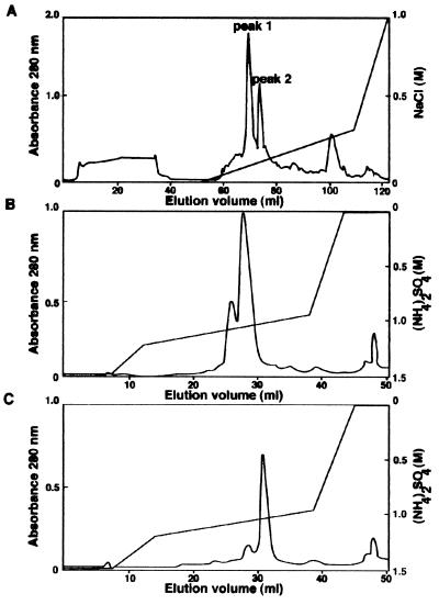
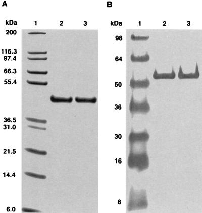
Fermentation conditions were worked out for optimum yield of product, and intracellular extraction was carried out with a Gaulin cell disrupter. The yeast cell extract was loaded directly onto a Streamline expanded-bed chromatography column, and the product was eluted by a sodium chloride step gradient. Product eluted from the expanded-bed chromatography column was estimated to be 10% pure, with a total protein concentration of 0.92 mg/ml. After dialysis, the material was loaded onto a Mono S cation-exchange column for further purification. Western blot and ELISA data indicated that BoNT/A(Hc) eluted from the column in two distinct peaks, at 110 and 130 mM NaCl (Fig. 2A). These two 50-kDa immunologically positive bands were indistinguishable by SDS-PAGE, ELISA, and Western blot analysis. However, given the fact that two peaks of BoNT/A(Hc) eluted from the cation-exchange column, fractions of each peak were pooled separately as peak 1 (protein that eluted at 110 mM NaCl) and peak 2 (protein that eluted at 130 mM NaCl). After Mono S chromatography, the peak 1 and peak 2 pools were subjected to HIC as a final purification step and adjusted to 1.5 M ammonium sulfate. Each protein peak was loaded onto an HIC column and eluted with a gradient of decreasing ammonium sulfate (Fig. 2B and C). Peak 1 eluted at 1.04 M ammonium sulfate, and peak 2 eluted at 1.02 M ammonium sulfate. BoNT/A(Hc) immunologically positive fractions were combined and dialyzed to remove ammonium sulfate. Only a 50-kDa BoNT/A(Hc) band was detected by SDS-PAGE and Western blot analysis for both peaks, and they were thus judged to be more than 95% pure after the final step (Fig. 3). Protein determination revealed that 1.64 mg of peak 1 was recovered at a concentration of 205 μg/ml and 0.77 mg of peak 2 was recovered at a concentration of 102 μg/ml.
FIG. 2.
Purification of BoNT/A(Hc) by sequential chromatography. (A) Mono S cation-exchange chromatography of the Streamline pool. Proteins were eluted with an increasing NaCl gradient, yielding two immunoreactive peaks. Peak 1 (B) and peak 2 (C) were pooled individually and subjected to HIC, and proteins were eluted with a decreasing ammonium sulfate gradient. Protein production was monitored by A280 (left axis); elution conditions are recorded on the right axis, with a gradient trace laid over the chromatogram.
FIG. 3.
(A) Reducing SDS-PAGE of FPLC-purified BoNT/A(Hc) separated on a 10% tricine gel and visualized by Coomassie blue. The gel shows Novex wide-range molecular weight markers (15 μl; lane 1), peak 1 (2 μg; lane 2), and peak 2 (2 μg; lane 3). (B) Western blot analysis of FPLC-purified BoNT/A(Hc) detected with MAb 6E9-11. The blot shows Novex prestained molecular weight markers (15 μl; lane 1), peak 1 (2 μg; lane 2), and peak 2 (2 μg; lane 3).
Potency study of purified BoNT/A(Hc).
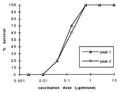
To assess the immunogenicity of purified recombinant BoNT/A(Hc), a potency study was carried out with mice. Because FPLC data suggested a possible chemical difference between peak 1 and peak 2, both proteins were assayed for potency. Mice were injected once with purified BoNT/A(Hc) at seven different doses ranging from 10 μg to 2.4 ng and then challenged with 1,000 mouse i.p. LD50 of BoNT/A. Results revealed that the proteins had similar potencies and were highly immunogenic (Fig. 4). The probit analysis ED50 for peak 1 was 0.089 μg/mouse, with a 95% confidence level of 0.049 to 0.162 μg/mouse. Similarly, for peak 2, the ED50 was 0.101 μg/mouse, with a 95% confidence level of 0.056 to 0.183 μg/mouse. These results compare favorably with those for the control, an E. coli-derived product having an ED50 of 0.278 μg/mouse, with a 95% confidence level of 0.100 to 0.795 μg/mouse. A single injection of the yeast-derived products was fully protective (10 survivors among 10 mice tested for both peak 1 and peak 2) at doses of 0.625 μg/mouse or higher. Conversely, there were no survivors among those animals that received a dose of 9.8 ng or below. The only difference between the two antigens in this potency assay was at the 0.156-μg/mouse dose level, where 7 of 10 mice injected with peak 1 survived and 6 of 10 mice injected with peak 2 survived.
FIG. 4.
Dependence of mouse survival on dose of purified BoNT/A(Hc) administered. Ten mice per group were vaccinated once with peak 1 and peak 2 at doses ranging from 2.4 ng to 10 μg per mouse. Mice were challenged with 1,000 i.p. LD50 of BoNT/A toxin 21 days after vaccination.
Efficacy study of purified BoNT/A(Hc).
Purified BoNT/A(Hc) was evaluated for dose response in an efficacy study. Because the immunogen protected mice challenged with 1,000 mouse i.p. LD50, we wanted to determine if the immunogen could protect against a higher challenge of BoNT/A. Mice were injected once, twice, or three times at five different doses with peak 1 and then challenged with 100,000 mouse i.p. LD50 of BoNT/A toxin. There were no survivors after a single immunization of 0.01 μg/mouse, but 8 of 10 animals survived challenge after a dose of 2.0 μg/mouse (Table 1). However, two injections partially or completely protected the animals at all dose levels, with complete protection observed at 1.0 μg/mouse or above. Three immunizations afforded even greater protection, as complete or nearly complete survival was provided by a dose of ≥0.1 μg/mouse.
TABLE 1.
Survival, group antibody ELISA titers, and serum neutralization titers of mice after vaccination with purified peak 1 BoNT/A(Hc)
| Vaccination dose (μg) | Survival (no. alive/10 tested)a
|
Group antibody ELISA titersb
|
Serum neutralization titer (IU/ml)c
|
||||||
|---|---|---|---|---|---|---|---|---|---|
| 1d | 2 | 3 | 1 | 2 | 3 | 1 | 2 | 3 | |
| 0.01 | 0 | 6 | 5 | 400 | 1,600 | 1,600 | <0.16 | <0.16 | 0.49 |
| 0.1 | 2 | 5 | 9 | 400 | 1,600 | 25,600 | <0.16 | 0.40 | 26.62 |
| 0.5 | 4 | 9 | 10 | 400 | 6,400 | 25,600 | <0.21 | 1.28 | 13.31 |
| 1.0 | 7 | 10 | 10 | 1,600 | 6,400 | 25,600 | <0.16 | 1.28 | 33.55 |
| 2.0 | 8 | 10 | 10 | 1,600 | 25,600 | 102,400 | <0.16 | 13.31 | 33.55 |
Mice were challenged with 100,000 i.p. LD50 of BoNT/A 21 days after the last injection.
Reciprocal of the highest dilution having an A405 greater than 0.2 after correction for background. P = 0.0001 in a test of association of group ELISA titers as a predictor of survival.
Pooled mouse serum (50 μl per mouse) from each group was diluted initially 1:8 and then fourfold for serum neutralization titers. P = 0.0554 in a test of association of serum neutralization titers as a predictor of survival.
Number of vaccinations.
Mouse serum ELISA and BoNT/A neutralization assay.
Two days before challenge, the 150 mice plus the 10 controls in the efficacy study were bled for ELISA titers. Individual serum samples were diluted fourfold, ranging from 1:100 to 1:1,600,000 (titers from 100 to 1,600,000). ELISA titers correlated well with survival (Table 2), as only 6 of 42 mice with antibody titers of ≤100 survived an BoNT/A challenge of 100,000 i.p. LD50. Conversely, 82 of 83 mice with titers of ≥1,600 survived the same challenge. An average ELISA titer was also determined for each group (Table 1). Group titers also correlated well with survival (P = 0.0001). If the group titer was ≥6,400, complete or nearly complete protection was observed, but if the group titer was ≤1,600, there was partial to complete lack of survival. The group titer increased with the number of injections at all five dose levels except 0.01 μg, where two and three injections both elicited titers of 1,600; at this dose level, 60 and 50% survival, respectively, was observed. It was also the only case where percent survival did not increase with increasing number of injections.
TABLE 2.
Correlation of individual ELISA titers with survival after vaccination with purified peak 1 BoNT/A(Hc)
| Individual ELISA titera | Survival (no. alive/total)b | % Survival |
|---|---|---|
| ≤100 | 6/42 | 14.3 |
| 400 | 17/25 | 68.0 |
| ≥1,600 | 82/83 | 98.8 |
Serum was bled from each mouse individually. Titer is the reciprocal of the highest dilution having an A405 of greater than 0.2 after correction for background.
Mice were challenged with 100,000 i.p. LD50 of BoNT/A 21 days after the last vaccination.
Serum from the ELISA study was used in a BoNT/A mouse neutralization assay. Due to the limited amount of serum available, serum from each group of 10 mice was pooled, and so only the average neutralization titer of the group could be determined. The lack of serum decreased the sensitivity of the assay from a possible detection limit of 0.01 IU/ml to a detection limit of 0.16 IU/ml. A titer of 0.02 IU/ml is considered to protect against botulinum type A challenge in humans (13). Since some serum pools had titers of <0.16 IU/ml but some of the mice survived individual challenge, these pools may have had a low level of neutralizing antibody not measurable in our assay. Even with this caveat, the data from serum neutralization titers moderately correlated with survival (P = 0.0554) (Table 1). In one group where 5 of 10 mice survived, the group serum neutralization titer was 0.49 IU/ml, while in another group where 8 out of 10 mice survived, the group serum neutralization titer was <0.16 IU/ml. On the other hand, there seems to be a threshold value for neutralizing titer where complete or nearly complete protection is observed. Groups with 9 or 10 survivors (n = 7) all had neutralizing titers of ≥1.28 IU/ml. Though the group neutralizing titer only moderately correlated with survival, it did correlate well with number of injections. At all five doses, the neutralizing antibody titers increased with the number of injections. In fact, at all dose levels, groups that received only a single injection had serum neutralization titers that were below detectable limits for this assay.
DISCUSSION
Recombinant BoNT/A(Hc) was purified to homogeneity from the yeast P. pastoris by conventional chromatography. Since this protein is a vaccine candidate, large quantities will ultimately need to be prepared. Therefore, it was envisioned that the resulting purification scheme should be scalable and include as few steps as possible. Also, the purification process should include only conventional separation methods to avoid altering the native structure of the immunogen. Affinity resins should also be avoided because when the target protein is eluted from the column, it may leach antibody from the column matrix. Thus, an appropriate first chromatographic step would be to use expanded-bed chromatography. This strategy eliminates the need for a centrifugation or clarifying step after cell disruption. Because the calculated isoelectric point of BoNT/A(Hc) is 9.2, the second chromatographic technique used in the process was Mono S cation exchange. Finally, HIC, which separates proteins by differences in surface hydrophobicity, was used to complete the purification. Two immunoreactive pools of BoNT/A(Hc) were produced with purities of >95%. Although these proteins were indistinguishable by SDS-PAGE and by such immunoreactive methods as ELISA and Western blot analyses, they separated on a cation-exchange column. Analysis of peak 1 and peak 2 by an isoelectric focusing gel (32) indicated that the two proteins had slightly different isoelectric points, 8.9 for peak 1 and 8.7 for peak 2, providing a plausible explanation for their chromatographic behavior on a cation-exchange column. It is possible the difference in isoelectric point between the proteins is due to C-terminal amidation, though amidation would not be expected to alter the isoelectric point of BoNT/A(Hc) by 0.2 pH unit. Another explanation for this difference could be that one of the proteins was truncated at the N or C terminus. However, these proteins still had the same molecular weights even when examined on a large SDS-polyacrylamide gel.
The aim of this study was to produce a recombinant vaccine that could protect against a high challenge level of BoNT/A. The purpose of conducting a potency study was to answer two questions. First, how effective is the purified antigen at eliciting protective immunity to a direct challenge with BoNT/A? Second, how do the potencies of peak 1 and peak 2 compare? A single immunization of 0.625 μg/mouse or higher fully protected the animals against an intermediate challenge level of 1,000 mouse i.p. LD50 of BoNT/A. Material from the two BoNT/A(Hc) peaks had similar ED50s, 0.089 and 0.101 μg/mouse, which compare favorably to the value for the control material expressed in E. coli. The ED50s reported here are quite impressive, revealing that these antigens were highly immunogenic.
We performed the efficacy study to understand the relationship between multiple immunizations and protection at various dose levels. The higher challenge level of toxin was designed to reveal the immunogen limits. Clearly, multiple injections of antigen protected mice better than a single injection. Mice given two injections survived 20 to 60% better at all dose levels assayed. However, three injections did not significantly improve survival. Only the 0.1-μg dose resulted in an increase in survival of more than 10%. Comparing the results of the efficacy study using one injection with the results of the potency study for peak 1 revealed similar ED50s, even though neurotoxin challenge levels were 100-fold different. This finding suggests there may be a threshold dose that could protect against a wide range of challenge levels. The efficacy study also addressed the question of how well the yeast-expressed purified BoNT/A(Hc) protected mice subjected to a high BoNT/A challenge. After mice were injected with ≤1 μg of antigen and challenged with 100,000 mouse i.p. LD50 of BoNT/A, there was significant protection.
Serum neutralization titers for each mouse group and ELISA titers for each mouse were evaluated in serum taken 2 days before efficacy challenge. These data indicate that ELISA titers for individual mice successfully predicted survival. When the titers were at least 1,600, 98.8% of the mice survived. When the titers were 100 or less, mice had only a 14.3% survival rate. As expected, group titers also correlated well with survival. However, serum neutralization titers only moderately correlated with survival. Serum neutralization titers are commonly used to assess the antibody titer of an animal and are excellent predictors of an animal’s ability to survive challenge. However, it appears that pooled serum titers in the neutralization assay were not as accurate as individual serum titers in predicting ability to survive challenge. This was probably due to variation in response to the vaccine regimen within the group. Although both group ELISA and serum neutralization titers correlate well with survival, group ELISA titer has a much greater statistical significance associated with its predictive power. Our results suggest that ELISA titer, which is simpler and less costly to measure than neutralization titer, may be an appropriate substitute measure for protection.
ACKNOWLEDGMENTS
We thank Michael Meagher, Mark Bevins, Vijay Chiruvolu, and Karen Potter for their work on the fermentation and cell cracking optimization at the University of Nebraska—Lincoln, Biological Process Development Facility. We also thank Paul Gibbs for providing statistical analyses and Lancer Cuddy, Matthew Hinz, and Brent Webb for technical assistance.
REFERENCES
- 1.Anderson J H, Lewis G E. Clinical evaluation of botulinum toxoids. In: Lewis G E, editor. Biomedical aspects of botulism. New York, N.Y: Academic Press; 1981. pp. 233–246. [Google Scholar]
- 2.Black J D, Dolly O. Interaction of 125I-botulinum neurotoxins with nerve terminals. I. Ultrastructural autoradiographic localization and quantitation of distinct membrane acceptors for types A and B on motor nerves. J Cell Biol. 1986;103:521–534. doi: 10.1083/jcb.103.2.521. [DOI] [PMC free article] [PubMed] [Google Scholar]
- 3.Bowmer E J. Preparation and assay of the international standards for Clostridium botulinum types A, B, C, D, and E antitoxins. Bull W H O. 1963;29:701–709. [PMC free article] [PubMed] [Google Scholar]
- 4.Chiruvolu V, Cregg J M, Meagher M M. Recombinant protein expression in an alcohol oxidase-defective strain of Pichia pastoris in feedbatch fermentations. Enzyme Microbiol Technol. 1997;21:277–283. [Google Scholar]
- 5.Clare J J, Rayment F B, Ballantine S P, Sreekrishna K, Romanos M A. High-level expression of tetanus toxin fragment C in Pichia pastoris strains containing multiple tandem integrations of the gene. Bio/Technology. 1991;9:455–460. doi: 10.1038/nbt0591-455. [DOI] [PubMed] [Google Scholar]
- 6.Clayton M A, Clayton J M, Brown D R, Middlebrook J L. Protective vaccination with a recombinant fragment of Clostridium botulinum neurotoxin serotype A expressed from a synthetic gene in Escherichia coli. Infect Immun. 1995;63:2738–2742. doi: 10.1128/iai.63.7.2738-2742.1995. [DOI] [PMC free article] [PubMed] [Google Scholar]
- 7.Cregg J M, Barringer K J, Hessler A Y, Madden K R. Pichia pastoris as a host system for transformations. Mol Cell Biol. 1985;5:3376–3385. doi: 10.1128/mcb.5.12.3376. [DOI] [PMC free article] [PubMed] [Google Scholar]
- 8.Cregg J M, Vedvick T S, Raschke W C. Recent advances in the expression of foreign genes in Pichia pastoris. Bio/Technology. 1993;11:905–909. doi: 10.1038/nbt0893-905. [DOI] [PubMed] [Google Scholar]
- 9.DasGupta B R, Sugiyama H. A common subunit structure in Clostridium botulinum type A, B, and E toxins. Biochem Biophys Res Commun. 1972;48:108–112. doi: 10.1016/0006-291x(72)90350-6. [DOI] [PubMed] [Google Scholar]
- 10.DasGupta B R. The structure of botulinum neurotoxins. In: Simpson L L, editor. Botulinum neurotoxin and tetanus toxin. New York, N.Y: Academic Press; 1989. pp. 53–67. [Google Scholar]
- 11.Ellis R J. Immunobiologic agents and drugs available from the Centers for Disease Control. Descriptions, recommendations, adverse reactions, and serologic response. 3rd ed. Atlanta, Ga: Centers for Disease Control; 1982. [Google Scholar]
- 12.Fairweather N F, Lyness V A, Maskell D J. Immunization of mice against tetanus with fragments of tetanus toxin synthesized in Escherichia coli. Infect Immun. 1987;55:2541–2545. doi: 10.1128/iai.55.11.2541-2545.1987. [DOI] [PMC free article] [PubMed] [Google Scholar]
- 13.Fiock M A, Devine L F, Gearinger N F, Duff J T, Wright G G, Kadull P J. Studies on immunity to toxins of Clostridium botulinum. VIII. Immunologic response of man to purified bivalent AB botulinum toxoid. J Immunol. 1962;88:277–283. [PubMed] [Google Scholar]
- 14.Fiock M A, Cardella M A, Gearinger N F. Studies of immunity to toxins of Clostridium botulinum. IX. Immunologic response of man to purified pentavalent ABCDE botulinum toxoid. J Immunol. 1963;90:697–702. [PubMed] [Google Scholar]
- 15.Habermann E, Dreyer F. Clostridial neurotoxins: handling and action at the cellular and molecular level. Curr Top Microbiol Immunol. 1986;129:93–179. doi: 10.1007/978-3-642-71399-6_2. [DOI] [PubMed] [Google Scholar]
- 16.Helting T B, Nau H H. Analysis of the immune response to papain digestion products of tetanus toxin. Acta Pathol Microbiol Scand Sect C. 1984;92:59–63. doi: 10.1111/j.1699-0463.1984.tb00052.x. [DOI] [PubMed] [Google Scholar]
- 17.LaPenotiere H F, Clayton M A, Middlebrook J L. Expression of a large, nontoxic fragment of botulinum neurotoxin serotype A and its use as an immunogen. Toxicon. 1995;33:1383–1386. doi: 10.1016/0041-0101(95)00072-t. [DOI] [PubMed] [Google Scholar]
- 18.Nishiki T-I, Kamata Y, Nemoto Y, Omori A, Ito T, Takahashi M, Kozaki S. Identification of protein receptor for Clostridium botulinum type B neurotoxin in rat brain synaptosomes. J Biol Chem. 1994;269:10498–10503. [PubMed] [Google Scholar]
- 19.Oguma K, Fujinaga Y, Inoue K. Structure and function of Clostridium botulinum toxins. Microbiol Immunol. 1995;39:161–168. doi: 10.1111/j.1348-0421.1995.tb02184.x. [DOI] [PubMed] [Google Scholar]
- 20.Poulain B, Mochida S, Weller U, Högy B, Habermann E, Wadsworth J D F, Shone C C, Dolly J O, Tauc L. Heterologous combinations of heavy and light chains from botulinum neurotoxin A and tetanus toxin inhibit neurotransmitter release in Aplysia. J Biol Chem. 1991;266:9580–9585. [PubMed] [Google Scholar]
- 21.Romanos M A, Scorer C A, Clare J J. Foreign gene expression in yeast: a review. Yeast. 1992;8:423–488. doi: 10.1002/yea.320080602. [DOI] [PubMed] [Google Scholar]
- 22.Schiavo G, Benfenati F, Poulain B, Rossetto O, Polverino de Laureto P, DasGupta B R, Montecucco C. Tetanus and botulinum-B neurotoxins block neurotransmitter release by proteolytic cleavage of synaptobrevin. Nature. 1992;359:832–835. doi: 10.1038/359832a0. [DOI] [PubMed] [Google Scholar]
- 23.Schiavo G, Poulain B, Rossetto O, Benfenati F, Tauc L, Montecucco C. Tetanus toxin is a zinc protein and its inhibition of neurotransmitter release and protease activity depend on zinc. EMBO J. 1992;11:3577–3583. doi: 10.1002/j.1460-2075.1992.tb05441.x. [DOI] [PMC free article] [PubMed] [Google Scholar]
- 24.Schiavo G, Rossetto O, Catsicas S, Polverino de Laureto P, DasGupta B R, Benfenati F, Montecucco C. Identification of the nerve terminal targets of botulinum neurotoxin serotypes A, D, and E. J Biol Chem. 1993;268:23784–23787. [PubMed] [Google Scholar]
- 25.Scorer C A, Clare J J, McCombie W R, Romanos M A, Sreekrishna K. Rapid selection using G418 of high copy number transformants of Pichia pastoris for high-level foreign gene expression. Bio/Technology. 1994;12:181–184. doi: 10.1038/nbt0294-181. [DOI] [PubMed] [Google Scholar]
- 26.Shone C C, Hambleton P, Melling J. Inactivation of Clostridium botulinum type A neurotoxin by trypsin and purification of two tryptic fragments. Proteolytic action near the COOH-terminus of the heavy subunit destroys toxin-binding activity. Eur J Biochem. 1985;151:75–82. doi: 10.1111/j.1432-1033.1985.tb09070.x. [DOI] [PubMed] [Google Scholar]
- 27.Shone C C, Hambleton P, Melling J. A 50-kDa fragment from the NH2-terminus of the heavy subunit of Clostridium botulinum type A neurotoxin forms channels in lipid vesicles. Eur J Biochem. 1987;167:175–180. doi: 10.1111/j.1432-1033.1987.tb13320.x. [DOI] [PubMed] [Google Scholar]
- 28.Shone C C, Quinn C P, Wait R, Hallis B, Fooks S, Hambleton P. Proteolytic cleavage of synthetic fragments of vesicle-associated membrane protein, isoform-2 by botulinum type B neurotoxin. Eur J Biochem. 1993;217:965–971. doi: 10.1111/j.1432-1033.1993.tb18327.x. [DOI] [PubMed] [Google Scholar]
- 29.Siegel L S. Human immune response to botulinum pentavalent (ABCDE) toxoid determined by a neutralization test and by an enzyme-linked immunosorbent assay. J Clin Microbiol. 1988;26:2351–2356. doi: 10.1128/jcm.26.11.2351-2356.1988. [DOI] [PMC free article] [PubMed] [Google Scholar]
- 30.Simpson L L. Molecular pharmacology of botulinum toxin and tetanus toxin. Annu Rev Pharmacol Toxicol. 1986;26:427–453. doi: 10.1146/annurev.pa.26.040186.002235. [DOI] [PubMed] [Google Scholar]
- 31.Smith, L. A. Development of molecularly engineered vaccines against botulinum toxins. Toxicon, in press.
- 32.Smith, L. A., M. M. Meagher, and M. P. Byrne. Unpublished data.
- 33.Sreekrishna K, Potenz R H B, Cruze J A, McCombie W R, Parker K A, Nelles L, Mazzaferro P K, Holden K A, Harrison R G, Wood P J, Phelps D A, Hubbard C E, Fuke M. High level expression of heterologous proteins in methylotrophic yeast Pichia pastoris. J Basic Microbiol. 1988;28:265–278. doi: 10.1002/jobm.3620280410. [DOI] [PubMed] [Google Scholar]


