



Abstract
Objectives
To evaluate the safety and efficacy of the port access approach for left atrial (LA) myxoma resection and to analyze the learning curve for this procedure.
Methods
Thirty-six consecutive patients with LA myxoma who underwent port access surgery between April 2018 and March 2023 were enrolled in this retrospective study. The procedure included (1) unilateral or bilateral femoral artery cannulation; (2) the use of three 5-mm trocars and a 20- to 30-mm port; (3) a transseptal, biatrial, or LA approach depending on the location of the tumor base; and (4) complete or subendocardial tumor resection. CUSUM analysis was used to evaluate the aortic cross-clamp (ACC) time and cardiopulmonary bypass (CPB) time learning curves. Variables among the learning curve phases were compared.
Results
The average ACC and CPB times were 49 (range, 45-79) minutes and 127 (range, 120-164) minutes, respectively. There was 1 case of conversion to sternotomy due to aortic root bleeding and 1 case of unilateral pulmonary edema. CUSUMACCtime analysis included 3 phases: phase I, the initial learning period (cases 1-11); phase II, the technical competence period (cases 12-23); and phase III, the challenging period (cases 24-36).
Conclusions
The port access approach is safe and feasible for LA myxoma resection. According to the learning curve analysis, 11 cases are required to achieve technical competence, and 23 cases are required to address highly challenging cases.
Key Words: minimally endoscopic cardiac surgery, port access, left atrial myxoma, learning curve
Graphical Abstract

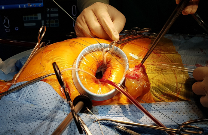
Tumor resection and removal through endoscopic surgery.
Central Message.
There are few reports on learning curves in endoscopic cardiac surgery. Results from these studies will be the basis for learners to determine a roadmap to achieve the training goals.
Perspective.
The port access approach for left atrial myxoma resection is safe and feasible. Learning curve studies will be the basis for learners to determine a roadmap to achieve the training goals.
Myxomas account for 50 to 80% of all adult benign cardiac tumors. Most myxomas occur sporadically, and they are more common in women compared to men.1,2 Myxomas usually originate in the left atrium, at a rate of 75%.3 In some cases, left atrial (LA) myxoma requires emergency surgery because of pulmonary edema due to mitral valve (MV) obstruction or a high risk of embolism.4,5 After more than 3 decades of advances in minimally invasive cardiac surgery, totally endoscopic surgery (TES) through a port/trocars is safe and feasible with some advantages, including pain relief, early recovery, cosmetic scars, and improved postoperative quality of life.6 However, TES has not been widely applied in LA myxoma resection, with the literature consisting predominately of single case reports or case series with few patients7, 8, 9 and no studies examining the learning curve and the appropriate approach for totally endoscopic LA myxoma resection.
In this study, we used statistical analysis to demonstrate the safety and effectiveness of the procedure. In addition, we used the cumulative sum (CUSUM) model to analyze the required learning curve with 3 phases based on the inflection points, the consecutive cases necessary for the learner to master this procedure.
Methods
Study Design
The criteria for patient selection included LA myxoma diagnosed by preoperative transthoracic echocardiography (TTE), which may include MV and tricuspid valve diseases. Exclusion criteria were a history of right lung surgery and peripheral arterial disease.
Thirty-six consecutive patients with LA myxoma who underwent port access surgery between April 2018 and March 2023 were enrolled in this retrospective study. Preoperative demographic data and pathophysiologic characteristics are presented in Table 1. The hospital’s Ethics Committee approved the study (approval 25/HĐĐĐĐHYHN, March 13, 2018). Before surgery, all the patients and their families were informed of the advantages, disadvantages, and possible complications of the operation. All the patients agreed to participate in the study and allowed the use of information, images, and videos during surgery for scientific purposes with written consent.
Table 1.
Preoperative patient characteristics (N = 36)
| Characteristic | Value |
|---|---|
| Age, y, mean ± SD | 52.4 ± 9.6 |
| Female sex, n (%) | 28 (77.8) |
| Body surface area, m2, mean ± SD | 1.55 ± 0.14 |
| History of embolism, n (%) | |
| Stroke | 1 (2.8) |
| Peripheral arterial embolism | 2 (5.6) |
| Emergency | 3 (8.3) |
| Symptoms, n (%) | |
| Chest pain | 13 (36.1) |
| Shortness of breath | 11 (30.6) |
| Fever | 2 (5.6) |
| Fatigue | 3 (8.3) |
| Syncope | 3 (8.3) |
| Dizziness | 2 (5.6) |
| Weight loss | 1 (2.8) |
| Palpitations | 1 (2.8) |
Endpoints
Primary endpoints included aortic cross-clamp (ACC) time, cardiopulmonary bypass (CPB) time, operation time, ventilation time, intensive care unit (ICU) length of stay, hospital length of stay, and ACC and CPB learning curve times. Operation time was defined as the skin to skin time. The learning curve is determined based on the 3-phase CUSUM model.
Secondary endpoints included the suitable approach depending on the tumor attachment, complications, and endoscopic failure. Endoscopic failure was defined as enlargement of the incision or conversion to sternotomy. Complications were defined and divided into minor complications (eg, postoperative bleeding requiring transfusion of blood products, pneumonia, postoperative atelectasis, infection, delayed wound healing) and major complications (eg, reoperation, in-hospital mortality, unilateral pulmonary edema, postoperative stroke, stenosis/obstruction of the iliac and femoral arteries). In-hospital mortality was defined as death occurring in the hospital or within 30 days after surgery.
Operative Procedures
A single surgeon performed all operations. General anesthesia was achieved with a single-lumen endotracheal tube.
Peripheral CPB installation
Peripheral cannulation was established with a Dacron graft (Uni-Graft K DV; B. Braun) anastomosed to the right common femoral artery (FA), superior vena cava cannula (Bio-Medicus jugular venous cannula; Medtronic) and inferior vena cava (IVC) cannula (DLP femoral venous cannula; Medtronic) through the right internal jugular and right femoral veins, respectively. In cases of arterial pressure ≥240 mm Hg under full-flow CPB, we inserted an FA cannula 2 to 4 Fr smaller than the patient's FA size into the left FA to reduce the pressure.
Port and trocar installation
The port and trocars in the right chest included (1) a 2- to 3-cm main working port at the fifth intercostal space (ICS) on the anterior axillary line, exposure achieved by the smallest size wound protector (SurgiSleeve wound protector extra small incision, size 2-4 cm; Covidien); (2) a 5.5-mm trocar (Thoracoport 5.5 mm; Covidien) at the fourth ICS on the midaxillary line for the left-hand instruments and Chitwood clamp; (3) a 10-mm trocar (Trocar sleeve 10/110 mm threaded with tap; Aesculap) or 5-mm trocar (Trocar sleeve 5/60 mm smooth with tap; Aesculap) at the fifth ICS on the midaxillary line for a 3-dimensional or 2-dimensional endoscopic camera, respectively, with CO2 insufflation; and (4) a 5-mm port at the sixth ICS on the midaxillary line for a left vent (Figure 1). Immediately after port and trocar installation, the pleural and pericardial spaces were filled with a CO2 flow of 2 L/min.
Figure 1.

Port and trocar installation. A, The midaxillary line and extended submammary fold (dotted lines) were drawn preoperatively. The red arrow indicates the site of superior vena cava cannulation. B, The port and trocars included (1) the main working port (red line in A) at the fifth intercostal space on the anterior axillary line, (2) a 5.5-mm trocar for the left-hand instruments and Chitwood clamp (yellow circle in A), (3) a 5-mm or 10-mm trocar for the endoscopic camera and CO2 insufflation (green circle in A), and (4) a 5-mm port for the left vent (black circle in A and white star in B). An antegrade cardioplegia needle was placed on the ascending aorta through the main working port (black arrow).
Resection procedure
The superior vena cava was snared in all cases, but the IVC was snared in some cases but not in others. The right atrium was opened, and some stay sutures were placed for exposure. After clamping the aorta using a Chitwood clamp through the trocar for left-hand instruments, the myocardial protection solution was infused through an aortic root cannula (Figure 1, B). Custodiol HTK solution or warm-blood cardioplegia was chosen depending on the expected ACC time. A left vent was placed into the superior right pulmonary vein (PV) through the most inferior trocar (Figure 1). When using Custodiol HTK, the myocardial protection solution was aspirated mainly at the coronary sinus opening.
After opening the interatrial septum (IAS) (Figure 2, A), tumor characteristics were determined, including (1) shape (soft, lobulated or solid, oval mass), (2) size, (3) pedunculated or not, and (4) location and diameter of the base attachment. Complete tumor resection was indicated when the tumor was located entirely in the IAS (Figure 2, B and C). After tumor resection, the IAS was closed using a direct suture or a patch. Subendocardial resection was applied for tumors partially adhering to the IAS (Videos 1 and 2).
Figure 2.

The operative steps. A, Opening of the interatrial septum (IAS). B, Complete tumor resection with a 5- to 10-mm rim. Subendocardial resection was indicated for myxoma not originating from the IAS. The cut surface was electrically burned and closed using a direct suture or autopericardium patch. C, Removal of the tumor from the left atrium and out of the chest wall using a retrieval bag. D, The soft, lobulated mass. SVC, Superior vena cava; IVC, inferior vena cava.
After tumor resection, the cut surface was electrically burned and closed using a direct suture or autologous pericardium patch (Video 1). In cases of a huge myxoma passing through the MV on preoperative TTE, MV regurgitation was always checked intraoperatively. Concomitant cardiac procedures included MV repair/replacement and tricuspid valve repair.
The transseptal approach was converted to a biatrial approach when the myxoma was determined intraoperatively to adhere to the IAS and the right PV (Green oval and gray oval in Figure 3). If it also originated from the posterior LA wall, the posterior LA wall between the inferior right PV and the IVC could be excised and reconstructed using bovine pericardium (gray oval in Figure 3). The LA approach could be indicated for LA myxoma located out of the IAS.
Figure 3.
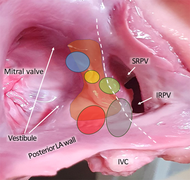
Tumor attachment sites on the interatrial septum (IAS). Blue oval, tumors attached to the IAS and the left atrial (LA) vestibule; yellow oval, tumors completely attached to the IAS; green oval, tumors attached to the IAS and right pulmonary vein (PV); red oval, tumors attached to the IAS and posterior LA wall; gray oval, tumors attached to the IAS, posterior LA wall, and inferior right pulmonary vein (IRPV). White dashed line, the LA approach through Waterston's groove. SRPV, Superior right pulmonary vein; IVC, inferior vena cava.
Perioperative Care and Follow-up
All patients routinely underwent TTE and standard electrocardiography (ECG) before the operation. Emergency surgery was indicated for patients with pulmonary edema due to MV obstruction or a high risk of embolism. Without local pain relief, strategies for drug administration included opioid use on the first day after surgery, followed by nonopioids. The numeric pain rating scale was used to assess patient pain level. The chest tube was removed as soon as possible, and the patient was encouraged to mobilize early and stop painkillers when only mild pain remained. Before discharge, all patients were monitored with TTE, ECG, and chest X-ray. Histopathologic findings in all patients confirmed the myxoma diagnosis.
All patients were followed up in the outpatient clinic at 1 month and 6 months after the operation, then every year thereafter. Standard ECG, peripheral vascular ultrasound, and TTE were included in the follow-up. Multi-slice computed tomography was indicated to confirm the diagnosis of suspected peripheral arterial disease. Transesophageal echocardiography (TEE) would identify recurrent myxoma.
Data Analysis
Statistical analysis
SPSS version 22.0 (IBM) was used for statistical analysis. Continuous variables were expressed as mean ± SD or median (IQR) depending on normal or non-normal distribution. Categorical variables were represented as percentage. Comparisons between groups were performed using the t test or nonparametric tests for continuous variables and the χ2 test or Fisher exact test for categorical variables. The results were statistically significant with a P value ≤ .05.
Learning curve analysis
ACC time is the most critical measure of the surgeon's training progress in totally endoscopic myxoma resection. Logarithmic curve analysis was used to determine whether the learning curve for the ACC time was achieved.
CUSUM analysis was applied to determine the learning curve of the ACC time and CPB time. CUSUM analysis detects the deviation between the data of individual cases and the average value of the overall data and then accumulates each deviation sequentially:
where Xi is an individual’s time, and μ is the mean overall time. The inflection points of the CUSUM curve identified 3 phases. Phase 1 showed a positive slope indicating an increasing trend: the initial learning curve. Phase 2 is presented as a plateau, which is the additional experience. Finally, Phase 3, the postlearning period, showed a negative slope.
Results
Preoperative TTE did not distinguish whether the tumor was entirely or partially attached to the IAS. For partially attached tumors, missed tumor attachment sites included the posterior LA wall, LA vestibule, and right PV (Table E1, Figure 3). A biatrial approach was used in 3 patients, 2 of whom had posterior LA wall reconstruction using bovine pericardium. There were 3 MV repairs, 1 MV replacement due to rheumatic disease, and 7 tricuspid valve repairs (Table E2). Conversion to sternotomy was recorded in 1 patient due to aortic root bleeding after weaning from CPB. One female patient underwent emergency surgery on the last day of her menstrual period; unilateral pulmonary edema occurred postoperatively and required a 7-day stay in the ICU (Table E3).
The average ACC and CPB times were 49 (IQR, 45-79) minutes and 127 (IQR, 120-164) minutes, respectively. All patients had an uneventful recovery, with little blood drainage and no or mild pain at discharge (Table 2). The patients and their families were satisfied with the cosmetic result of surgical scars (Figure E1). No major complications were recorded during a mean follow-up of 27.5 ± 15.0 months.
Table 2.
Perioperative parameters (N = 36)
| Variable | Value |
|---|---|
| CPB installation time, min, median (IQR) | 40 (32.5-40) |
| CPB time, min, median (IQR) | 127 (120-164) |
| ACC time, min, median (IQR) | 49 (45-79) |
| Operation time, min, median (IQR) | 200 (190-235) |
| Ventilation time, h, median (IQR) | 10 (7-14) |
| ICU length of stay, d, median (IQR) | 3 (3-3.5) |
| Drainage volume, mL, mean ± SD | 230.7 ± 65.6 |
| Freedom from postoperative analgesics, d, median (IQR) | 4 (4-4) |
| Postoperative hospital length of stay, d, median (IQR) | 7 (6-9) |
CPB, Cardiopulmonary bypass; IQR, interquartile range; ACC, aortic cross-clamp; ICU, intensive care unit.
Figure E1.

Surgical scars at discharge.
The logarithmic curve analysis showed a learning curve for ACC time with P = .001 (Figure E2). Based on CUSUMACCtime analysis, phase 1 was the initial learning period (cases 1-11), phase 2 represented the technical competence period (cases 12-23), and phase 3 was the challenging period (cases 24-36) (Figure 4, A).
Figure E2.

The learning curve for aortic cross-clamp time.
Figure 4.

CUSUM (cumulative sum) analysis of aortic cross-clamp (ACC) time (A) and cardiopulmonary bypass (CPB) time (B).
There were no differences in patient age and body surface area, postoperative recovery times (ventilation time, postoperative hospital length of stay), and drainage volume among the 3 phases. Meanwhile, the parameters reflecting the learning process of the new technique, including ACC time, CPB time, and operation time, showed significant progress through each stage (Table 3). The research highlights are illustrated in Figure 5.
Table 3.
Interphase comparisons of perioperative characteristics for LA myxoma (N = 36)
| Characteristic | Phase I (N = 11) | Phase II (N = 12) | Phase III (N = 13) | P value | p1 | p2 | p3 |
|---|---|---|---|---|---|---|---|
| Age, y, mean ± SD | 56.3 ± 8.0 | 51.9 ± 10.0 | 49.6 ± 10.1 | .239 | |||
| BSA, m2, mean ± SD | 1.49 ± 0.09 | 1.62 ± 0.14 | 1.54 ± 0.14 | .077 | |||
| CPB installation time, min, median (IQR) | 40 (30-40) | 40 (32.5-40) | 40 (40-40) | .391 | |||
| CPB time, min, median (IQR) | 170 (156-177) | 120 (115-146) | 126.2 ± 13.2 | .002 | .009 | .001 | .743 |
| ACC time, min, median (IQR) | 92.6 ± 33.6 | 46.5 (39.5-71.5) | 46.8 ± 7.7 | .002 | .018 | .001 | .806 |
| Operative time, min, median (IQR) | 241.8 ± 39.2 | 200 (190-225) | 193.1 ± 12.2 | .001 | .035 | <.001 | .110 |
| Ventilation time, h, mean ± SD | 10.3 ± 4.3 | 8.8 ± 3.5 | 12 (9-15) | .105 | |||
| Drainage volume, mL, mean ± SD | 241.4 ± 96.8 | 223.3 ± 58.1 | 228.5 ± 38.5 | .805 | |||
| Postop hospital stay, d, median (IQR) | 7 (6-7) | 9.4 ± 4.3 | 7 (6-7) | .364 |
p1, Comparison between phases 1 and 2; p2, comparison between phases 1 and 3; p3, comparison between phases 2 and 3; BSA, body surface area; CPB, cardiopulmonary bypass; IQR, interquartile range; ACC, aortic cross-clamp; LA, left atrial.
Figure 5.

Research highlights. SRPV, Superior right pulmonary vein; IRPV, inferior right pulmonary vein; IVC, inferior vena cava; LA, left atrial; CUSUM, cumulative sum; ACC, aortic cross-clamp; CPB, cardiopulmonary bypass.
Discussion
The main working port in port access endoscopic surgery needs to be selected according to tumor size. The small port/trocar makes manipulating and removing the tumor challenging.10 The surgeon sometimes needs to enlarge the incision to remove the tumor to avoid perforating the retrieval bag. Kronenberger and colleagues10 suggested that the 12-mm port was unsuitable for large tumors. Meanwhile, a 30-mm port has been commonly used in published studies.11, 12, 13 This study found that the 20- to 30-mm port was suitable for tumors of all sizes, including giant tumors. With such a port size, the tumor can easily be removed or divided into pieces and safely removed in a retrieval bag. Moreover, instead of using multiple ports, including a port on the anterior chest wall.7,12,13 we used only 1 port and 3 small trocars in the midaxillary line, making the scars less noticeable (Figures 1 and E1). Using multiple ports makes the manipulation more straightforward and the operation times shorter, because 2 or 3 instruments can work together through 1 port. In contrast, a trocar lets only 1 instrument through. Therefore, using small trocars requires the surgeons to reasonably arrange the order of using instruments through each trocar.
If the myxoma is a round mass located in the familiar site of the LA thrombus (LA appendage or posterior LA wall), especially accompanied by rheumatic MV disease, distinguishing the myxoma from thrombosis preoperatively is tricky. The definitive diagnosis is based primarily on histopathologic findings.14,15 We recorded 2 patients with LA appendage masses and 1 patient with an LA mass in the background of rheumatic MV disease. Histopathologic results helped confirm the diagnosis of LA myxoma in these cases (Tables 2 and 3).
Determining the base attachment is vital when choosing the approach for LA myxoma resection. The majority (80%) of LA myxomas are determined to originate from the border of the fossa ovalis,3,16 but they can arise from any location in the LA, including the posterior wall, appendage, roof, and right PV. Tumors that are entirely attached to the IAS can be wholly resected; however, there are many cases in which the tumor is partially attached to the IAS and partly to the surrounding structures (posterior LA wall, LA vestibule, and the right PVs) (Figure 3). In these cases, the tumor can be resected nearly completely, including resection of the IAS containing the myxoma and subendocardial resection for the attachment out of the IAS (Videos 1 and 2). Although complete tumor resection is believed to reduce the risk of recurrence, the association between the recurrence and the type of tumor resection is not well established, because the recurrence rate is low and the tumors usually do not recur at the same original position.1 Furthermore, subendocardial resection is unavoidable when the tumor is outside the IAS.17 In their study, Shah and colleagues18 noted that subendocardial resection accounted for two-thirds of their cases, but this was not a factor increasing the risk of tumor recurrence.
Three main approaches used for LA myxoma resection include transseptal, biatrial, and LA approaches. The choice of the approach depends on the surgeon's habits and experience, tumor size, and the base attachment seen on preoperative TTE.19 The LA approach is commonly used in TES for LA myxoma resection owing to its advantages in controlling the tumor efficiently, but subendocardial resection is mainly used.7,10,11,20 The transseptal and biatrial approaches increase the possibility of complete tumor resection13 but have not been widely applied in TES.13,21
We believe that in cases in which the myxoma does not originate from the IAS (LA roof, LA appendage, or MV annulus) identified on preoperative TTE, TEE, multi-slice computed tomography, or magnetic resonance imaging, the LA approach is appropriate because the myxoma will be almost subendocardially resected. A vast or giant myxoma close to the LA wall makes it difficult for diagnostic imaging methods to accurately identify a mass attached to the IAS entirely or partially. Our study determined that 86.1% of myxomas preoperatively originate from the IAS; however, this group was determined intraoperatively to include (1) entire IAS attachment (27.8%), (2) partial IAS and posterior LA wall attachment (36.1%), (3) partial IAS and LA vestibule attachment (11.1%), (4) partial IAS and PV attachment (2.8%), and (5) partial IAS, posterior LA wall, and inferior right PV attachment (8.3%) (Table 2, Figure 3). In scenarios 2 and 3 outlined above, complete or nearly complete tumor resection could be done without difficulty through the transseptal approach (Videos 1 and 2).
Scenarios 4 and 5 are the most complex cases for port-access endoscopic surgery because the entire tumor base attachment cannot not be seen through the transseptal approach, whereas the LA approach would cross the middle of the tumor base (white dashed line in Figure 3). Therefore, the biatrial approach, which should be indicated for these exceptional cases, will help control the entire base attachment. Bortolotti and colleagues22 noted some postoperative atrial arrhythmias in the biatrial approach group, but no correlation was identified. This conclusion was also recorded in some studies with a follow-up period of 20 to 50 years.17,19,22 We used the biatrial approach in only 3 patients, and no cases of postoperative atrial arrhythmia were recorded.
The ACC time, CPB time, ventilation time, drainage volume, and postoperative hospital length of stay in our study were similar to the results of published reports on totally endoscopic LA myxoma resection, with or without robotic assistance.12,13,20
Factors known to affect a learner’s ability to master new techniques include age23 and familiarity with the technique. This familiarity is obtained from practice in the laboratory or from participating in surgery as an assistant surgeon.24 In addition, video games are a factor outside the operating room that improve the surgeon’s endoscopic skill.23
We used a stepwise approach consisting of 3 steps for training surgeons in the endoscopic LA myxoma resection procedure: step 1, remove LA myxomas through mini-thoracotomy with video assistance; step 2, practice totally endoscopic atrial septal defect or MV surgery; step 3, apply port access endoscopic surgery in LA myxoma resection. The more step 2 was performed, the shorter the learning curve. We achieved the phase 1 and 2 milestones in totally endoscopic atrial septal defect repair at the 34th and 55th patients, respectively. Meanwhile, we achieved these milestones with the same surgeon in totally endoscopic LA myxoma resection at the 11th and 23rd patients. The learning curve was shortened on the same surgeon because of the difference in step 2 of the 2 learning processes.
In port access endoscopic surgery for repairing atrial septal defects, we did not go through step 2, because we were the first to implement this technique. While performing port access endoscopic surgery for LA myxoma resection, we performed step 2 to improve skills over the long term.
Conclusions
In our experience, the port access approach is safe and feasible for LA myxoma resection. According to the learning curve analysis, experience with 11 cases is required to achieve technical competence, and experience with 23 cases is required to address highly challenging cases.
Conflict of Interest Statement
The authors reported no conflicts of interest.
The Journal policy requires editors and reviewers to disclose conflicts of interest and to decline handling or reviewing manuscripts for which they may have a conflict of interest. The editors and reviewers of this article have no conflicts of interest.
Acknowledgments
We thank our surgical team for their contribution to this study.
Supplementary Data
Totally endoscopic myxoma resection with the base on the interatrial septum and posterior left atrial wall. Video available at: https://www.jtcvs.org/article/S2666-2507(23)00465-0/fulltext.

Totally endoscopic myxoma resection with the base on the interatrial septum and the left atrial vestibule. Video available at: https://www.jtcvs.org/article/S2666-2507(23)00465-0/fulltext.

Appendix E1
Table E1.
Parameters for TTE and operation (N = 36)
| Parameter | TTE | Operation |
|---|---|---|
| Number of masses (%) | ||
| One mass | 36 (100) | 35 (97.2) |
| Two masses | 0 | 1 (2.8) |
| Shape characteristics, n (%) | ||
| Soft, lobulated tumor | 21 (58.3) | |
| Solid, oval tumor | 15 (41.7) | |
| Tumor size, mean ± SD/median (IQR) | ||
| Diameter in length, mm | 45.6 ± 19.6 | 50 (30-60) |
| Diameter in width, mm | 26.8 ± 10.1 | 30 (20-42.5) |
| Base and peduncle, n (%) | ||
| Pedicle | 27 (75) | 19 (52.8) |
| Sessile | 9 (25) | 17 (47.2) |
| Diameter of base attachment, mm, median (IQR) | 15 (10-20) | |
| Mobile mass, n (%) | 29 (80.6) | |
| Protrusion into LV, n (%) | 20 (69) | |
| Location of base attachment, n (%) | ||
| IAS | 31 (86.1) | 10 (27.8) |
| IAS and posterior LA wall | 0 | 13 (36.1) |
| IAS and the LA vestibule | 0 | 4 (11.1) |
| IAS and right PV | 1 (2.8) | 1 (2.8) |
| IAS, posterior LA wall and IRPV | 0 | 3 (8.3) |
| MV annulus | 0 | 1 (2.8) |
| LA roof | 2 (5.6) | 2 (5.6) |
| Base of LA appendage | 2 (5.6) | 2 (5.6) |
TTE, Transthoracic echocardiography; IQR, interquartile range; LV, left ventricle; IAS, interatrial septum; LA, left atrial; PV, pulmonary vein; IRPV, inferior right pulmonary vein; MV, mitral valve.
Table E2.
Operative data (N = 36)
| Variable | Value |
|---|---|
| Bilateral femoral arterial cannulation, n (%) | 2 (5.6) |
| Approach, n (%) | |
| Transseptal | 32 (88.9) |
| Biatrial | 3 (8.3) |
| Left atrial | 1 (2.8) |
| Myocardial protection, n (%) | |
| Custodiol HTK | 16 (44.4) |
| Warm blood | 20 (55.6) |
| MV surgery, n (%) | |
| MV repair | 3 (8.3) |
| MV replacement | 1 (2.8)∗ |
| Tricuspid valve repair, n (%) | |
| Posterior annuloplasty | 2 (5.6) |
| Ring annuloplasty | 5 (13.9) |
| IAS reconstruction, n (%) | |
| Direct suture | 19 (52.8) |
| Patch closure | 16 (44.4) |
| None | 1 (2.8) |
| Posterior left atrial wall reconstruction, n (%) | 2 (5.6) |
MV, Mitral valve; IAS, interatrial septum.
One MV replacement due to rheumatic disease.
Table E3.
Postoperative data (N = 36)
| Variable | Value |
|---|---|
| Follow-up, mo, mean ± SD | 27.5 ± 15.0 |
| Recurrence, n (%) | 0 |
| Complications, n (%) | |
| In-hospital mortality | 0 |
| Death during follow-up | 0 |
| Stroke | 0 |
| Reoperation | 0 |
| Unilateral pulmonary edema | 1 (2.8) |
| Conversion to sternotomy | 1 (2.8) |
| Postoperative blood transfusion | 3 (8.3) |
| Atelectasis | 0 |
| Postoperative pneumonia | 0 |
| Nonhealing surgical wound | 1 (2.8) |
| Surgical wound infection | 0 |
References
- 1.Cohn L.H. 3rd ed. McGraw-Hill; 2007. Cardiac Surgery in the Adult. [Google Scholar]
- 2.Islam A.K. Cardiac myxomas: a narrative review. World J Cardiol. 2022;14:206–219. doi: 10.4330/wjc.v14.i4.206. [DOI] [PMC free article] [PubMed] [Google Scholar]
- 3.Grysman N.H., Watad A., Ofek E., Tzur B., Amital H. Rare myxoma arising from posterior wall of left atrium. Isr Med Assoc J. 2016;18:370–371. [PubMed] [Google Scholar]
- 4.Brant L.C., Mitu O., Gomide L., Bráulio R., Nunes M.C. Large atrial myxoma causing mitral obstruction and severe pulmonary hypertension. J Heart Valve Dis. 2011;20:357–359. [PubMed] [Google Scholar]
- 5.Bahloul A., Sarray H., Kammoun Y., Charfeddine S., Gueldich M., Dammak A., et al. Giant left atrial myxoma with mitral valve obstruction. J Cardiovasc Echogr. 2021;31:110–112. doi: 10.4103/jcecho.jcecho_111_20. [DOI] [PMC free article] [PubMed] [Google Scholar]
- 6.Ma Z.S., Yin Q.Y., Dong M.F., Feng Z.Y., Wang L.X. Quality of life in patients undergoing totally thoracoscopic closure for atrial septal defect. Ann Thorac Surg. 2011;92:2230–2234. doi: 10.1016/j.athoracsur.2011.07.053. [DOI] [PubMed] [Google Scholar]
- 7.Shi J., Wang Y., Wang Q., Bing X., Ma Z. Simultaneously performed, totally endoscopic left atrial myxoma resection and lobectomy. Heart Surg Forum. 2020;23:E292–E294. doi: 10.1532/hsf.2839. [DOI] [PubMed] [Google Scholar]
- 8.Dang Q.H., Le N.T., Tran D.D., Ngo T.H. Totally endoscopic resection of giant left atrial myxoma without robotic assistance. Innovations (Phila) 2018;13:136–139. doi: 10.1097/IMI.0000000000000487. [DOI] [PubMed] [Google Scholar]
- 9.Hassan M., Smith J.M. Robotic assisted excision of a left ventricular myxoma. Interact Cardiovasc Thorac Surg. 2012;14:113–114. doi: 10.1093/icvts/ivr021. [DOI] [PMC free article] [PubMed] [Google Scholar]
- 10.Kronenberger R., Bentala M., Devos A., de Asmundis C., La Meir M. Excision of atrial myxoma through a totally thoracoscopic approach on the fibrillating heart. Innovations (Phila) 2023;18:289–291. doi: 10.1177/15569845231178014. [DOI] [PubMed] [Google Scholar]
- 11.Zhao F., Chen T., Tang Y., Chen Q., Jiang N., Guo Z. Totally thoracoscopic surgery for treating left atrial myxoma. Medicine (Baltimore) 2021;100 doi: 10.1097/MD.0000000000027819. [DOI] [PMC free article] [PubMed] [Google Scholar]
- 12.Deng L., Zhang G.W., Liu Z.H., Meng W.X., Liu H.Y. Totally thoracoscopic surgery for atrial myxomas resection and atrial septal defect repair. Eur Rev Med Pharmacol Sci. 2017;21:569–575. [PubMed] [Google Scholar]
- 13.Yu S., Xu X., Zhao B., Jin Z., Gao Z., Wang Y., et al. Totally thoracoscopic surgical resection of cardiac myxoma in 12 patients. Ann Thorac Surg. 2010;90:674–676. doi: 10.1016/j.athoracsur.2009.08.078. [DOI] [PubMed] [Google Scholar]
- 14.Kate Y., Syed M.P., Doshi A., Patil S., Kumar D. Atrial mass versus thrombus. Balkan Med J. 2020;37:166–167. doi: 10.4274/balkanmedj.galenos.2019.2019.11.79. [DOI] [PMC free article] [PubMed] [Google Scholar]
- 15.Park H., Jo S., Cho Y.K., Kim J., Cho S., Kim J.H., et al. Differential diagnosis of a left atrial mass after surgical excision of myxoma: a remnant or a thrombus? Korean Circ J. 2016;46:875–878. doi: 10.4070/kcj.2016.46.6.875. [DOI] [PMC free article] [PubMed] [Google Scholar]
- 16.Cottini M., Pergolini A., Zampi G., Buffa V., Pino P.G., Polizzi V., et al. Posterior wall as atypical localization of left atrial myxoma: diagnosis and management. Herz. 2017;42:390–394. doi: 10.1007/s00059-016-4480-y. [DOI] [PubMed] [Google Scholar]
- 17.Yüksel A., Saba D., Velioğlu Y., Ener S., Özkan H. Biatrial approach provides better outcomes in the surgical treatment of cardiac myxomas. Braz J Cardiovasc Surg. 2016;31:309–317. doi: 10.5935/1678-9741.20160066. [DOI] [PMC free article] [PubMed] [Google Scholar]
- 18.Shah I.K., Dearani J.A., Daly R.C., Suri R.M., Park S.J., Joyce L.D., et al. Cardiac myxomas: a 50-year experience with resection and analysis of risk factors for recurrence. Ann Thorac Surg. 2015;100:495–500. doi: 10.1016/j.athoracsur.2015.03.007. [DOI] [PubMed] [Google Scholar]
- 19.Lee K.S., Kim G.S., Jung Y., Jeong I.S., Na K.J., Oh B.S., et al. Surgical resection of cardiac myxoma-a 30-year single institutional experience. J Cardiothorac Surg. 2017;12:18. doi: 10.1186/s13019-017-0583-7. [DOI] [PMC free article] [PubMed] [Google Scholar]
- 20.Gao C., Yang M., Wang G., Wang J., Xiao C., Wu Y., et al. Excision of atrial myxoma using robotic technology. J Thorac Cardiovasc Surg. 2010;139:1282–1285. doi: 10.1016/j.jtcvs.2009.09.013. [DOI] [PubMed] [Google Scholar]
- 21.Kadiroğulları E., Onan B., Aydın Ü., Başgöze S., Şen O. A comparison of robotically-assisted endoscopic versus sternotomy approach for myxoma excision: a single-center experience. Turk Gogus Kalp Damar Cerrahisi Derg. 2020;28:450–459. doi: 10.5606/tgkdc.dergisi.2020.19278. [DOI] [PMC free article] [PubMed] [Google Scholar]
- 22.Bortolotti U., Maraglino G., Rubino M., Santini F., Mazzucco A., Milano A., et al. Surgical excision of intracardiac myxomas: a 20-year follow-up. Ann Thorac Surg. 1990;49:449–453. doi: 10.1016/0003-4975(90)90253-3. [DOI] [PubMed] [Google Scholar]
- 23.Lynch J., Aughwane P., Hammond T.M. Video games and surgical ability: a literature review. J Surg Educ. 2010;67:184–189. doi: 10.1016/j.jsurg.2010.02.010. [DOI] [PubMed] [Google Scholar]
- 24.Bonaros N., Schachner T., Oehlinger A., Ruetzler E., Kolbitsch C., Dichtl W., et al. Robotically assisted totally endoscopic atrial septal defect repair: insights from operative times, learning curves, and clinical outcome. Ann Thorac Surg. 2006;82:687–693. doi: 10.1016/j.athoracsur.2006.03.024. [DOI] [PubMed] [Google Scholar]
Associated Data
This section collects any data citations, data availability statements, or supplementary materials included in this article.
Supplementary Materials
Totally endoscopic myxoma resection with the base on the interatrial septum and posterior left atrial wall. Video available at: https://www.jtcvs.org/article/S2666-2507(23)00465-0/fulltext.
Totally endoscopic myxoma resection with the base on the interatrial septum and the left atrial vestibule. Video available at: https://www.jtcvs.org/article/S2666-2507(23)00465-0/fulltext.