

Introduction
Trichoepitheliomas (TEs) are uncommon benign tumors derived from the hair follicles. They most often occur in women, and present as asymptomatic translucent, round papules or nodules mainly localized on the face or scalp [1]. They may appear as solitary or multiple lesions, presenting as familial or non-familial forms, sometimes associated with genetic syndromes [2].
So far, in literature TEs have been widely described as translucent non-pigmented papules with mostly milia-like cysts, shiny-white background, short and thin telangiectasias on dermoscopy [3].
Case Presentation
We analyzed all histologically confirmed TEs arising over a 17-year period (January 2005–December 2021). Clinical and dermoscopic images of histologically-confirmed TEs were retrospectively collected from the database of our Dermatology Unit. Our database included 86 TEs in 80 patients in the considered period. Among these ones, 77 lesions could be clinically and dermoscopically evaluated.
The average age of our patients at diagnosis was 59 years (range, 26–84 years). A large predominance for female gender was found (63.7%). TEs were found in the following areas: head (82.6% of the lesions), trunk (12.8%), neck (3.5%), upper limbs (1.1%).
The dermoscopic features of all histologically confirmed TEs were evaluated by 3 dermatologists independently and the final results were taken from the mean of their observations.
The major part [47/77 (61.0%)] of these TEs showed to be (more or less) pigmented. We sub-classified pigmented TEs based on the percentage of their pigmentation, expressed as higher than 50% of the lesion area, between 50% and 10% and less than 10%. Pigmentation percentage >50% was found in 42.6% (20) of TEs. Pigmentation percentage among 50 and 10% was found in 12 cases, and <10% in 15 cases.
Figure 1.
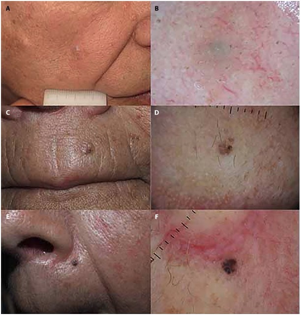
Three examples of trichoepitheliomas classified based on the percentage of pigmentation. (A,B) Clinical picture and dermoscopy of < 10% pigmented trichoepithelioma of the right cheek; (C,D) trichoepithelioma of the left supralabial region showing 10%-50% pigmentation expressed mostly as brown globules; (E,F) trichoepithelioma of the left nasolabial fold expressing >50% pigmentation as brown globules and blue/black blotches.
The patterns of pigmentation were mainly characterized by brown globules, observed in 64% of pigmented TEs (30/47), brown-blue nests in 34% (16/47), black-blue blotches in 17% (8/47), and leaf-like areas in 8.5% (4/47).
Arborizing vessels (76.6%) were overall the most common dermoscopic features (seen in 66% of pigmented and in 90% of non-pigmented TEs), followed by shiny white lines (32% among pigmented TEs and 40% among non-pigmented TEs). Milia-like cysts were found in 43.3% of non-pigmented TEs, while only 17% of pigmented TEs showed this feature. A whitish background was found in 50% of non-pigmented TEs.
Literature data regarding TEs describe them as mainly non-pigmented papule, but as we experienced in our patients, most of TEs were instead pigmented, with a high percentage of pigmentation (> 50%) in the majority of cases. Milia-like cysts and whitish background, which are described in literature as the main dermoscopic features, were found, instead, to be much less prevalent among pigmented TEs.
Conclusions
Differential diagnosis with basal cell carcinoma (BCC) without histological confirmation remains difficult. However, pigmented papules, especially localized on the face, that show dermoscopic features like brown globules, brown-blue nests, and arborizing vessels should let the dermatologists think, especially in young women, to pigmented trichoepitheliomas and not only to BCCs.
Footnotes
Funding: None.
Competing Interests: None.
Authorship: All authors have contributed significantly to this publication.
References
- 1.Kim SH, Kim DW, Hwang JH, Kim KS, Lee SY. A rare development of basal cell carcinoma on trichoepithelioma in a chemical burn scar tissue: A case report. Medicine (Baltimore) 2018;97(38):e12252. doi: 10.1097/MD.0000000000012252. [DOI] [PMC free article] [PubMed] [Google Scholar]
- 2.Guardoli D, Argenziano G, Ponti G, et al. A novel CYLD germline mutation in Brooke-Spiegler syndrome. J Eur Acad Dermatol Venereol. 2015;29(3):457–462. doi: 10.1111/jdv.12578. [DOI] [PubMed] [Google Scholar]
- 3.Sławińska M, Płaszczyńska A, Lakomy J, et al. Significance of Dermoscopy in Association with Clinical Features in Differentiation of Basal Cell Carcinoma and Benign Trichoblastic Tumours. Cancers (Basel) 2022;14(16):3964. doi: 10.3390/cancers14163964. [DOI] [PMC free article] [PubMed] [Google Scholar]