Abstract
The omp1 genes encoding the major outer membrane proteins (MOMPs) of avian Chlamydia psittaci serovar A and D strains were cloned and sequenced. The nucleotide sequences of the avian C. psittaci serovar A and D MOMP genes were found to be 98.9 and 87.8% identical, respectively, to that of the avian C. psittaci serovar A strain 6BC, 84.6 and 99.8% identical to that of the avian C. psittaci serovar D strain NJ1, 79.1 and 81.1% identical to that of the C. psittaci guinea pig inclusion conjunctivitis strain, 60.9 and 62.5% identical to that of the Chlamydia trachomatis L2 strain, and 57.5 and 60.4% identical to that of the Chlamydia pneumoniae IOL-207 strain. The serovar A or D MOMPs were cloned in the mammalian expression plasmid pcDNA1. When pcDNA1/MOMP A or pcDNA1/MOMP D was introduced into COS7 cells, a 40-kDa protein that was identical in size, antigenicity, and electrophoretic mobility to native MOMP was produced. Recombinant MOMP (rMOMP) was located in the cytoplasm of transfected COS7 cells as well as in the plasma membrane and was immunoaccessible. Intramuscular administration of pcDNA1/MOMP in specific-pathogen-free turkeys resulted in local expression of rMOMP in its native conformation, after which anti-MOMP antibodies appeared in the serum.
Chlamydia trachomatis and Chlamydia pneumoniae, two obligate intracellular bacteria, are important human pathogens that cause infections which are generally restricted to mucosal epithelial cells of the conjunctiva or the urogenital and respiratory tracts, respectively. Considerable efforts are being made to produce a human chlamydia vaccine. However, all attempts to induce chlamydia immunity to date have failed to produce a solid long-lasting immunity, and prophylactics against human chlamydial infections are as yet nonexistent.
Chlamydia psittaci, another member of the genus Chlamydia, is an important turkey pathogen that causes infections of mucosal epithelial cells and macrophages of the respiratory tract followed by septicemia and localization in epithelial cells and macrophages in various organs (25). C. psittaci can be a primary respiratory pathogen as well as an important complicating agent in any outbreak of respiratory disease in turkeys. Chlamydial infections in turkeys not only present significant economical problems but also threaten public health, since veterinary surgeons and poultry workers are at high risk of becoming infected by this zoonotic agent. A C. psittaci vaccine would significantly enhance efforts to prevent respiratory infections in turkeys and would diminish the zoonotic risk. However, as for humans, chlamydial vaccines for poultry are nonexistent.
The only protective chlamydial antigen which has been unambiguously identified is the major outer membrane protein (MOMP). This protein, discovered independently by two groups in the United States (3, 8) and one in the United Kingdom (11), represents the majority of the surface-exposed protein of members of the genus Chlamydia. It is a protein of around 40 kDa and is characterized by four variable sequences (I to IV) and five intervening constant regions of conserved structure and function. MOMP is an immunodominant protein that carries genus-, species- and, interestingly, serovar-specific epitopes eliciting neutralizing antibodies (10, 16, 31).
Prior vaccination experiments conducted in animal models of either C. trachomatis or C. psittaci infections have used purified inactivated elementary bodies (EB), purified MOMP or recombinant MOMP (rMOMP) expressed by Escherichia coli, and subfraction or subunit preparations of MOMP (2, 5, 9, 12, 17, 19). However, these methods had substantial limitations that would be overcome if the outer membrane protein 1 (omp1) gene encoding the MOMP could be expressed in the host cell itself. A chlamydia DNA vaccine could offer this opportunity, since rMOMP is produced inside host cells and is presented in the context of major histocompatibility complex class I and II molecules to elicit both CD4 and CD8 T-cell responses.
The purpose of the present study was to determine whether chlamydial rMOMP expressed by plasmid DNA assembles into a native conformation in eukaryotic cells and whether rMOMP was localized to the host cell membrane. Therefore the omp1 genes of two strains belonging to the avian C. psittaci serovars A and D, respectively, were cloned into the mammalian expression plasmid pcDNA1. High-level expression was obtained from a cytomegalovirus promoter, providing an efficient and simple system for assaying the localization and immunological properties of the expressed MOMP.
MATERIALS AND METHODS
C. psittaci strains.
The following C. psittaci strains were used: strain 84/55, isolated from the lungs of a diseased parakeet (obtained from J. W. Frost, Staatliches Medizinal-Lebensmittel und Veterinär Untersuchungsambt, Frankfurt am Main, Germany), and strain 92/1293, isolated from a pooled homogenate of the lungs, the cloacae and the spleens of diseased turkeys obtained from a C. psittaci outbreak on a turkey broiler farm in The Netherlands (22). Both strains were previously characterized by using serovar-specific monoclonal antibodies in a microimmunofluorescence test and by restriction fragment length analysis of the omp1 gene. Strain 84/55 was classified as avian serovar A and genotype A, while strain 92/1293 was classified as avian serovar D and genotype E (23, 27).
Plasmid construction.
Chlamydia isolates were grown and purified as described previously (21, 27). Genomic DNA was purified from 108 chlamydia inclusion-forming units of purified serovar A and D elementary bodies. Pure genomic DNA was obtained by the QIAGEN Genomic DNA Purification Procedure in accordance with the standard protocol for bacteria (QIAGEN GmbH, Hilden, Germany).
The serovar A and D omp1 genes were obtained by polymerase chain reaction (PCR) amplification from genomic DNA. The amplification primers (Table 1) were chosen from the highly conserved regions of the published omp1 sequences of C. trachomatis and C. psittaci (15, 32). The oligonucleotide primers (Pharmacia, Uppsala, Sweden) flanked both ends of the omp1 gene open reading frame and provided EcoRI restriction sites for subsequent cloning. The amplification reaction was carried out in a 50-μl volume containing 12.5 μl of genomic DNA extract, 25 μl of MQ water, 5 μl of SuperTaq buffer (10×), 1 μl of each deoxynucleoside triphosphate (10 mM), 2.5 μl of each primer (DV-1 and DV-2) (20 pmol/μl), and 1 μl of SuperTaq polymerase (15 U/μl) (1/50 dilution in SuperTaq buffer). A DNA Thermal Cycler (Biometra, Göttingen, Germany) was used with 30 s of melting at 95°C, 2 min of annealing at 50°C, and 1 min of polymerization at 72°C for 30 cycles. To assess the amplification, 5 μl of the PCR mixture was subjected to electrophoresis on a 1.2% agarose (Biozyme, Landgraaf, The Netherlands) gel stained with ethidium bromide and photographed under UV illumination. PstI (Boehringer, Mannheim, Germany)-digested lambda DNA (Life Technologies, Merelbeke, Belgium) was used as a molecular size marker. Amplification products of the appropriate size (∼1,200 bp) were obtained, and to further confirm omp1 amplification, AluI restriction patterns were analyzed as previously described and displayed the correct serovar-specific restriction patterns (22).
TABLE 1.
Oligonucleotides used in DNA amplification and sequencing of avian C. psittaci serovar A and D omp1 genes
| Oligonucleotide | Length (bp) | Oligonucleotide sequence (5′ to 3′) |
|---|---|---|
| DV-1 | 30 | CGGAATTCATGAAAAAACTCTTGAAATCGG |
| DV-2 | 36 | CGGAATTCAATGTTCGAAAAGACTRAAGTARAACAA |
| 55G2-F | 18 | ATTTGGGATCGCTTCGAC |
| 55G2-R | 23 | CCTTTATAGCCTCTTGGTTTGTG |
| 1293A5-F | 18 | ACCAATGCAGCTTTCCTC |
| 1293A5-R | 23 | CCTTTATAGCCTCTTGGTTTGTG |
The amplified serovar A and D omp1 genes were cloned into the mammalian expression plasmid pcDNA1 (Invitrogen, Leek, The Netherlands) by insertion of the amplified omp1 genes into the dephosphorylated EcoRI site of pcDNA1. E. coli MC1061/P3 cells were transfected by electroporation (Gene Pulser; Bio-Rad, Nazareth, Belgium), and clones were selected on medium containing ampicillin plus tetracycline and grown in microtiter plates.
The presence of inserts was confirmed by EcoRI restriction enzyme analysis of plasmid mini preparations (QIAGEN) and by PCR clone analysis using Sp6 and T7 primers which flanked the cloning site. PCR clone analysis was performed in microtiter plates with the BioMek Thermal Cycler (Perkin-Elmer Cetus, Zaventem, Belgium). First, 5 μl of each clone was subjected to PCR in a 50-μl final reaction mixture containing 50 mM KCl, 20 mM Tris-HCl (pH 8.3), 2 mM MgCl2, 0.1% Tween 20, 200 μM each deoxynucleoside triphosphate, 20 μM each primer, and 0.1 U of SuperTaq (15 U/μl) polymerase. Samples were subjected to 25 cycles of amplification. Cycling conditions were as follows: denaturation for 30 s at 95°C, primer annealing for 1 min at 55°C, and primer extension for 2 min at 72°C.
Sequencing.
The sequences of the omp1 inserts were determined by the dideoxynucleotide chain termination method (13) using pcDNA1 T7 (5′) and Sp6 (3′) priming sites and thereafter specific 18- and 23-mer oligonucleotides (Table 1) (Pharmacia) at approximately 300-bp intervals on both strands. Sequencing samples were analyzed on the ABI PRISM 377 DNA sequencer (Perkin-Elmer Cetus). Sequences were translated into amino acid sequences with DNA Strider computer software, and interspecies and serovar alignment was performed by using FASTA and SeqVu 1.1 computer software.
Following sequencing, two plasmids designated pcDNA1/MOMP A and pcDNA1/MOMP D were selected for subsequent analyses. pcDNA1 was used as a control. Plasmids were grown in MC1061/P3 and purified by the Tip 2500 plasmid preparation method (QIAGEN). DNA concentration was determined by measuring the optical density at 260 nm and was confirmed by comparing intensities of ethidium bromide-stained EcoRI restriction endonuclease fragments with standards of known concentrations. DNA was stored at −20°C in 1 mM Tris (pH 7.8)-0.1 mM EDTA.
Transfections.
COS7 cells (kindly provided by R. Contreras, Laboratory for Molecular Biology, University of Gent, Belgium) were cultured in Dulbecco’s modified Eagle’s medium supplemented with 3.7 g of sodium bicarbonate/liter, 1 mM l-glutamine, and 10% fetal calf serum (Life Technologies). Transfection with plasmid DNA was performed by using DEAE dextran as described by Tregaskes and Young (18). Forty-eight hours posttransfection, tissue culture flasks were stored at −70°C.
For intramuscular administration, DNA was diluted in saline (0.9% NaCl). Subsequently, the quadriceps muscles of five 1-day-old specific-pathogen-free (SPF) turkeys (Centre National d’Etude Vétérinaire et Alimentaire, Ploufragan, France) were injected with either 100 μg of pcDNA1/MOMP A (n = 2), 100 μg of plasmid pcDNA1/MOMP D (n = 2), or 100 μg of pcDNA1 control (n = 1), and India ink was used as an injection site marker. Seven days after a single injection of 100 μg of DNA into the musculus quadriceps, blood was collected by venipuncture of the vena ulnaris, and serum samples were stored at −20°C. Subsequently, turkeys were euthanized and muscle tissue was excised around the area stained with India ink, snap frozen in liquid-nitrogen-cooled isopropanol, and stored at −70°C.
MAbs.
The following MOMP-specific monoclonal antibodies (MAbs) were used to analyze the expressed MOMP: a MAb against a genus-specific epitope (immunoglobulin G1 [IgG1]), two MAbs against an avian C. psittaci serovar A-specific epitope (both IgM), and a MAb against an avian C. psittaci serovar D-specific epitope (IgG1) (1, 24). MOMP-specific MAb reactivities are summarized in Table 2. Except for the reactivity on live EBs in immunofluorescence, all MOMP-specific MAb reactivity testing methods have been described elsewhere (23, 24). MAb reactivity on unfixed EBs was evaluated by immunofluorescence staining of unfixed Buffalo Green Monkey (BGM) cells, 1 h after inoculating the serovar A or D strain. Unfixed and noninoculated BGM cells served as a control. Two MAbs against ampicillin, one IgG1 and one IgM (20), served as negative controls in all analyses.
TABLE 2.
Reactivities of MOMP-specific MAbs
| Method (reference) | MOMP-specific MAb reactivity
|
|||
|---|---|---|---|---|
| CA18 (genus specific) | CA2 (serovar A specific) | VS1 (serovar A specific) | NJD3 (serovar D specific) | |
| Immunofluorescence | ||||
| Methanol-fixed EBs (24) | + | + | + | + |
| Acetone-fixed EBs (22) | + | + | + | + |
| Unfixed EBs (this study) | + | + | + | + |
| Western blotting (24) | − | + | + | + |
| Immunoprecipitation (24) | + | + | + | + |
| Neutralization (24) | − | + | + | + |
Western blot analysis of the expressed product.
The expression of rMOMP by pcDNA1/MOMP A and pcDNA1/MOMP D in COS7 cells was evaluated by Western blotting. Frozen transfected COS7 cells were lysed by freezing and thawing. The cell lysate was separated from culture supernatant by centrifugation and was then solubilized by boiling in sample buffer. Polypeptides were separated by sodium dodecyl sulfate-polyacrylamide gel electrophoresis and blotted onto a polyvinylidone membrane as described by Vanrompay et al. (28). The membrane was blocked with phosphate-buffered saline (PBS) (pH 7.4) supplemented with 0.2% Tween 20 and 10% γ-globulin-free horse serum and then probed for MOMP with MAbs CA2, VS1, and NJD3. Antibody binding was detected by use of polyclonal peroxidase-labelled anti-mouse conjugate and amino-ethyl carbazole. Purified EBs from strains 84/55 and 92/1293 were used for the detection of authentic MOMP.
Localization of rMOMP in COS7 cells.
The expression of rMOMP was examined by viewing methanol-fixed fluorescence-stained cells with a confocal laser scanning microscope (CLSM) (Zeiss, Brussels, Belgium). Nontransfected as well as pcDNA1/MOMP A-, pcDNA1/MOMP D-, or pcDNA1-transfected COS7 cells were grown on cover slips (12-mm diameter) in flat-bottomed Chlamydia Trac Bottles (International Medical, Brussels, Belgium). Transfected COS7 cells were incubated for 48 h at 37°C in 5.0% CO2. Subsequently an indirect immunofluorescence staining was performed in Chlamydia Trac Bottles. All dilutions were made in PBS (pH 7.3). The monolayers were fixed in methanol at −20°C for 10 min. Thereafter, transfected and nontransfected monolayers were incubated for 30 min at 37°C in a moist chamber with 25 μl of 1/200 dilutions of the MOMP-specific MAbs. Furthermore, transfected cells were incubated for 30 min at 37°C in a moist chamber with 25 μl of 1/30 diluted rabbit anti-mouse Ig labelled with fluorescein isothiocyanate (FITC) (Nordic Immunological Laboratories, The Netherlands) or two MAbs against ampicillin, one IgG1 and one IgM (20). The latter served as negative controls. The monolayers were washed twice for 5 min with PBS and were incubated thereafter for 60 min at 37°C in a moist chamber with 25 μl of FITC-labelled rabbit anti-mouse Ig diluted 1/30. Finally, the monolayers were washed twice for 5 min with PBS and for 30 s with distilled water and then air dried. The monolayers were mounted with a glycerine solution containing DABCO and examined with a CLSM.
The cell surface expression of rMOMP was analyzed by using flow cytometric analysis of live fluorescence stained cells. Nontransfected COS7 cells as well as pcDNA1/MOMP A-, pcDNA1/MOMP D-, and pcDNA1-transfected COS7 cells were grown in 75-cm2 tissue culture flasks for 48 h at 37°C in 5.0% CO2 in air, after which flow cytometry was performed. One day before flow cytometric analysis, nontransfected and transfected monolayers were treated with trypsin (2.5%) containing 0.2% EDTA and returned to tissue culture flasks. The next day, individual cells could be obtained by simply washing the monolayers with PBS (pH 7.3) supplemented with 0.02% verseen buffer. Individual nontransfected and transfected COS7 cells were suspended in staining medium consisting of RPMI 1640 medium (Life Technologies) at 4°C with 0.02% sodium azide (Sigma, Antwerp, Belgium) and 1% fetal calf serum (Life Technologies). Subsequently, live cells were stained by indirect immunofluorescence in microtiter plates as previously described (26). Flow cytometry was performed by a FACScan cell sorter (Becton Dickinson, Aalst, Belgium).
Demonstration of rMOMP in turkey muscle cells.
Transfected turkey muscle cells were analyzed for the presence of rMOMP by in situ immunohistochemical staining. Longitudinal as well as serial cryostat sections of the injected turkey muscle tissues (10 μm) were examined by using genus- and serovar-specific MAbs (Table 2) in an indirect immunofluorescence staining. All dilutions were made in PBS (pH 7.3). Briefly, acetone-fixed cryostat tissue sections were washed in PBS for 5 min. The slides were then incubated with 25 μl of undiluted chlamydia-negative goat serum for 1 h at 37°C. Subsequently, the slides were washed in PBS (twice for 5 min) and incubated for 45 min at 37°C with 25 μl of either a 1/200 dilution of genus-specific MAb or a 1/200 dilution of serovar-specific MAb. The sections were washed in PBS (twice for 5 min) and incubated for 30 min at 37°C with 25 μl of a 1/30 dilution of goat anti-mouse Ig labelled with FITC. Finally, the slides were washed in PBS (twice for 5 min) and in distilled water (twice for 30 s). The slides were air dried, mounted, and examined with a fluorescence microscope (Leitz DMRB, Germany).
Detection of anti-MOMP antibodies in turkey sera.
Anti-MOMP antibody titers were determined by an enzyme-linked immunosorbent assay with rMOMP as antigen (29).
Nucleotide sequence accession numbers.
The deduced peptide sequences of avian C. psittaci 84/55 (serovar A) and 92/1293 (serovar D) have been submitted to the DDBJ/EMBL/GenBank databases under accession no. Y16561 and Y16562, respectively.
RESULTS AND DISCUSSION
Plasmid construction.
The omp1 genes, obtained by PCR amplification from C. psittaci 84/55 (serovar A) and 92/1293 (serovar D) genomic DNA, were cloned into the mammalian expression plasmid pcDNA1. Several of the resulting MC1061/P3 clones were shown to contain a plasmid with omp1 downstream from the cytomegalovirus promoter. Two plasmids, designated pcDNA1/MOMP A (serovar A) and pcDNA1/MOMP D (serovar D), were used in all subsequent analyses.
MOMP sequencing.
Little information on the sequence of the omp1 gene of strains belonging to different avian C. psittaci serovars is available. Only the sequence of the omp1 gene of the avian C. psittaci serovar A strain 6BC has been published (6) (GenBank accession no. X56980). In databases, an unpublished sequence of an uncharacterized strain is the only other available avian C. psittaci omp1 sequence (EMBL accession no. L25436). Recently, Everett analyzed the omp1 sequence of the avian C. psittaci serovar D strain NJ1 (7). The cloned omp1 genes and deduced peptide sequences from the present study were compared to the omp1 sequences of the characterized avian C. psittaci strains 6BC and NJ1, the C. psittaci guinea pig inclusion conjunctivitis strain (32), and the human strains C. trachomatis L2 (14) and C. pneumoniae IOL-207 (4).
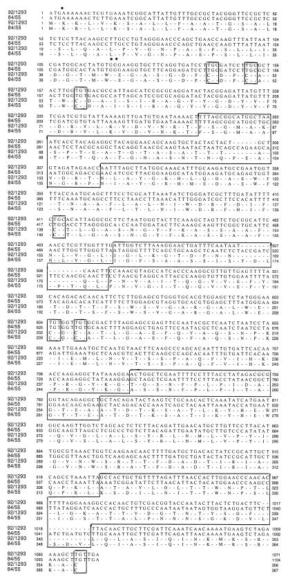
The omp1 genes and deduced peptide sequences of the avian C. psittaci 84/55 (serovar A) and 92/1293 (serovar D) are presented in Fig. 1. The cloned 84/55 omp1 gene, from the translational start ATG to the stop codon TGA, was 33 bp longer than the 92/1293 serovar D omp1 gene. The reading frames consist of 1,101 bp encoding a 367-amino-acid pre-MOMP of strain 84/55 and of 1,068 bp encoding a 356-amino-acid pre-MOMP of strain 92/1293. The mature MOMPs of 84/55 and 92/1293 contain 345 and 334 amino acid residues, respectively. As for other known chlamydia MOMP sequences, the mature N terminus of the C. psittaci MOMPs analyzed in this study is designated leucine and is preceded by a leader peptide of 22 residues displaying significant interspecies homology.
FIG. 1.
Avian C. psittaci serovar A strain 84/55 and avian C. psittaci serovar D strain 92/1293 MOMP structural genes and deduced peptide sequences. The variable domains and conserved cysteines are boxed. The translational initiation codon ATG is marked with ∗. The N terminus of mature MOMP is marked by ∗∗.
Both avian serovar A and D omp1 genes are interspersed symmetrically with four variable domains (VDs) (VD I to IV) at exactly the same positions (Fig. 1), and all insertions and deletions are restricted to these VDs. Seven cysteines were observed at exactly the same position as in all other known MOMPs, emphasizing their importance in the structure and function of the protein.
Computer alignment (Table 3) of the C. psittaci 84/55 and 92/1293 omp1 genes revealed 84% nucleotide identity, demonstrating strong sequence conservation outside the VDs. When compared to known omp1 sequences of other avian C. psittaci strains, the degree of similarity was significantly higher within strains of the same serovar. Indeed, the nucleotide sequences of the avian C. psittaci serovar A and D omp1 genes were found to be 98.9 and 87.8% identical, respectively, to that of the avian C. psittaci serovar A strain 6BC and 84.6 and 99.8% identical, respectively, to that of the avian C. psittaci serovar D strain NJ1. This is in agreement with the observed intra- and interserovar conservation described for C. trachomatis (95 and 83%, respectively) (32). Compared to mammalian chlamydia strains, the nucleotide sequences of the avian C. psittaci serovar A and D omp1 genes were 79.1 and 81.0% identical, respectively, to that of the C. psittaci guinea pig inclusion conjunctivitis strain, 60.9 and 62.5% identical to that of the C. trachomatis L2 strain, and 57.5 and 60.4% identical to that of the C. pneumoniae IOL-207 strain.
TABLE 3.
Nucleotide and amino acid homologies of MOMP genes of Chlamydia strains
| Strain (avian serovar) | % Nucleotide (amino acid) identity
|
|||||
|---|---|---|---|---|---|---|
| 6BC (serovar A) | 92/1293 (serovar D) | NJ1 (serovar D) | GPIC | L2 | IOL-207 | |
| 84/55 (A) | 98.9 (98.0) | 84.1 (85.5) | 84.6 (86.1) | 79.1 (80.2) | 60.9 (62.3) | 57.5 (69.7) |
| 92/1293 (D) | 87.8 (83.7) | 100 (100) | 99.8 (99.4) | 81.0 (83.0) | 62.5 (64.6) | 60.4 (73.9) |
MAbs.
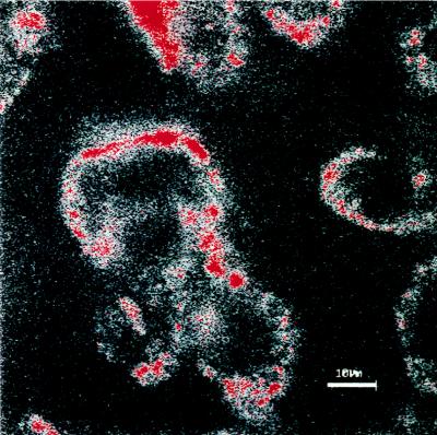
The usefulness of rMOMP expressed by pcDNA1 as a MOMP-based DNA vaccine depends on its ability to assemble into a structure which resembles the conformation of the native molecule as it occurs on the surface of the chlamydial elementary bodies. Therefore, MAbs which recognized either native or denatured MOMP were used (Table 2). The genus-specific MAb CA18 recognized native MOMP on live elementary bodies by indirect immunofluorescence but not denatured MOMP by Western blotting. Therefore, the epitope recognized by CA18 is most likely conformational in nature, since it was shown that heat-induced conformational changes in MOMP destroy the antigenicity of the epitope (31). Conversely, the serovar-specific MAbs CA2, VS1, and NJD3 recognized native MOMP on the surface of elementary bodies (Fig. 2) as well as denatured MOMP in Western blots, suggesting that these MAbs most likely identify linear epitopes on the MOMP.
FIG. 2.
CLSM examination of BGM cells infected with the avian C. psittaci serovar A strain 84/55. Unfixed BGM cells were stained with the serovar-specific MAb (VS1). Note the extracellular elementary bodies (red) adhered to the host cell membrane. Bar = 10 μm.
Expression in transfected COS7 cells.
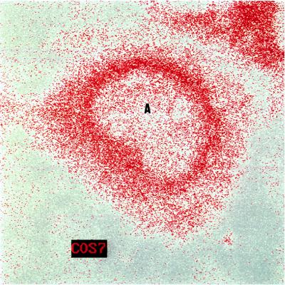
It was important to evaluate whether the rMOMP expressed in transfected COS7 assembled into a native conformation as observed in elementary bodies. pcDNA1/MOMP A- and pcDNA1/MOMP D-transfected COS7 cells were prepared for CLSM examination by fixing them in methanol. Under these conditions, all four MAbs readily detected rMOMP, indicating that rMOMP had a conformationally correct structure (Fig. 3).
FIG. 3.
CLSM examination of COS7 cells transfected with pcDNA1 encoding the MOMP of the avian C. psittaci serovar D strain 92/1293. Fixed cells were stained with a serovar-specific MAb (NJD3) by indirect immunofluorescence staining. Note the nucleus (A) of the COS7 cell and the presence of rMOMP (red) inside the cytoplasm. Bar = 10 μm.
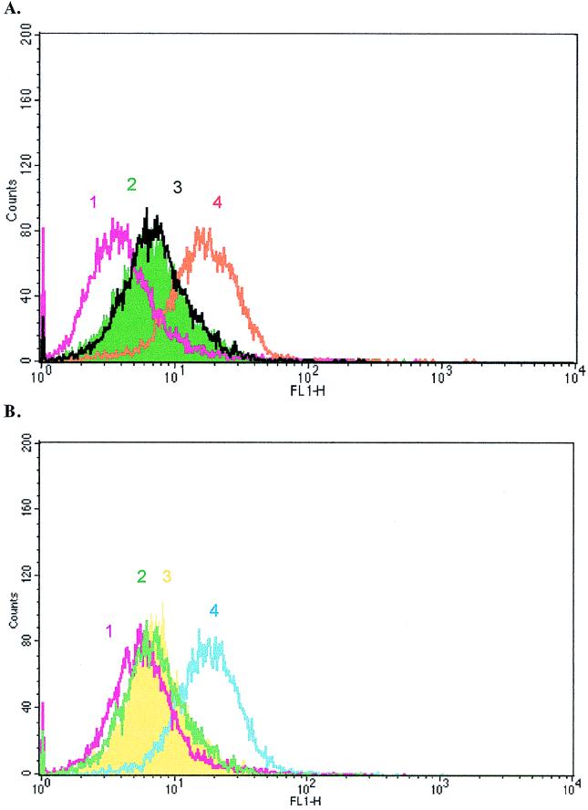
Additionally, cell surface localization of rMOMP was determined on unfixed transfected COS7 cells (Fig. 4). CA18 recognized the surface of all pcDNA1/MOMP-transfected COS7 cells but did not react with the surface of cells transfected with the control plasmid pcDNA1. Moreover, the serovar D-specific MAb and the two serovar A-specific MAbs were reactive only with pcDNA1/MOMP A- and pcDNA1/MOMP D-transfected cells, respectively (Fig. 4). The capacity of rMOMP to successfully assemble into a native conformation on the surface of eukaryotic cells was somewhat surprising since there are no reports of host cell surface localization of C. psittaci MOMP. In fact, previous flow cytometric analysis of unfixed avian C. psittaci serovar A-infected BGM cells, using polyclonal and anti-MOMP MAbs at 32 h postinfection, revealed the absence of chlamydial antigens on cell surfaces (26). Conversely, using high-resolution postembedding staining immunoelectron microscopy, Wyrick et al. (30) were able to demonstrate the presence of MOMP, lipopolysaccharide, and an exoglycolipid on the surface of C. trachomatis-infected human endometrial epithelial cells (HEC-1B). The finding that surface localization of MOMP was drastically reduced or eliminated by exposure of the infected cells to brefeldin A, an inhibitor of anterograde vesicular traffic from the Golgi apparatus, led these investigators to conclude that C. trachomatis MOMP has access to the Golgi network and can reach the eukaryotic cell surface by the secretory pathway. However, the appearance of C. trachomatis MOMP and lipopolysaccharide on the infected HEC-1B cell surfaces was time course dependent and was qualitatively not as dramatic as the amount of exoglycolipid. In fact, escape of C. trachomatis MOMP from inclusions was noticed at about 24 h postinfection and was detectable only in the late stage of the developmental cycle (at 48 h postinfection). Possibly, at 32 h postinfection, flow cytometry could not discriminate C. psittaci-infected BGM cells expressing undetectable levels of rMOMP but at 48 h postinfection was able to detect rMOMP on the surfaces of COS7 cells in which the recombinant protein was highly expressed.
FIG. 4.
Histogram of fluorescence intensities of unfixed COS7 cells transfected with pcDNA1 encoding the MOMP of the avian C. psittaci serovar A strain 84/55 following staining with different FITC-labelled antibodies. (A) 1, nontransfected COS7 cells stained with the polyclonal anti-mouse antibody labelled with FITC; 2, transfected COS7 cells stained with the polyclonal anti-mouse antibody labelled with FITC; 3, transfected COS7 cells stained with the irrelevant IgM MAb; 4, transfected COS7 cells stained with the serovar A-specific MAb VS1 (IgM). (B) 1 and 2, same as in panel A; 3, transfected COS7 cells stained with the irrelevant IgG1 MAb; 4, transfected COS7 cells stained with the genus-specific MAb CA18 (IgG1).
The expression of rMOMP in COS7 cells was also analyzed by Western blotting. A highly antigenic protein of 40 kDa, identical in size and mobility to MOMP of C. psittaci elementary bodies, was identified.
Thus, the 40-kDa polypeptide expressed by pcDNA1/MOMP A- and pcDNA1/MOMP D-transfected COS7 cells is identical in size, antigenicity, and electrophoretic mobility to MOMP of native elementary bodies. Furthermore, avian C. psittaci serovar A and D rMOMPs are located in the cytoplasm as well as on the plasma membrane of transfected COS7 cells, where they are immunoaccessible. High-level expression of conformationally correct rMOMP in COS7 cells offers the opportunity of purification, allowing the potential use of rMOMP in serodiagnosis of avian chlamydial infections.
Expression in turkey skeletal muscle.
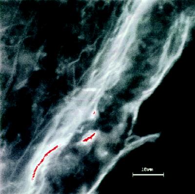
Expression of rMOMP in its native conformation was also observed in pcDNA1/MOMP A- and pcDNA1/MOMP D-transfected musculus quadriceps cells of specific-pathogen-free turkeys (Fig. 5). Recombinant MOMP expression was still detectable at 7 days posttransfection, indicating that there is high stability of the plasmid and of the rMOMP expressed in these cells. Interestingly, while no anti-MOMP antibodies were present before inoculation of plasmid DNA, anti-MOMP antibody titers (1/32) could be demonstrated in turkeys 7 days postinjection. The latter experiment underlined the potential use of plasmid-expressed MOMP as a chlamydia DNA vaccine. At present, experiments are under way to evaluate MOMP-based DNA vaccination in turkeys.
FIG. 5.
CLSM examination of turkey muscle tissue transfected with pcDNA1 encoding the MOMP of the avian C. psittaci serovar A strain 84/55. Tissue was stained with a serovar-specific MAb (VS1) in an indirect immunofluorescence assay. Note the presence of rMOMP (red). Bar = 10 μm.
ACKNOWLEDGMENTS
We gratefully thank A. A. Andersen for the serovar-specific MAbs and K. Everett for sending us the omp1 sequence of the C. psittaci NJ1 strain (National Animal Disease Center, U.S. Department of Agriculture, Ames, Iowa). A. Vanlommel (School of Medicine, Department of Histopathology, University of Leuven, Belgium) is acknowledged for assisting with CLSM analysis. We also thank M. Voet and K. Myelemans for their technical assistance.
The IWT (Flemish Institute for the promotion of Scientific-Technological Research in Industry, Belgium) and FWO (Fund for Scientific Research, Flanders, Belgium) are acknowledged for providing both postdoctoral grants and financial support to D. Vanrompay.
REFERENCES
- 1.Andersen A A, Van Deusen R A. Production and partial characterization of monoclonal antibodies to four Chlamydia psittaci isolates. Infect Immun. 1988;56:2075–2079. doi: 10.1128/iai.56.8.2075-2079.1988. [DOI] [PMC free article] [PubMed] [Google Scholar]
- 2.Anderson I E, Tan T W, Jones G E, Herring A J. Efficacy against ovine enzootic abortion of an experimental vaccine containing purified elementary bodies of Chlamydia psittaci. Vet Microbiol. 1990;24:21–27. doi: 10.1016/0378-1135(90)90047-y. [DOI] [PubMed] [Google Scholar]
- 3.Caldwell H D, Kromhout J, Schachter J. Purification and partial characterization of the major outer membrane protein of Chlamydia trachomatis. Infect Immun. 1981;31:1161–1176. doi: 10.1128/iai.31.3.1161-1176.1981. [DOI] [PMC free article] [PubMed] [Google Scholar]
- 4.Carter M W, Al-Mahdawi S A H, Giles I G, Treharne J D, Ward M E, Clarke I N. Nucleotide sequence and taxonomic value of the major outer membrane protein gene of Chlamydia pneumoniae IOL-207. J Gen Microbiol. 1991;137:466–475. doi: 10.1099/00221287-137-3-465. [DOI] [PubMed] [Google Scholar]
- 5.Conlan J W, Ferris S, Clarke I N, Ward M E. Isolation of recombinant fragments of the major outer membrane protein of Chlamydia trachomatis: their use as potential subunit vaccines. J Gen Microbiol. 1990;136:2013–2020. doi: 10.1099/00221287-136-10-2013. [DOI] [PubMed] [Google Scholar]
- 6.Everett K D, Andersen A A, Plaunt M R, Hatch T P. Cloning and sequence analysis of the major outer membrane protein gene of Chlamydia psittaci 6BC. Infect Immun. 1991;59:2853–2855. doi: 10.1128/iai.59.8.2853-2855.1991. [DOI] [PMC free article] [PubMed] [Google Scholar]
- 7.Everett, K. D. 1998. Personal communication.
- 8.Hatch T P, Vance D W, Al-Hossainey E. Identification of a major outer envelope protein in Chlamydia spp. J Bacteriol. 1981;146:426–429. doi: 10.1128/jb.146.1.426-429.1981. [DOI] [PMC free article] [PubMed] [Google Scholar]
- 9.Knight S C, Iqball S, Woods C, Stagg A, Ward M E, Tuffrey M. A peptide of Chlamydia trachomatis shown to be a primary T-cell epitope in vitro induces cell-mediated immunity in vivo. Immunology. 1995;85:8–15. [PMC free article] [PubMed] [Google Scholar]
- 10.Lucero M E, Kuo C C. Neutralization of Chlamydia trachomatis cell culture infection by serovar-specific monoclonal antibodies. Infect Immun. 1985;50:595–597. doi: 10.1128/iai.50.2.595-597.1985. [DOI] [PMC free article] [PubMed] [Google Scholar]
- 11.Salari S H, Ward M E. Polypeptide composition of Chlamydia trachomatis. J Gen Microbiol. 1981;123:197–207. doi: 10.1099/00221287-123-2-197. [DOI] [PubMed] [Google Scholar]
- 12.Sandbulte J, TerWee J, Wigington K, Sabara M. Evaluation of Chlamydia psittaci subfraction and subunit preparations for their protective capacities. Vet Microbiol. 1996;48:269–282. doi: 10.1016/0378-1135(95)00166-2. [DOI] [PubMed] [Google Scholar]
- 13.Sanger F, Nicklen S, Coulson A R. DNA sequencing with chain-terminating inhibitors. Proc Natl Acad Sci USA. 1977;74:5463–5467. doi: 10.1073/pnas.74.12.5463. [DOI] [PMC free article] [PubMed] [Google Scholar]
- 14.Stephens R S, Mullenbach G, Sanchez-Pescador R, Agablan N. Sequence analysis of the major outer membrane protein gene from Chlamydia trachomatis serovar L2. J Bacteriol. 1986;168:1277–1282. doi: 10.1128/jb.168.3.1277-1282.1986. [DOI] [PMC free article] [PubMed] [Google Scholar]
- 15.Stephens R S, Sanchez-Pescador R, Wagar E A, Inouye C, Urdea M S. Diversity of Chlamydia trachomatis major outer membrane protein genes. J Bacteriol. 1987;169:3879–3885. doi: 10.1128/jb.169.9.3879-3885.1987. [DOI] [PMC free article] [PubMed] [Google Scholar]
- 16.Su H, Caldwell H D. In vitro neutralization of Chlamydia trachomatis by monovalent Fab antibody specific to the major outer membrane protein. Infect Immun. 1991;59:2843–2845. doi: 10.1128/iai.59.8.2843-2845.1991. [DOI] [PMC free article] [PubMed] [Google Scholar]
- 17.Tan T W, Herring A J, Anderson I E, Jones G E. Protection of sheep against Chlamydia psittaci infection with a subcellular vaccine containing the major outer membrane protein. Infect Immun. 1990;58:3101–3108. doi: 10.1128/iai.58.9.3101-3108.1990. [DOI] [PMC free article] [PubMed] [Google Scholar]
- 18.Tregaskes C A, Young J R. Cloning of chicken lymphocyte marker cDNAs from eukaryotic expression libraries. In: Lefkovits I, editor. Immunology methods manual. London, England: London Academic Press; 1997. pp. 2295–2314. [Google Scholar]
- 19.Tuffrey M, Alexander F, Conlan W, Woods C, Ward M E. Heterotypic protection of mice against chlamydial salpingitis and colonization of the lower genital tract with a human serovar F isolate of Chlamydia trachomatis by prior immunization with recombinant serovar L1 major outer membrane protein. J Gen Microbiol. 1992;138:1707. doi: 10.1099/00221287-138-8-1707. [DOI] [PubMed] [Google Scholar]
- 20.Vandorpe C, Cox E, Goddeeris B M. Proceedings of the EuroResidue III Conference, Veldhoven, The Netherlands. 1996. Induction of group-specific monoclonal antibodies for penicillins; pp. 387–391. [Google Scholar]
- 21.Vanrompay D, Ducatelle R, Haesebrouck F. Diagnosis of avian chlamydiosis: specificity of the modified Giménez staining on smears and comparison of the isolation in eggs and three different cell cultures. J Vet Med B. 1992;39:105–112. doi: 10.1111/j.1439-0450.1992.tb01144.x. [DOI] [PubMed] [Google Scholar]
- 22.Vanrompay D, Ducatelle R, Haesebrouck F, Hendrickx W. Primary pathogenicity of an European isolate of Chlamydia psittaci for turkey poults. Vet Microbiol. 1993;38:103–113. doi: 10.1016/0378-1135(93)90078-l. [DOI] [PubMed] [Google Scholar]
- 23.Vanrompay D, Andersen A A, Ducatelle R, Haesebrouck F. The serotyping of European isolates of Chlamydia psittaci from poultry and birds. J Clin Microbiol. 1993;31:134–137. doi: 10.1128/jcm.31.1.134-137.1993. [DOI] [PMC free article] [PubMed] [Google Scholar]
- 24.Vanrompay D. Avian pathogenicity strains and their pathogenicity for turkeys. Ph.D. thesis. Merelbeke, Belgium: University of Gent; 1994. [Google Scholar]
- 25.Vanrompay D, Mast J, Ducatelle R, Haesebrouck F, Goddeeris B M. Chlamydia psittaci infections in turkeys: pathogenesis of infections in avian serovar A, B and D. Vet Microbiol. 1995;47:245–256. doi: 10.1016/0378-1135(95)00125-5. [DOI] [PubMed] [Google Scholar]
- 26.Vanrompay D, Charlier G, Ducatelle R, Haesebrouck F. Ultrastructural changes in avian serovar A-, B-, and D-infected Buffalo Green Monkey cells. Infect Immun. 1996;64:1265–1271. doi: 10.1128/iai.64.4.1265-1271.1996. [DOI] [PMC free article] [PubMed] [Google Scholar]
- 27.Vanrompay D, Sayada C, Butaye P, Ducatelle R, Haesebrouck F. Characterization of European avian Chlamydia psittaci strains using omp1 gene restriction mapping and serovar-specific monoclonal antibodies. Res Microbiol. 1997;148:327–333. doi: 10.1016/S0923-2508(97)81588-4. [DOI] [PubMed] [Google Scholar]
- 28.Vanrompay D, Butaye P, Van Nerom A, Ducatelle R, Haesebrouck F. The prevalence of Chlamydia psittaci infections in Belgian commercial turkey poults. Vet Microbiol. 1997;54:85–93. doi: 10.1016/s0378-1135(96)01224-2. [DOI] [PubMed] [Google Scholar]
- 29.Vanrompay D, Cox E, Goddeeris B M, Volckaert G. Abstracts of the XIth International Congress of the World Veterinary Poultry Association, Budapest, Hungary. 1997. Application of Chlamydia psittaci recombinant major outer membrane protein in an enzyme immunoassay for determination of chlamydia specific antibodies in turkeys; p. 54. [Google Scholar]
- 30.Wyrick P B, Choong J, Knight S T, Goyeau D, Stuart E S, MacDonald A B. Chlamydia trachomatis antigens on the surface of infected human endometrial epithelial cells. Immunol Infect Dis. 1994;4:131–141. [Google Scholar]
- 31.Zhang Y-X, Stewart S, Joseph T, Taylor H R, Caldwell H D. Protective monoclonal antibodies recognize epitopes located on the major outer membrane protein of C. trachomatis. J Immunol. 1987;138:575–581. [PubMed] [Google Scholar]
- 32.Zhang Y-X, Morrison S G, Caldwell H D, Baehr W. Cloning and sequence analysis of the major outer membrane protein genes of two Chlamydia psittaci strains. Infect Immun. 1989;57:1621–1625. doi: 10.1128/iai.57.5.1621-1625.1989. [DOI] [PMC free article] [PubMed] [Google Scholar]


