Abstract
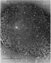
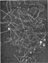
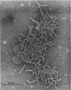
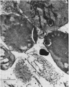
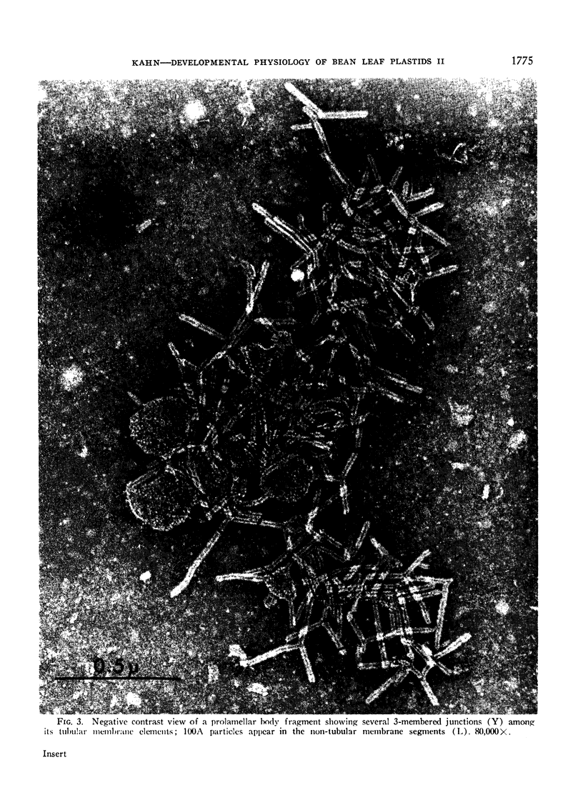
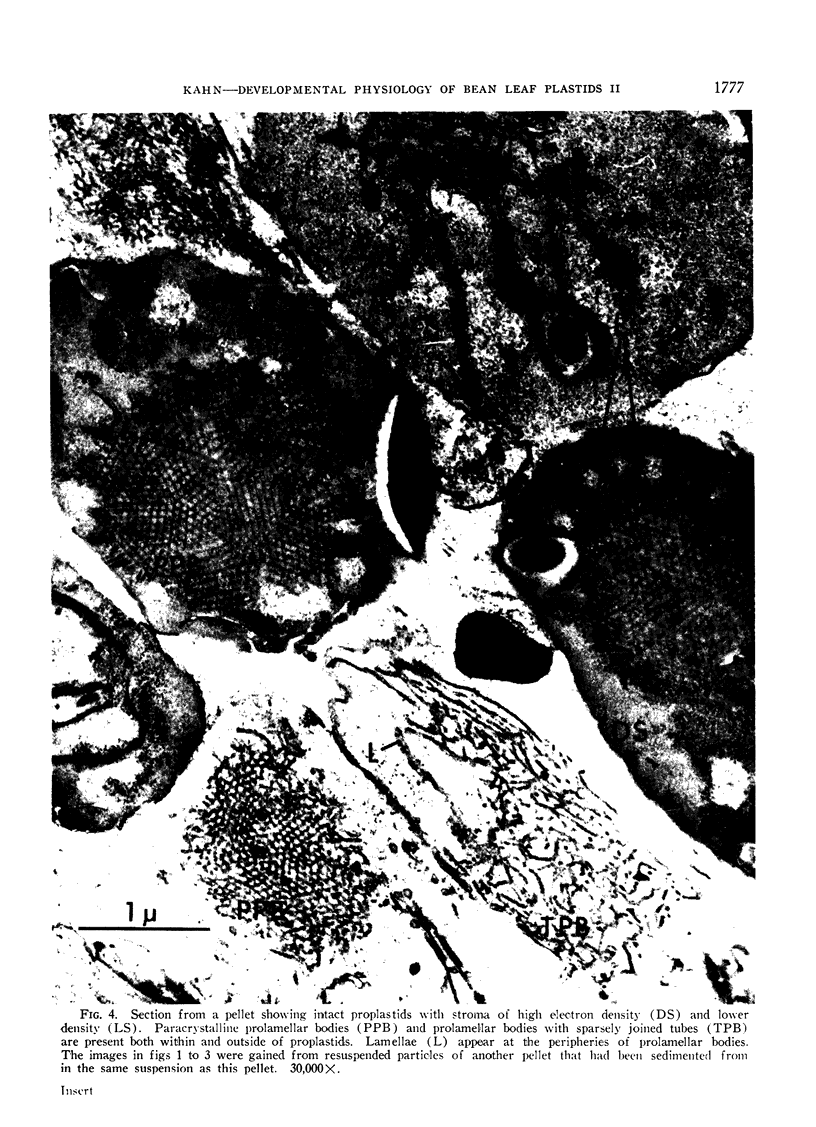
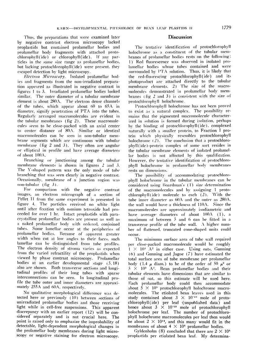
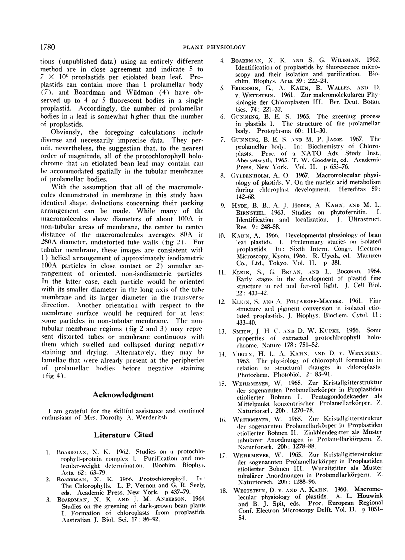
Proplastids and prolamellar bodies with tubular membranes were isolated from the dark grown primary leaves of bean seedlings (Phaseolus vulgaris L.). The combination of fluorescence microscopy and negative contrast electron microscopy provided the tentative identification of protochlorophyll holochrome as a constituent of prolamellar body membranes and new evidence for solution-filled channels within the tubular membrane systems of prolamellar bodies.
Full text
PDF
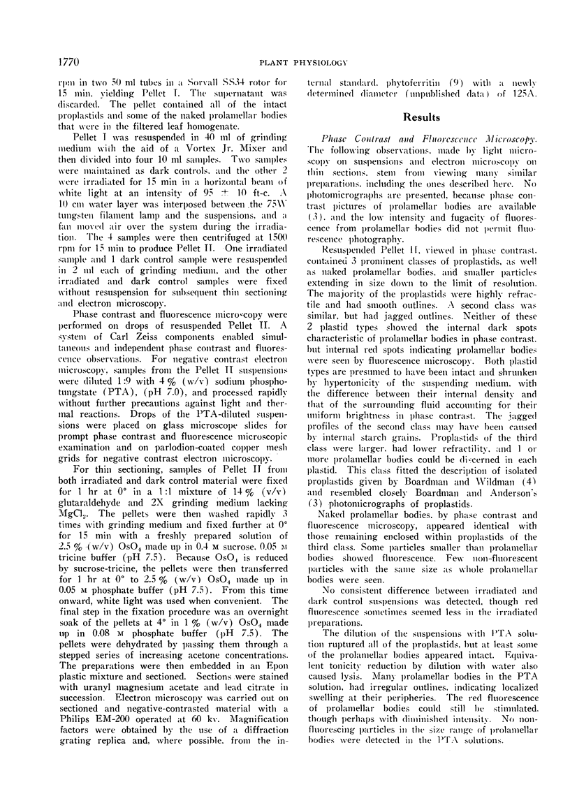
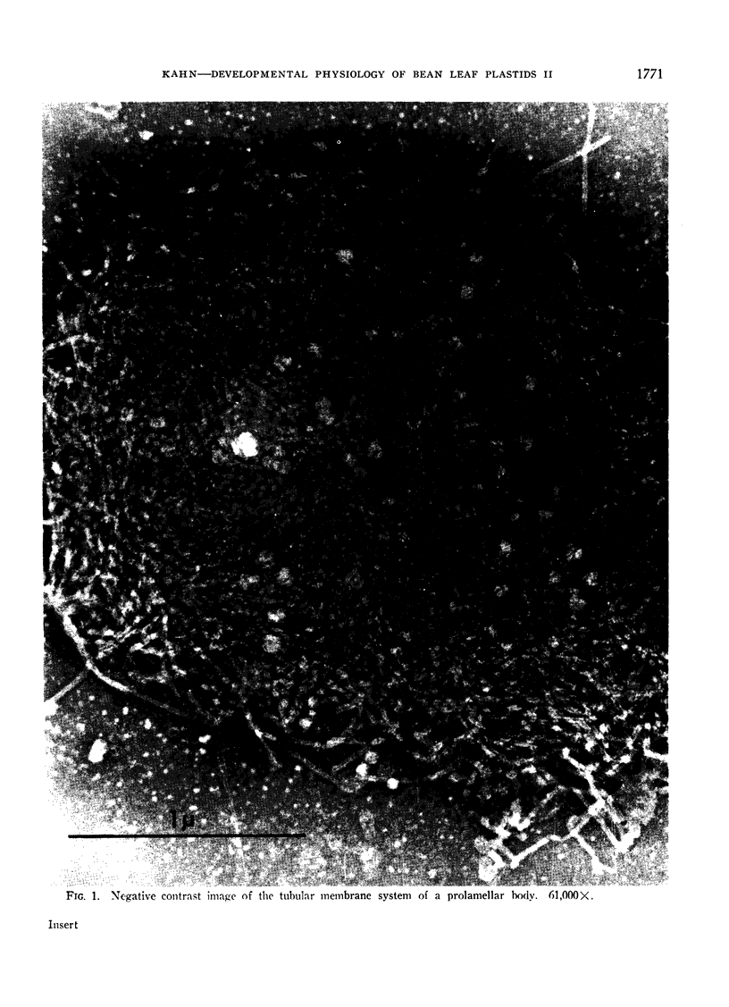
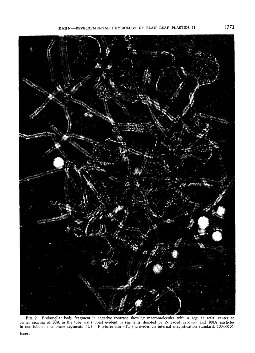
Images in this article
Selected References
These references are in PubMed. This may not be the complete list of references from this article.
- BOARDMAN N. K., WILDMAN S. G. Identification of proplastids by fluorescence microscopy and their isolation and purification. Biochim Biophys Acta. 1962 May 7;59:222–224. doi: 10.1016/0006-3002(62)90717-5. [DOI] [PubMed] [Google Scholar]
- HYDE B. B., HODGE A. J., KAHN A., BIRNSTIEL M. L. STUDIES ON PHYTOFERRITIN. I. IDENTIFICATION AND LOCALIZATION. J Ultrastruct Res. 1963 Oct;59:248–258. doi: 10.1016/s0022-5320(63)80005-2. [DOI] [PubMed] [Google Scholar]
- KLEIN S., POLJAKOFF-MAYBER A. Fine structure and pigment conversion in isolated etiolated proplastids. J Biophys Biochem Cytol. 1961 Nov;11:433–440. doi: 10.1083/jcb.11.2.433. [DOI] [PMC free article] [PubMed] [Google Scholar]


