Abstract
Granulomatous amoebic encephalitis due to Acanthamoeba castellanii is a serious human infection with fatal consequences, but it is not clear how the circulating amoebae interact with the blood-brain barrier and transmigrate into the central nervous system. We studied the effects of an Acanthamoeba encephalitis isolate belonging to the T1 genotype on human brain microvascular endothelial cells, which constitute the blood-brain barrier. Using an apoptosis-specific enzyme-linked immunosorbent assay, we showed that Acanthamoeba induces programmed cell death in brain microvascular endothelial cells. Next, we observed that Acanthamoeba specifically activates phosphatidylinositol 3-kinase. Acanthamoeba-mediated brain endothelial cell death was abolished using LY294002, a phosphatidylinositol 3-kinase inhibitor. These results were further confirmed using brain microvascular endothelial cells expressing dominant negative forms of phosphatidylinositol 3-kinase. This is the first demonstration that Acanthamoeba-mediated brain microvascular endothelial cell death is dependent on phosphatidylinositol 3-kinase.
Acanthamoeba spp. are opportunistic protozoan parasites that are widely distributed throughout the environment (12, 18). The genus Acanthamoeba consists of both pathogenic and nonpathogenic species. Given the host susceptibility and correct environmental conditions, Acanthamoeba can cause granulomatous amoebic encephalitis (GAE), a fatal central nervous system (CNS) infection that occurs in immunocompromised patients (7-10, 11, 19). Several lines of evidence suggest that hematogenous spread is a prerequisite in Acanthamoeba encephalitis (19-21), but it is not clear how circulating amoebae cross the blood-brain barrier and gain access to the CNS to produce disease. We have demonstrated that pathogenic Acanthamoeba exhibits more than 60% binding to human brain microvascular endothelial cells (HBMEC), which constitute the blood-brain barrier (2). Acanthamoeba binding to HBMEC is mediated by a mannose-binding protein expressed on the surface of Acanthamoeba cells (2). Moreover, we showed that Acanthamoeba produces severe HBMEC cytotoxicity by secreting extracellular proteases, as well as using contact-dependent mechanisms such as phagocytosis (12), which may play an important role in blood-brain barrier perturbations. However, the host intracellular signaling pathways and the molecular mechanisms associated with Acanthamoeba-mediated HBMEC cytotoxicity have not been determined.
Lipid second messengers, such as those derived from the polyphosphoinositide cycle, play a central role in many signaling networks. The majority of inositol lipids reside in membranes and serve as substrates for kinases, phosphatases, and phospholipases. Phosphatidylinositol 3-kinases (PI3Ks) are important signaling molecules that phosphorylate the 3′ OH position of the inositol ring of phosphoinositides (PIs), generating the second messengers PI(3)P, PI(3,4)P2, and PI(3,4,5)P3 (4, 17). These second messengers recruit the downstream effector molecules. More specifically, substrates with certain FYVE domains (named after the first four proteins in this motif, Fab1, YotB, Vac1p, and EEA1) bind PIP, and these pathways have been implicated in vesicular trafficking or receptor-mediated endocytosis (5). In contrast, proteins containing pleckstrin homology (PH) domains bind PIP2 and PIP3 and have been shown to have important cellular functions, including cell proliferation, cell survival, cellular differentiation, metabolism, and cytoskeletal rearrangements (17). However, most of the studies have been focused on growth hormones, mitogenic cytokines, inflammatory cytokines, chemoattractants, oncogenesis, or intracellular pathogens. In this study, we determined HBMEC responses to the extracellular parasite Acanthamoeba and the role of PI3K in Acanthamoeba-mediated HBMEC death.
MATERIALS AND METHODS
All chemicals were purchased from Sigma Laboratories (Poole, Dorset, United Kingdom) unless stated otherwise.
Acanthamoeba culture.
Acanthamoeba castellanii ATCC 50494 was isolated from a human brain necropsy from a patient who died of GAE, as previously described (29). The genus Acanthamoeba is divided into 15 different genotypes based on rRNA gene sequencing; the isolate used in the present study belongs to the T1 genotype (12, 27). Acanthamoeba was routinely grown in PYG medium (0.75% [wt/vol] proteose peptone, 0.75% [wt/vol] yeast extract, and 1.5% [wt/vol] glucose) at 30°C in tissue culture flasks, and the medium was refreshed 17 to 20 h prior to experiments as previously described (14). Unbound amoebae were removed by several washes. The Acanthamoeba cells that remained bound to flasks represented the trophozoite form and were used in all subsequent assays.
Human brain microvascular endothelial cell culture and transfection.
Primary brain microvascular endothelial cells were isolated from human tissue and cultured as previously described (2, 30). Briefly, HBMEC were purified by fluorescence-activated cell sorting and tested for endothelial characteristics, such as expression of endothelial markers, F-VIII, carbonic anhydrase IV, and uptake of acetylated low-density lipoprotein, which resulted in cultures that were more than 99% pure. HBMEC were routinely grown on rat tail collagen-coated dishes in RPMI 1640 containing 10% heat-inactivated fetal bovine serum, 10% NuSerum, 2 mM glutamine, 1 mM pyruvate, penicillin (100 U/ml), streptomycin (100 U/ml), nonessential amino acids, and vitamins (2, 30).
In some experiments, HBMEC were transfected with dominant negative mutants with mutations in p110 (p110K) subunits of PI3K. The cDNAs of p110 mutants were cloned under the control of cytomegalovirus promoter with an amino-terminal FLAG epitope tag in the eukaryotic expression vector pcDNA3, which also conferred G418 resistance. The p110 construct was a kinase inactive mutant, had a CAAX motif, and was constitutively translocated to the membrane. HBMEC transfected with the expression vector pcDNA3 were used as negative controls (Invitrogen, Paisley, United Kingdom). The pcDNA3 expression vectors with or without dominant negative p110 constructs were transfected into the HBMEC using LipofectAMINE as previously described (15). Briefly, a DNA-LipofectAMINE complex in RPMI 1640 was added to 50% confluent HBMEC monolayers. After 6 h of incubation, the cells were washed and grown in complete medium for 3 days, followed by selection with G418 (400 μg/ml). Antibiotic-resistant colonies were pooled and confirmed using Western blotting assays (26).
Cytotoxicity assays.
To determine the pathogenic potential of the Acanthamoeba used in this study, cytotoxicity assays were performed as previously described (13). Briefly, HBMEC were grown to monolayers in 24-well plates. Acanthamoeba isolates (5 × 10⁵ amoebae/well) were incubated with HBMEC monolayers in serum-free medium (RPMI 1640 containing 2 mM glutamine, 1 mM pyruvate, and nonessential amino acids) at 37°C in 5% CO2 for 24 h. At the end of this incubation period, supernatants were collected, and cytotoxicity was determined by measuring lactate dehydrogenase (LDH) release (cytotoxicity detection kit; Roche Applied Science, Lewes, East Sussex, United Kingdom). Briefly, conditioned media of cocultures of Acanthamoeba and HBMEC were collected, and the percentage of LDH was determined as follows: percentage of cytotoxicity = (test value − control value/total amount of LDH released − control value) × 100). Control values were obtained from HBMEC incubated alone, and the total amount of LDH released was measured with HBMEC treated with 1% Triton X-100 for 1 h at 37°C.
Apoptosis assays.
To determine the ability of Acanthamoeba to produce host cell apoptosis using mouse monoclonal antibodies showing DNA and histone fragmentation, an apoptosis-specific enzyme-linked immunosorbent assay (ELISA) was performed according to the manufacturer's instructions (Roche Applied Science). This allowed specific determination of mono- and oligonucleosomes in the cytoplasmic fraction of cell lysates. Briefly, HBMEC were grown to monolayers in 96-well plates and incubated with 105 amoebae for 3 h in serum-free medium at 37°C in 5% CO2. Following this, unbound parasites were removed, and HBMEC with bound amoebae were lysed and centrifuged (750 × g, 10 min) to remove cellular debris according to the manufacturer's instructions. The apoptotic DNA fragments in the supernatants were determined using anti-histone-biotin and anti-DNA-peroxidase antibodies. Antigen-antibody complexes were collected using streptavidin-coated plates, followed by determination using the 2,2′-azinobis(3-ethylbenzthiazolinesulfonic acid) (ABTS) substrate. Finally, plates were read at 405 nm using 492 nm as a reference. HBMEC incubated alone were used as negative controls, while HBMEC incubated with etoposide (an apoptosis inducer) were used as positive controls. The percentage of apoptosis was calculated as follows: percentage of apoptosis = (test value − negative control value) × 100. In some experiments, apoptosis assays were performed in the presence of the selective PI3K inhibitor LY294002 [2-(4-morpholinyl)-8-phenylchromone (2-morpholino-8-phenyl-4H-1-benzopyran-4-one)].
Western blotting assays.
Western blotting assays were performed to assess Akt phosphorylation as previously described (15). Briefly, HBMEC were grown to monolayers in 60-mm dishes, and cells were incubated with Acanthamoeba (5 × 10⁶ amoebae) for various times. Unbound amoebae were removed by several washes, and cells were lysed with RIPA lysis buffer (50 mM Tris-HCl [pH 7.4], 0.1% sodium dodecyl sulfate, 0.5% Na deoxycholate, 10 mM Na pyrophosphate, 25 mM β-glycerophosphate, 150 mM NaCl, 2 mM EDTA, 2 mM EGTA, 1% Triton X-100, 1 mM Na3VO4, 50 mM NaF, 1 mM phenylmethylsulfonyl fluoride, 1 μg/ml aprotonin, 1 μg/ml leupeptin, 1 μg/ml pepstatin). For immunoprecipitation, equal amounts of cell lysates (500 μg to 1 mg) were incubated with primary monoclonal antibody against phospho-Akt residues (Cell Signaling Tech, Beverly, MA) overnight at 4°C. Following this, 50 μl of protein A agarose beads (QIAGEN, Crawley, England) was added for 1 h to collect antigen-antibody complexes. The immunocomplexes were washed three times and electrophoresed using 10% sodium dodecyl sulfate-polyacrylamide gel electrophoresis under denaturing conditions. Proteins were transferred onto nitrocellulose membranes and blocked in 4% blocking buffer (blotting grade nonfat milk solution; Bio-Rad, Hemel Hempstead, England). Membranes were blotted with anti-phospho-Akt antibody in blocking buffer overnight at 4°C with gentle shaking. On the next day, the membranes were washed and subsequently incubated (60 min, 4°C) with horseradish peroxidase-linked secondary antibody. Protein bands were visualized using an enhanced chemiluminescence detection kit (Amersham Biosciences, Chalfont St. Giles, England).
RESULTS
Acanthamoeba GAE isolate belonging to the T1 genotype exhibits severe HBMEC cytotoxicity.
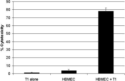
In this study, we used a clinical isolate of Acanthamoeba from a GAE patient and determined its effects on HBMEC, demonstrating the biological relevance of Acanthamoeba interactions with the blood-brain barrier (2, 12, 18). However, previous studies have shown that long-term axenic cultivation of Acanthamoeba could result in the loss of Acanthamoeba virulence (23). To determine the pathogenic potential of the Acanthamoeba isolate used in this study, cytotoxicity assays were performed. We determined that the Acanthamoeba isolate belonging to the T1 genotype exhibited more than 70% HBMEC cytotoxicity (Fig. 1), further confirming its pathogenic potential. Acanthamoeba incubated alone did not exhibit LDH release (Fig. 1).
FIG. 1.
Acanthamoeba encephalitis isolate belonging to the T1 genotype exhibits severe HBMEC cytotoxicity. Acanthamoeba (5 × 10⁵ amoebae) was added to confluent monolayers of HBMEC in 24-well plates and incubated at 37°C for 24 h. After this, supernatants were collected, and cytotoxicity was determined by measuring lactate dehydrogenase release as described in Materials and Methods. Endothelial cells without parasites were used as negative controls. Acanthamoeba exhibited more than 70% HBMEC cytotoxicity. The error bars indicate standard deviations. The data represent three independent experiments performed in triplicate.
GAE isolate belonging to the T1 genotype induces HBMEC apoptosis.
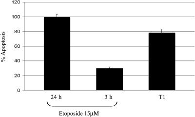
Acanthamoeba-mediated HBMEC cytotoxicity is a result of degradation of intracellular constituents and the buildup of toxins and toxic products of parasite metabolism, ultimately leading to host cell death (1, 22). However, the immediate effects of Acanthamoeba may involve cytokine release that leads to an inflammatory response (22), host cell cycle arrest (29), or induction of apoptosis. Apoptosis or programmed cell death involves active participation of the host cells regulated by intracellular signaling pathways (24, 25). For a positive control, we tested various concentrations of etoposide (an apoptosis inducer) and determined that treatment with a concentration of 15 μM for 24 h induced maximum HBMEC apoptosis; the apoptosis obtained with these values was defined as 100% apoptosis. To determine the ability of Acanthamoeba to induce HBMEC intracellular signaling pathways leading to programmed cell death, an apoptosis-specific ELISA was used. We observed that Acanthamoeba induced up to 70% HBMEC apoptosis within 3 h (Fig. 2). This is the first study demonstrating that Acanthamoeba induces apoptosis in HBMEC, implicating the role of host intracellular pathways in the response to Acanthamoeba. Interestingly, formalin- and/or acetone-fixed amoebae were unable to produce HBMEC apoptosis and exhibited no binding to HBMEC or human corneal epithelial cells (HCEC), indicating that live amoebae are required (data not shown).
FIG. 2.
Acanthamoeba encephalitis isolate belonging to the T1 genotype induces apoptosis in HBMEC. Acanthamoeba (105 amoebae/well) was added to confluent monolayers of HBMEC in 96-well plates and incubated at 37°C for 3 h. Following this, HBMEC were lysed, and the supernatants were collected. The DNA fragments in the supernatants were determined with anti-histone-biotin and anti-DNA-peroxidase antibodies using an apoptosis-specific ELISA as described in Materials and Methods. Endothelial cells without parasites were used as negative controls, while HBMEC incubated with etoposide (an apoptosis inducer; 15 μM) were used as positive controls. Treatment with etoposide (15 μM) for 24 h induced the maximum HBMEC apoptosis, and the value obtained was defined as 100% apoptosis. The percentage of apoptosis for test samples was calculated as follows: percentage of apoptosis = (test value − negative control value) × 100. The error bars indicate standard deviations. The data represent three independent experiments performed in triplicate.
Acanthamoeba induces PI3K activation in HBMEC.
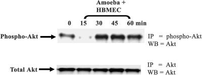
PI3K molecules directly phosphorylate the 3′ OH position of the inositol ring of PIs, generating the second messengers PI(3)P, PI(3,4)P2. and PI(3,4,5)P3(12,13). The 3′-phosphorylated PIP2 and PIP3 bind to the PH domain of various substrates. Among other effectors, Akt is a major downstream effector of PI3K. Stimulation of PI3K results in Akt recruitment to the membrane, and the Akt is subsequently phosphorylated at Thr308 by phosphoinositide-dependent kinase 1, followed by phosphorylation at Ser473 by phosphoinositide-dependent kinase 2, resulting in enhanced activity. Therefore, we used Akt phosphorylation as an indicator of PI3K activation. To determine whether Acanthamoeba activates PI3K, Western blotting assays were performed using anti-phospho-Akt for immunoprecipitation, followed by total Akt antibody for Western blotting assays. We observed that coincubation of Acanthamoeba with HBMEC resulted in Akt dephosphorylation within 15 min (Fig. 3). A more-than-sixfold decrease in Akt phosphorylation was observed (Fig. 3). HBMEC incubated alone for similar times without serum exhibited no changes in Akt phosphorylation compared with the zero-time phosphorylation (data not shown). Since Akt is well known to promote cell proliferation and/or antiapoptotic effects, our results suggest that the initial response of HBMEC to Acanthamoeba may inhibit cell survival pathways. However, further incubation of Acanthamoeba with HBMEC resulted in stimulation of Akt phosphorylation (a more-than-twofold increase) (Fig. 3). Interestingly, Akt antibodies are specific to human Akt and did not react with Acanthamoeba Akt (data not shown). Overall, these data indicate that the HBMEC response to Acanthamoeba involves PI3K-mediated signaling pathways.
FIG. 3.
Acanthamoeba encephalitis isolate belonging to the T1 genotype induces Akt phosphorylation in HBMEC. To determine whether Acanthamoeba activates PI3K, HBMEC were grown to monolayers and then incubated with Acanthamoeba for various times. The cell lysates were prepared and immunoprecipitated using phospho-Akt antibodies (IP = phospo-Akt and IP = Akt), which was followed by Western blotting with total Akt antibody (WB = Akt). The zero-time lane contained HBMEC without Acanthamoeba. Note that Acanthamoeba abolished Akt phosphorylation within 15 min. However, longer incubation times resulted in stimulation of Akt phosphorylation, suggesting that the HBMEC response to Acanthamoeba involves PI3K-mediated signaling pathways. The results are representative of three independent experiments.
Acanthamoeba-mediated HBMEC death is dependent on PI3K activation.
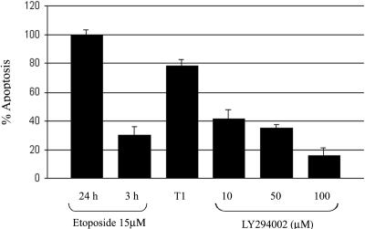
The finding described above that Acanthamoeba dephosphorylates Akt within 15 min, which is followed by increased Akt phosphorylation with longer incubation times (Fig. 3), suggests the involvement of PI3K-mediated signaling but does not clarify the role of PI3K in Acanthamoeba-mediated HBMEC death. To precisely determine the role of PI3K activation in Acanthamoeba-mediated HBMEC death, we used the specific PI3K inhibitor LY294002. LY294002 selectively binds to the ATP binding site of PI3K, thereby inhibiting its catalytic activity, and has no inhibitory effect on other ATP-requiring enzymes, such as protein kinase, lipid kinase, and ATPase (32, 33). Apoptosis-specific ELISAs were performed in the presence of LY294002. We observed that LY294002 significantly inhibited HBMEC death in a concentration-dependent manner (Fig. 4) (P < 0.05), indicating that Acanthamoeba-induced HBMEC death is dependent on PI3K-mediated pathways.
FIG. 4.
PI3K inhibitor blocks Acanthamoeba-induced HBMEC apoptosis. To precisely determine the role of PI3K in Acanthamoeba-mediated HBMEC death, apoptosis-specific ELISAs were performed in the presence various concentrations of LY294002, a selective PI3K inhibitor, as described in Materials and Methods. LY294002 inhibits Acanthamoeba-induced apoptosis in a concentration-dependent manner. The error bars indicate standard deviations. The data represent three independent experiments performed in triplicate.
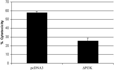
To further confirm the role of PI3K in Acanthamoeba-mediated host cell death, HBMEC were transfected with inactive forms of catalytic PI3K subunit p110 (ΔPI3K). We observed that Acanthamoeba produced significantly less cytotoxicity in HBMEC expressing ΔPI3K than in pcDNA3-transfected HBMEC (Fig. 5). Taken together, these findings show for the first time that Acanthamoeba-mediated HBMEC death is dependent on the PI3K signaling pathway.
FIG. 5.
HBMEC expressing dominant negative forms of PI3K are less susceptible to Acanthamoeba-mediated cell death. HBMEC were transfected with the eukaryotic expression vector pcDNA3 cloned with mutant PI3K subunit p110 (ΔPI3K) or with the vector alone. The HBMEC transfected with pcDNA3 or ΔPI3K were used in Acanthamoeba cytotoxicity assays as described in Materials and Methods. Note that Acanthamoeba-mediated HBMEC death was significantly inhibited in HBMEC expressing dominant negative PI3K. The error bars indicate standard deviations. The data represent three independent experiments performed in triplicate.
DISCUSSION
GAE due to Acanthamoeba is a serious infection that almost always is fatal; however, the pathogenesis and pathophysiology of Acanthamoeba encephalitis are not known. Several lines of evidence suggest that hematogenous spread is a primary requirement in GAE (19-21), but how circulating amoebae cross the blood-brain barrier remains to be determined. We recently isolated HBMEC, which constitute the blood-brain barrier, and developed an in vitro model to study host-parasite interactions. Previous studies using such models have focused on intracellular pathogens, such as meningitis-causing bacteria. For example, it has been shown that Escherichia coli K1 can invade brain microvascular endothelial cells on the apical side, traffic through the cytoplasm in a membrane-bound vacuole, and exit on the basolateral side to produce meningitis without affecting the blood-brain barrier function, as determined by HBMEC monolayer integrity, HBMEC permeability, and high electrical resistance (16, 28). In contrast, extracellular pathogens, such as Acanthamoeba, are unlikely to use similar mechanisms due to size limitations and must develop strategies to produce blood-brain barrier perturbations to gain access to the CNS to produce disease. In support of this, we show for the first time that Acanthamoeba produces programmed cell death in HBMEC. Findings of this study are of physiological relevance as we used a clinical isolate of Acanthamoeba from a deceased GAE patient. Moreover, our finding that Acanthamoeba-mediated HBMEC death is dependent on PI3K activation is novel. Previous reports have extensively shown the role of PI3K-mediated signaling in cell proliferation or antiapoptotic effects in an Akt-dependent fashion. For example, activated PI3K phosphorylates the 3′ OH position of the inositol ring of PIs, generating the second messengers PIP2 and PIP3 (17). These PI3K lipid products interact with proteins containing a PH domain. Among other proteins, Akt is a major downstream effector of PI3K that interacts with PIP2 or PIP3 through its PH domain and is phosphorylated at residues Ser473 (carboxy-terminal domain) and Thr308 (catalytic domain), resulting in activated Akt kinase. Phosphorylated and activated Akt stimulates cell proliferation or cytoprotective effects by modulating the function of a variety of downstream molecules, including BAD, caspase-9, CREB Forkhead, MDM2, NF-κB, cyclin D1, and p53 (3, 6). Overall, these findings suggest that Akt-mediated signaling pathways result in cell survival and/or cytoprotective effects. In support of this, our results show that Acanthamoeba interactions with HBMEC show a biphasic response. First, we found that Acanthamoeba incubation resulted in Akt dephosphorylation (inactivation) within 15 min. However, longer incubation of Acanthamoeba with HBMEC resulted in a more-than-twofold increase in Akt phosphorylation. This raises the question of why Acanthamoeba induces Akt activation, which almost always results in cytoprotective effects. One explanation is that Akt activation is a stress compensation response of HBMEC and may be independent of PI3K activation. This may involve direct phosphorylation of Akt by integrin-linked kinase 1 (35) or by Ca2+/calmodulin-dependent kinase kinase, leading to cell survival pathways (34). Additional studies are needed to determine the precise role of Akt activation in response to Acanthamoeba.
To determine the role of PI3K in Acanthamoeba-mediated HBMEC death, apoptosis assays were performed in the presence of LY294002, a selective PI3K inhibitor. We observed that direct inhibition of PI3K significantly reduced Acanthamoeba-mediated HBMEC death. This is somewhat surprising, as PI3K-mediated signaling has been traditionally involved in cell proliferation and cell survival effects. To further confirm these findings, we used HBMEC expressing dominant negative forms of PI3K and used for cytotoxicity assays. We observed that Acanthamoeba exhibited significantly less cytotoxicity with HBMEC expressing dominant negative forms of PI3K than with HBMEC transfected with the vector alone. Overall, these findings indicate that Acanthamoeba-mediated host cell death is dependent on PI3K activation. In support of this, Thyrell et al. (31) have shown that alpha interferon induces PI3K-mediated apoptosis in myeloma cells without Akt phosphorylation. It was further shown that downstream effectors of PI3K-mediated apoptosis involve activation of the proapoptotic molecules Bak and Bax, loss of mitochondrial membrane potential, and release of cytochrome c, all well-known mediators of apoptosis. Similar mechanisms may exist in Acanthamoeba-mediated host cell death, and studies are in progress to address these issues. In addition, previous studies have shown that Acanthamoeba induces increased cytosolic free calcium, resulting in apoptosis (1, 22); however, the relationships between calcium fluxes and PI3K and whether pathways involving calcium fluxes and PI3K are dependent or independent of each other remain to be determined.
Other pathologies due to Acanthamoeba include sight-threatening keratitis associated with severe pain due to radial neuritis, inflammation with redness, and photophobia. We determined whether Acanthamoeba uses similar signaling pathways to produce corneal epithelial cell death. For this, we used a clinical isolate of Acanthamoeba from a keratitis patient belonging to the T4 genotype and determined its effects on HCEC (29). Similar host responses were observed (data not shown). More interestingly, Acanthamoeba (T4 isolate) abolished HCEC Akt activation within 15 min of incubation, but longer incubation times resulted in a more-than-sixfold increase in Akt activation, a response similar to that of HBMEC stimulated with T1 amoebae, clearly indicating that Acanthamoeba induces similar mechanisms to produce host cell death. These findings may have important clinical implications in our search for drugs against infections due to Acanthamoeba. Overall, these studies suggest that Acanthamoeba-mediated HBMEC cytotoxic effects are dependent on PI3K signaling pathways. A complete understanding of mechanisms associated with Acanthamoeba-mediated host cell death may help us develop strategies to treat these serious infections.
Acknowledgments
This work was partially supported by grants from the British Council for Prevention of Blindness, the Central Research Fund, the University of London, and The Royal Society.
Editor: J. F. Urban, Jr.
REFERENCES
- 1.Alizadeh, H., M. S. Pidherney, J. P. McCulley, and J. Y. Niederkorn. 1994. Apoptosis as a mechanism of cytolysis of tumor cells by a pathogenic free-living amoeba. Infect. Immun. 62:1298-1303. [DOI] [PMC free article] [PubMed] [Google Scholar]
- 2.Alsam, S., K. S. Kim, M. Stins, A. O. Rivas, J. Sissons, and N. A. Khan. 2003. Acanthamoeba interactions with human brain microvascular endothelial cells. Microb. Pathog. 35:235-241. [DOI] [PubMed] [Google Scholar]
- 3.Brazil, D. P., J. Park, and B. A. Hemmings. 2002. PKB binding proteins: getting in on the Akt. Cell 111:293-303. [DOI] [PubMed] [Google Scholar]
- 4.Carpenter, C. L., and L. C. Cantley. 1996. Phosphoinositide kinases. Curr. Opin. Cell Biol. 8:153-158. [DOI] [PubMed] [Google Scholar]
- 5.Corvera, S., and M. P. Czech. 1998. Direct targets of phosphoinositide 3-kinase products in membrane traffic and signal transduction. Trends Cell Biol. 8:442-446. [DOI] [PubMed] [Google Scholar]
- 6.Datta, S. R., A. Brunet, and M. E. Greenberg. 1999. Cellular survival: a play in three Akts. Genes Dev. 13:2905-2927. [DOI] [PubMed] [Google Scholar]
- 7.Di Gregorio, C., F. Rivasi, N. Mongiardo, B. De Rienzo, S. Wallace, and G. S. Visvesvara. 1992. Acanthamoeba meningoencephalitis in an AIDS patient: first report from Europe. Arch. Pathol. Lab. Med. 116:1363-1365. [PubMed] [Google Scholar]
- 8.Friedland, L. R., S. A. Raphael, E. S. Deutsch, J. Johal, L. J. Martyn, G. S. Visvesvara, and H. Lischner. 1992. Disseminated Acanthamoeba infection in a child with symptomatic human immunodeficiency virus infection. Pediatr. Infect. Dis. J. 11:404-407. [DOI] [PubMed] [Google Scholar]
- 9.Gonzalez, M. M., E. Gould, G. Dickinson, A. J. Martinez, G. S. Visvesvara, T. J. Cleary, and G. T. Hensley. 1986. Acquired immunodefiency syndrome associated with Acanthamoeba infection and other opportunistic organisms. Arch. Pathol. Lab. Med. 110:749-751. [PubMed] [Google Scholar]
- 10.Gordon, S. M., J. P. Steinberg, M. DuPuis, P. Kozarsky, J. F. Nickerson, and G. S. Visvesvara. 1992. Culture isolation of Acanthamoeba species and leptomyxid amoebas from patients with amebic meningoencephalitis, including two patients with AIDS. Clin. Infect. Dis. 15:1024-1030. [DOI] [PubMed] [Google Scholar]
- 11.Jones, D. B., G. S. Visvesvara, and N. M. Robinson. 1975. Acanthamoeba polyphaga keratitis and Acanthamoeba uveitis associated with fatal meningoencephalitis. Trans. Ophthalmol. Soc. U. K. 95:221-232. [PubMed] [Google Scholar]
- 12.Khan, N. A. 2003. Pathogenesis of Acanthamoeba infections. Microb. Pathog. 34:277-285. [DOI] [PubMed] [Google Scholar]
- 13.Khan, N. A. 2001. Pathogenicity, morphology, and differentiation of Acanthamoeba. Curr. Microbiol. 43:391-395. [DOI] [PubMed] [Google Scholar]
- 14.Khan, N. A., E. L. Jarroll, N. Panjwani, Z. Cao, and T. A. Paget. 2000. Proteases as markers of differentiation of pathogenic and non-pathogenic Acanthamoeba. J. Clin. Microbiol. 38:2858-2861. [DOI] [PMC free article] [PubMed] [Google Scholar]
- 15.Khan, N. A., Y. Wang, K. J. Kim, J. Woong Chung, C. A. Wass, and K. S. Kim. 2002. Cytotoxic necrotizing factor-1 contributes to Escherichia coli K1 invasion of the central nervous system. J. Biol. Chem. 277:15607-15612. [DOI] [PubMed] [Google Scholar]
- 16.Kim, K. J., S. J. Elliott, F. Di Cello, M. F. Stins, and K. S. Kim. 2003. The K1 capsule modulates trafficking of E. coli-containing vacuoles and enhances intracellular bacterial survival in human brain microvascular endothelial cells. Cell. Microbiol. 5:245-252. [DOI] [PubMed] [Google Scholar]
- 17.Leevers, S., B. Vanhaesebroeck, and M. D. Waterfeld. 1999. Signalling through phosphoinositide 3-kinases: the lipids take centre stage. Curr. Opin. Cell Biol. 11:219-225. [DOI] [PubMed] [Google Scholar]
- 18.Marciano-Cabral, F., and G. Cabral. 2003. Acanthamoeba spp. as agents of disease in humans. Clin. Microbiol. Rev. 16:273-307. [DOI] [PMC free article] [PubMed] [Google Scholar]
- 19.Martinez, A. J.. 1987. Clinical manifestations of free-living amoebic infections, p. 161-177. In E. G. Rondanelli (ed.), Amphizoic amoebae: human pathology. Piccin, Pauda, Italy.
- 20.Martinez, A. J.. 2001. Free-living amebas and the immune deficient host, p. 1-12. In IX International Meeting on the Biology and Pathogenicity of Free-living Amoebae Proceedings. Editions John Libbey Eurotext, Montrouge, France.
- 21.Martinez, A. J., and G. S. Visvesvara. 1997. Free-living, amphizoic and opportunistic amebas. Brain Pathol. 7:583-598. [DOI] [PMC free article] [PubMed] [Google Scholar]
- 22.Mattana, A., V. Cappai, L. Alberti, C. Serra, P. L. Fiori, and P. Cappuccinelli. 2002. ADP and other metabolites released from Acanthamoeba castellanii lead to human monocytic cell death through apoptosis and stimulate the secretion of proinflammatory cytokines. Infect. Immun. 70:4424-4432. [DOI] [PMC free article] [PubMed] [Google Scholar]
- 23.Mazur, T., E. Hadas, and I. Iwanicka. 1995. The duration of the cyst stage and the viability and virulence of Acanthamoeba isolates. Trop. Med. Parasitol. 46:106-108. [PubMed] [Google Scholar]
- 24.Mizukami, Y., T. Okamura, T. Miura, M. Kimura, K. Mogami, N. Todoroki-Ikeda, S. Kobayashi, and M. Matsuzaki. 2001. Phosphorylation of proteins and apoptosis induced by c-Jun N-terminal kinase1 activation in rat cardiomyocytes by H2O2 stimulation. Biochim. Biophys. Acta 1540:213-220. [DOI] [PubMed] [Google Scholar]
- 25.Mutoh, T., T. Kumano, H. Nakagawa, and M. Kuriyama. 1999. Involvement of tyrosine phosphorylation in HMG-CoA reductase inhibitor-induced cell death in L6 myoblasts. FEBS Lett. 444:85-89. [DOI] [PubMed] [Google Scholar]
- 26.Reddy, M. A., N. V. Prasadarao, C. A. Wass, and K. S. Kim. 2000. Phosphatidylinositol 3-kinase activation and interaction with focal adhesion kinase in Escherichia coli K1 invasion of human brain microvascular endothelial cells. J. Biol. Chem. 275:36769-36774. [DOI] [PubMed] [Google Scholar]
- 27.Schuster, F. L., and G. S. Visvesvara. 2004. Free-living amoebae as opportunistic and non-opportunistic pathogens of humans and animals. Int. J. Parasitol. 34:1001-1027. [DOI] [PubMed] [Google Scholar]
- 28.Shin, H. J., M. S. Cho, S. Y. Jung, H. I. Kim, S. Park, J. H. Seo, J. C. Yoo, and K. I. Im. 2001. Cytopathic changes in rat microglial cells induced by pathogenic Acanthamoeba culbertsoni: morphology and cytokine release. Clin. Diagn. Lab. Immunol. 8:837-840. [DOI] [PMC free article] [PubMed] [Google Scholar]
- 29.Sissons, J., S. Alsam, S. Jayasekera, K. S. Kim, M. Stins, and N. A. Khan. 2004. Acanthamoeba induces cell-cycle arrest in the host cells. J. Med. Microbiol. 53:711-717. [DOI] [PubMed] [Google Scholar]
- 30.Stins, M. F., F. Gilles, and K. S. Kim. 1997. Selective expression of adhesion molecules on human brain microvascular endothelial cells. J. Neuroimmunol. 76:81-90. [DOI] [PubMed] [Google Scholar]
- 31.Thyrell, L., L. Hjortsberg, V. Arulampalam, T. Panaretakis, S. Uhles, B. Zhivotovsky, I. Leibiger, D. Grander, and K. Pokrovskaja. 2004. Interferon-alpha induced apoptosis in tumor cells is mediated through PI3K/mTOR signaling pathway. J. Biol. Chem. 279:24152-24162. [DOI] [PubMed] [Google Scholar]
- 32.Vlahos, C. J., W. F. Matter, K. Y. Hui, and R. F. Brown. 1994. A specific inhibitor of phosphatidylinositol 3-kinase, 2-(4-morpholinyl)-8-phenyl-4H-1-benzopyran-4-one (LY294002). J. Biol. Chem. 269:5241-5248. [PubMed] [Google Scholar]
- 33.Walker, E. H., M. E. Pacold, O. Perisic, L. Stephens, P. T. Hawkins, M. P. Wymann, and R. L. Williams. 2000. LY294002 structural determinants of phosphoinositide 3-kinase inhibition by wortmannin, quercetin, myricetin, and staurosporine. Mol. Cell 6:909-919. [DOI] [PubMed] [Google Scholar]
- 34.Yano, S., H. Tokumitsu, and T. R. Soderling. 1998. Calcium promotes cell survival through CaM-K kinase activation of the protein-kinase-B pathway. Nature 396:584-587. [DOI] [PubMed] [Google Scholar]
- 35.Yoganathan, T. N., P. Costello, X. Chen, M. Jabali, J. Yan, D. Leung, Z. Zhang, A. Yee, S. Dedhar, and J. Sanghera. 2000. Integrin-linked kinase (ILK): a “hot” therapeutic target. Biochem. Pharmacol. 60:1115-1119. [DOI] [PubMed] [Google Scholar]


