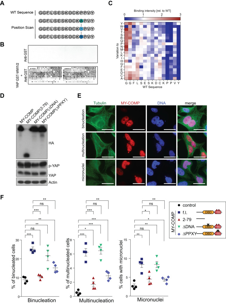
Fig. 2. MY-COMP leads to errors in cell division.
A Scheme for a full positional peptide array scan for the most prominent binder from the overlapping B-MYB peptide library reported in [18]. B The MY-COMP positional scan library was probed with either anti-GST (control) or with purified recombinant GST-WW1/2 and Anti-GST. Binding was detected by chemiluminescence. C Heat map representation of relative YAP binding affinity for MY-COMP peptides used in full positional scan. D Expression of the indicated MY-COMP constructs was verified by immunoblotting with an HA-antibody. MY-COMP constructs did not affect the expression of YAP and YAP phosphorylated on S127 (p-YAP). Actin served as a control. E Microphotographs showing examples of the different phenotypes following expression of MY-COMP. Bar: 25 μm. F Quantification of the fraction of binucleated and multinucleated cells and cells with micronuclei following expression of the indicated MY-COMP constructs. N = 4 biological replicates. Error bars indicate SEMs. Student’s t test. *p < 0.05, **p < 0.01, ***p < 0.001, ns not significant.

