Abstract
Mycobacterium bovis, a member of the Mycobacterium tuberculosis complex, has a particularly wide host range and causes tuberculosis in most mammals, including humans. A signature tag mutagenesis approach, which employed illegitimate recombination and infection of guinea pigs, was applied to M. bovis to discover genes important for virulence and to find potential vaccine candidates. Fifteen attenuated mutants were identified, four of which produced no lesions when inoculated separately into guinea pigs. One of these four mutants had nine deleted genes including mmpL4 and sigK and, in guinea pigs with aerosol challenge, provided protection against tuberculosis at least equal to that of M. bovis BCG. Seven mutants had mutations near the esxA (esat-6) locus, and immunoblot analysis of these confirmed the essential role of other genes at this locus in the secretion of EsxA (ESAT-6) and EsxB (CFP10). Mutations in the eight other attenuated mutants were widely spread through the chromosome and included pks1, which is naturally inactivated in clinical strains of M. tuberculosis. Many genes identified were different from those found by signature tag mutagenesis of M. tuberculosis by use of a mouse infection model and illustrate how the use of different approaches enables identification of a wider range of attenuating mutants.
Tuberculosis exacts enormous human suffering and death on a global basis and is also a widespread cause of animal morbidity and mortality. The primary human pathogen is Mycobacterium tuberculosis, although in many regions a significant amount of human disease results from infection with the very closely related organisms Mycobacterium africanum and the major animal pathogen Mycobacterium bovis. Together with a few less important species, these organisms are collectively referred to as the M. tuberculosis complex. The high degree of similarity in the pathogenesis of disease caused by these pathogens and the recent findings from genomic studies that the vast majority of genes within these species are identical or nearly so (16, 22) clearly indicate that the majority of their virulence genes are shared in common. There is particular interest in discovering the identity of these genes, so that new strategies including improved vaccines and antibacterials can be developed for combating tuberculosis.
Over the last 15 years, there has been continual development of molecular genetic techniques that can be applied to the M. tuberculosis complex (2). This enabled the discovery of individual tuberculosis virulence genes from 1995 onwards (7, 38) and led to the application over the last 5 years of a number of global techniques for identifying large numbers of genes that are essential for virulence or whose expression is altered during different stages of infection and disease. Among these global techniques, signature tag mutagenesis (18) is widely recognized as a powerful tool for investigating pathogenic organisms because of its ability to directly identify mutants that have reduced virulence (24). This identification is achieved by inoculating pools of mutants of a pathogen into a susceptible host. Each member of the pool is labeled with a different genetic tag that can be detected using a combination of PCR and DNA hybridization. Comparison of results from the inoculated pool and from the pool of organisms recovered from the host enables any mutant that is absent from the recovered pool to be identified, an absence that relates directly to its inability to grow and cause disease in the host. Two groups independently used this technique to screen a mutant library of M. tuberculosis (6, 11), each library generated by a different transposon derived from IS1096 (23). In both cases, similar numbers of organisms were injected intravenously into mice, the animals were sacrificed after 3 weeks, and organisms from the lungs were examined. The major result from both studies was the identification of mutations near the beginning and the end of the same set of 13 genes (Rv2930 to Rv2942) involved in the synthesis and transport of a complex lipid, phthiocerol dimycocerosate. While one of the studies also reported the identification of attenuating mutations at nine other genetic loci (6), all but one of the seven most attenuated strains in the study had mutations in the Rv2930-to-Rv2942 locus. Proposed explanations for the similarity of the results from these two studies were either that the number of virulence factors that can be detected in M. tuberculosis by this method is low or, alternatively, that the libraries created by IS1096-derived transposons are limited in their degree of complexity (23). Another contributing factor could have been the use of similar short-term mouse models.
One feature that emerged from early attempts to perform allelic exchange in strains of the M. tuberculosis complex was that these species have an efficient illegitimate recombination system (20). We have utilized this system extensively to produce illegitimate recombinants of M. bovis that were subsequently screened using surrogate markers of avirulence before testing the selected strains in a guinea pig model of virulence (8, 39). Moderate numbers of avirulent strains were identified by this approach, and some of these strains, when tested for vaccine efficacy in a guinea pig model with aerosol challenge, were found to produce protection against virulent M. bovis that was at least as good as that provided by M. bovis BCG (8, 13). In this study, in order to broaden the range of avirulent mutants of M. bovis for investigation, we combined the use of illegitimate recombination with signature tag mutagenesis. While this approach has been used for studying pathogenic fungi (5, 10), it does not appear to have been used previously in bacteria, where transposon mutagenesis has been preferred.
MATERIALS AND METHODS
Strains and growth conditions.
A wild-type M. bovis strain isolated from cattle in New Zealand was used. The strain was fully virulent in guinea pigs. The M. bovis strain and recombinants derived from it were cultured in Middlebrook 7H9 (Difco) liquid medium supplemented with albumin-dextrose complex (Difco), 0.05% Tween 80, and 0.4% sodium pyruvate and on Middlebrook 7H11 (Difco) solid medium supplemented with albumin-dextrose complex and 0.4% sodium pyruvate. Recombinants were cultured on medium supplemented with 20 μg of kanamycin/ml. For culture of M. bovis from guinea pig spleens, half the spleen was homogenized with 20 ml of water. This was filtered through sterile cheesecloth and centrifuged at 3,500 × g for 20 min. The pellet was resuspended in 0.5 ml of water, and 0.1-ml aliquots were plated onto Middlebrook 7H11 medium supplemented with 0.6 ml of oleic acid/liter, 50 g of bovine serum albumin/liter, 20 g of glucose/liter, 7.7 g of NaCl/liter, 0.4% sodium pyruvate, 0.5% lysed sheep red blood cells, 10% bovine serum, 10 μg of Fungizone/ml, 200 IU of polymyxin B sulfate, 100 μg of tarcarcillin/ml, and 10 μg of trimethoprim/ml. All plasmids were propagated in Escherichia coli XL1-Blue MR cultured on Luria-Bertani medium, containing 50 μg of kanamycin/ml.
Construction of signature-tagged mutant libraries.
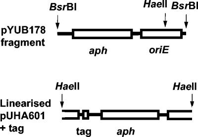
Plasmid pUHA601 (Fig. 1) was formed by ligating a 1,989-bp BsrBI fragment of pYUB178 (28) containing oriE and a kanamycin resistance gene (aph) to a 30-bp linker containing BssHII and BglII sites (made from annealing DMC132 and DMC133, Table 1) and electroporating the ligation mixture into E. coli. Signature tags were incorporated into pUHA601 by using an approach conceptually similar to that described previously (25). Briefly, an 89-bp linker containing signature tags was generated by using the oligonucleotide DMC120 (Table 1), which contained a 40-bp variable region, [NW]20 (N = A/T/G/C, W = A/T), as a template and the PCR primers DMC121 and DMC123, under standard PCR conditions. This linker was digested with BssHII and BglII, ligated to the matching sites in pUHA601, and electroporated into E. coli. Individual plasmid extracts were separated from remaining E. coli genomic DNA by electrophoresis, transferred to nylon filters by Southern blotting, and hybridized against a pooled probe of all the plasmid tags on the filter. The probe was produced from the combined plasmids by PCR labeling (33) with [α-32P]dCTP and the primers DMC122 and DMC124. Forty-five plasmids were obtained, each of which hybridized well. A pool of tags from the first 20 plasmids gave negligible hybridization with the remaining 25 plasmids, and a pool of tags from the remaining 25 plasmids gave negligible hybridization with the first 20 plasmids, indicating that unique plasmid tags had likely been selected. Individual pUHA601 plasmids containing tags were linearized by digestion with HaeII to produce the fragment shown in Fig. 1. The fragment was electroporated into M. bovis (36) and plated onto solid medium containing kanamycin. Individual colonies were subcultured in liquid medium and stored frozen at −70°C before use.
FIG. 1.
Construction of pUHA601 used to make tagged, illegitimate recombinants of M. bovis. For details see Materials and Methods.
TABLE 1.
Oligonucleotides used in this study
| Name | Sequence, 5′-3′ | Purpose |
|---|---|---|
| DMC132 | TTGGCGCGCCAAGGAATTCCGAAGATCTTC | Making linker with BssHII and BglII sites |
| DMC133 | GAAGATCTTCGGAATTCCTTGGCGCGCCAA | Making linker with BssHII and BglII sites |
| DMC120 | TTGGCGCGCTACAACCTCAAGCTT [NW]20- AAGCTTGGTTAGAATGAGATCTTCA | Making signature tags |
| DMC121 | TTGGCGCGCTACAACCT | Making signature tags |
| DMC123 | TGAAGATCTCATTCTAACC | Making signature tags |
| DMC124 | ATCTCATTCTAACCAAGCT | Making tag probes |
| DMC122 | CGCTACAACCTCAAGCT | Making tag probes and sequencing insertion |
| DMC201 | GCTCCCTCACTTTCTGG | Sequencing insertion sites |
| DMC483 | GGCAGAGATGAAGACCGATG | esxB PCR primer |
| DMC484 | GTCGAGTTCCTGCTTCTGCT | esxB PCR and reverse transcription primer |
| DMC549 | AGAGCAGCAGTGGAATTTCG | esxA PCR primer |
| DMC550 | GTCCCATTTTTGCTGGACAC | esxA PCR and reverse transcription primer |
Infection studies.
Forty-five M. bovis recombinants each containing a different tag were subcultured separately, and approximately equal amounts of each subculture were pooled together to form an inoculated pool. The optical density of the pooled cultures was used to estimate the number of organisms present, and approximately 106 CFU of each pool was inoculated subcutaneously into the flank of three Dunkin-Hartley guinea pigs. After 8 weeks, the animals were sacrificed and their spleens were cultured on solid medium for M. bovis. Between 500 and 2,000 colonies recovered from each of the three spleens was combined to form the recovered pool. DNA was extracted from both inoculated and recovered pools (35). The tags present in both pools were amplified by PCR with DMC122 and DMC124, and the gel-purified PCR products were labeled by a second PCR with [α-32P]dCTP. The radioactive products were digested with HindIII to remove the common sequences flanking the unique tags and used as probes to hybridize against all 45 plasmid tags that had been Southern blotted onto nylon filters. Recombinants whose level of tag hybridization had been reduced to near background in the recovered pool were individually inoculated into three guinea pigs in the same way as the original pools. Procedures used in all experiments on animals were approved by the Institute's Animal Ethics Committee.
Mapping of illegitimate insertion sites and sequence analysis.
To identify the sites of illegitimate recombination, chromosomal DNA was prepared from recombinants that had reduced virulence in guinea pigs. The DNA was digested with a restriction enzyme that did not have a site in pUHA601, ligated into pBluescript, electroporated into E. coli, and selected with kanamycin. Plasmid DNA was prepared from a kanamycin-resistant clone and subjected to DNA sequencing, reading through the HaeII site from both sides with primers DMC122 and DMC201 (Table 1). In some cases where a plasmid dimer had inserted into the M. bovis genome, the pBluescript constructs were sequenced using T3 and T7 primers followed by specific primers designed to walk along the mycobacterial DNA sequence until a fragment of pUHA601 was reached. DNA sequences were analyzed by comparison to GenBank (www.ncbi.nlm.nih.gov) and Sanger (www.sanger.ac.uk) databases and by using the programs of the Genetics Computer Group.
Analysis of gene function in the esxA (esat-6) region.
RNA was prepared by adding 4 volumes of guanidinium thiocyanate solution directly to broth cultures, harvesting the cells by centrifugation (approximately 108 cells/extraction), and disrupting the cells in Lysing Matrix B tubes (Qbiogene) with a FastPrep cell disruptor (Qbiogene) in the presence of 1 ml of Trizol (Invitrogen). After Trizol extraction, residual DNA was removed by incubation in DNase I (Invitrogen), and RNA was further purified using an RNeasy minikit (Qiagen). Reverse transcription was performed using gene-specific primers (Table 1) and either C. therm or Transcriptor reverse transcriptase (Roche) according to the manufacturer's instructions. PCR was performed using primers specific to individual genes (Table 1). Proteins were extracted from early-log-phase cultures (mean optical density at 600 nm = 0.171 ± 0.009 [standard error (SE)]) of the wild-type strain and recombinants and their culture media. Briefly, to prepare cellular protein extracts, cells were collected by centrifugation from 70 ml of culture. The cell pellet was resuspended in 1 ml of phosphate-buffered saline (pH 7.4; PBS), added to Lysing Matrix B tubes, and disrupted twice for 20 s (speed 6) in a FastPrep cell disruptor, with 2 min on ice between the two disruptions. Following addition of PBS to 4.5 ml and double-filter sterilization, cell proteins were concentrated (3,400 × g) using Ultrafree 4 (Millipore/Amicon) centrifugal filter units (Biomax 5000 NMWL membrane), followed by two centrifugal washes with 4 ml of PBS. To prepare secreted protein extracts, 70 ml of medium was double filter sterilized and then concentrated (3,400 × g) using Centricon Plus-80 (Millipore/Amicon) centrifugal filter units (Biomax 5000 NMWL membrane), followed by two washes with 70 ml of PBS. Samples were stored at −70°C. Proteins were separated by 16.5% Tris-Tricine sodium dodecyl sulfate-polyacrylamide gel electrophoresis and silver stained (1). Proteins on unstained duplicate polyacrylamide gels were transferred to nylon filters by Western blotting, and separate blots were subjected to immunoblot analysis with a polyclonal rabbit antiserum to EsxB (CFP10) and a mouse monoclonal antibody to EsxA provided by Peter Andersen, Statens Serum Institut. Antibody binding was detected using ECL detection system antibodies and reagents (Amersham Pharmacia) according to the manufacturer's instructions.
Determination of vaccine efficacy.
Recombinants that gave no spleen lesions when inoculated into guinea pigs were tested for their vaccine efficacy as described previously (8). Briefly, groups of six Dunkin-Hartley guinea pigs were vaccinated subcutaneously with 105 CFU of one of the avirulent recombinant strains or BCG. A control group of six animals was not vaccinated. Eight weeks postvaccination, all animals were challenged by aerosol with 2 to 10 CFU of wild-type M. bovis. Five weeks after challenge (8 weeks for WAg585), the animals were sacrificed and autopsied, and body weight and gross pathology were recorded. Samples of spleen and lung were subjected to mycobacterial culture and enumeration. Statistical analyses by analysis of variance were performed on spleen weights and on log10 transformations of spleen and lung bacterial counts and numbers of macroscopic lesions in the spleen.
RESULTS
Production and selection of signature-tagged mutants of M. bovis.
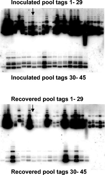
Plasmid pUHA601 was constructed for the production of M. bovis mutants by illegitimate recombination. The linearized form electroporated into M. bovis is shown in Fig. 1. The key components of this suicide fragment are a selective kanamycin resistance gene (aph) and a unique tag sequence, both situated over 300 bp from the ends of the linearized pUHA601. In M. bovis, deletion of the terminal ends of illegitimately recombining fragments frequently occurs (8), and so to achieve efficient production of antibiotic-resistant mutants containing signature tags, neither the resistance gene nor the tag was sited at the ends of the fragment. The 45 separate plasmids that were used hybridized well when probed with a pool of all 45 tags and did not hybridize when pools of half the tags were hybridized to a probe made from the remaining tags. Forty-five separate small libraries of tagged mutants of M. bovis were produced, and one mutant from each library was combined to form each pool that was inoculated into guinea pigs. A total of 1,215 mutants were screened. Previous studies with signature tag mutagenesis carried out dot blot hybridizations with probes made from mutant pools (6, 11, 25), but we obtained unacceptably high backgrounds using this approach. Blots for hybridization in this study were made by electrophoresing the plasmids on agarose gels and transferring them to nylon filters by Southern blotting. Figure 2 shows two blots of the 45 plasmids, one hybridized with a probe made from an inoculated pool and the other hybridized with a probe from a matching recovered pool, with arrows indicating the signal for a mutant that was highly underrepresented in the recovered pool.
FIG. 2.
DNA hybridization analysis of the 45 different tags from inoculated and recovered pools on filters amplified by PCR. A mutant underrepresented in the recovered pool is indicated by arrows.
Identification and characterization of attenuated mutants.
Thirty-six mutants that gave hybridization results indicating that they were highly underrepresented in recovered pools were tested individually for virulence in guinea pigs. Half of the mutants had wild-type levels of virulence, and three had a level of virulence that was substantial but less than that of the wild type. The level of virulence for the remaining 15 mutants is shown compared to their wild-type parent strain in Table 2. For comparison purposes, the virulence of an allelic-exchange mutant of M. bovis (WAg756ko) in which all of esxA (Mb3905, esat-6) and parts of esxB (Mb3904) and Mb3906 had been deleted in an earlier study (37) is also shown in Table 2. Four of the signature tag mutants gave no visible lesions in guinea pigs, a level of attenuation that we have used as a criterion for selecting strains as vaccination candidates (8). The remaining 11 mutants have been ordered in Table 2 on the basis of total number of spleen lesions. The sites of insertion in the genome for these 15 mutants were determined and are shown in Table 2. In all but one mutant, there had been a chromosomal deletion at the site of insertion, and in six mutants the deletion was substantial, ranging from 2.3 to 14.3 kb. Seven of the mutants had mutations at or near the esxA locus, while mutations in the remaining eight strains were all in different parts of the chromosome.
TABLE 2.
Analysis of attenuated strains of M. bovis produced by signature tag mutagenesisa
| Mutant strain | Size of deletion (bp) | Gene(s) disrupted or deleted | Gene information | No. of animals with lesions in:
|
Total no. of spleen lesions in 3 animals | |
|---|---|---|---|---|---|---|
| Spleen | Other organs | |||||
| WAg539 | 10,141 | Mb0453c-Mb0461 | mmpL4, mmpS4 (membrane proteins), ufaA1 (lipid, metabolism), sigK (σ factor) and unknown function | 0/3 | 0/3 | 0 |
| WAg567 | 14,310 | Mb3908-Mb3918c | Genes downstream of esxA | 0/3 | 0/3 | 0 |
| WAg568 | 3 | Mb0345c | Unknown function, upstream of aspC | 0/3 | 0/3 | 0 |
| WAg585 | 13,941 | Mb0198-Mb0207c | Unknown function | 0/3 | 0/3 | 0 |
| WAg570 | 0 | Mb3575c | Unknown function, immediately upstream of fadE28 | 1/3 | 0/3 | 1 |
| WAg572 | 34 | Mb3900 | Gene upstream of esxA | 1/3 | 0/3 | 2 |
| WAg571 | 132 | Mb2960-Mb2961 | ppsE (phenolphthiocerol synthesis) and drrA (daunorubicin resistance) | 1/3 | 0/3 | 3 |
| WAg573 | 6,876 | Mb3898-Mb3903 | Genes upstream of esxA | 2/3 | 0/3 | 5 |
| WAg574 | 5,906 | Mb3899-Mb3902 | Genes upstream of esxA | 2/3 | 0/3 | 8 |
| WAg579 | 2 | Mb2971c | pks1 (polyketide synthase) | 3/3 | 0/3 | 38 |
| WAg576 | 2,318 | Mb3897-Mb3899 | Genes upstream of esxA | 3/3 | 0/3 | 50 |
| WAg578 | 2,337 | Mb3905-Mb3907 | esxA and two downstream genes | 2/3 | 1/3 | 51 |
| WAg575 | 145 | Mb2280c | Unknown function | 2/3 | 0/3 | 52 |
| WAg540 | 33 | Mb3901 | Gene upstream of esxA | 1/3 | 1/3 | 100 |
| WAg577 | 7,678 | Mb1848-Mb1852 | secA2 (protein translocase), ilvG (acetolactate synthase), PE_PGRS, and unknown function | 3/3 | 0/3 | 125 |
| Wild type | 3/3 | 3/3 | >300 | |||
| WAg756ko | 574 | Mb3904-Mb3906 | Allelic-exchange knockout of esxA | 1/3 | 0/3 | 10 |
Mutants with interrupted genes at or near esxA are shown in boldface.
Analysis of strains with mutations at or near esxA.
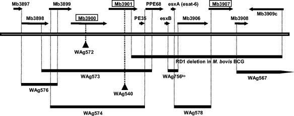
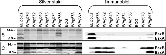
In earlier work, we showed that inactivation by allelic exchange of the esxA locus in M. bovis produced a mutant (WAg756ko, Table 2) that had greatly reduced virulence for guinea pigs (37). In this study, seven of the mutants had mutations at or near the esxA locus and varied from being moderately attenuated (WAg540) to completely avirulent (WAg567). The sites of mutation and sizes of chromosomal deletions in these seven mutants as well as in WAg756ko are shown in Fig. 3, relative to the genomic annotation of M. bovis and the RD1 deletion found in M. bovis BCG. These strains were further characterized by semiquantitative determination of their production of RNA transcripts for esxA and esxB and by determination of exported and cellular levels of EsxA and EsxB. Silver-stained polyacrylamide gels of secreted and cellular proteins are shown in Fig. 4 as well as corresponding immunoblots for EsxA (ESAT-6) and EsxB (CFP10). Immunoblot assays carried out separately for EsxA and EsxB, showed that the antibodies did not significantly cross-react with each other or other proteins (results not shown). The immunoblot results are summarized together with the results of RNA transcript analysis of esxA and esxB in Table 3. RNA transcript analysis and production of cellular EsxA and EsxB were closely related. As expected, wild-type M. bovis had both cellular and secreted EsxA and EsxB, while WAg756ko and BCG that do not contain esxA or esxB did not produce either protein. EsxA and EsxB accumulated beyond the levels found for wild-type M. bovis in the cellular fractions of WAg540, WAg567, WAg572, and WAg576, and all these strains exported little or no EsxA or EsxB. In all cases, the results for esxB RNA transcripts were similar to those for esxA, except for WAg756ko, in which esxA is completely deleted and the partial deletion of esxB is downstream of the region amplified for the RNA transcription analysis.
FIG. 3.
Annotation of genes around the esxA locus of M. bovis showing the positions of insertion and size of deletion in signature tag mutants of M. bovis. For comparison, the RD1 deletion in M. bovis BCG and the deletion of the esxA locus in an allelic-exchange mutant of M. bovis (WAg756ko) from an earlier study are also shown. Genes encoding proteins known to be involved in secretion of EsxA and EsxB are boxed.
FIG. 4.
Silver-stained polyacrylamide gels of secreted (S) and cellular (C) proteins from wild-type and mutant M. bovis strains as well as corresponding immunoblots for EsxA (ESAT-6) and EsxB (CFP10). Numbers at left are molecular masses in kilodaltons.
TABLE 3.
Levels of RNA transcription of esxA and esxB and cellular and secreted EsxA and EsxB for M. bovis strains with mutations at or near esxAa
| Strain | esxA (esat-6) RNA transcript | esxB RNA tran- script | EsxA (ESAT-6)
|
EsxB (CFP10)
|
||
|---|---|---|---|---|---|---|
| Cellular | Secreted | Cellular | Secreted | |||
| M. bovis wild type | ++ | ++ | ++ | +++ | ++ | +++ |
| WAg576 | ++ | ++ | +++ | ± | +++ | ± |
| WAg572 | ++ | ++ | +++ | − | +++ | ± |
| WAg540 | ++ | ++ | +++ | ± | +++ | ± |
| WAg573 | + | + | − | − | − | − |
| WAg574 | + | + | ± | − | + | − |
| WAg756ko | − | ++ | − | − | − | − |
| BCG | − | − | − | − | − | − |
| WAg578 | + | + | − | − | ++ | − |
| WAg567 | ++ | ++ | +++ | + | +++ | + |
Relative levels were recorded as high (+++), moderate (++), low (+), trace (±), or not detectable (−).
Vaccination efficacy of avirulent mutants.
The four mutants that gave no visible lesions when inoculated into guinea pigs were tested for their vaccine efficacy. The tests were carried out at different times, and the results for each mutant are given in Table 4 compared to control animals and animals inoculated with BCG in the same experiment. In the vaccine testing of WAg539, WAg567, and WAg568 the animals were sacrificed 5 weeks after challenge, while in the experiment with WAg585 they were sacrificed 8 weeks after challenge. WAg539 gave the best vaccination result of the four avirulent mutants and induced better protection than did BCG in all measures of vaccine efficacy. However, the differences between it and BCG were small and not significant and should be regarded as preliminary until the vaccine efficacy of WAg539 is further evaluated. At the other extreme, WAg568 gave less protection than did BCG on all measures of vaccine efficacy, and this was significant for both the numbers of spleen lesions and lung CFU. All four of these mutants grew at a rate similar to that of wild-type M. bovis in vitro, but they may have different rates of growth and survival in vivo that could be a contributing factor to these differences in vaccine efficacy.
TABLE 4.
Vaccination results of four selected M. bovis mutantsa
| Expt | M. bovis strain | No. of spleen lesions (mean ± SE) | Log10 CFU (±SE) of M. bovis in:
|
|
|---|---|---|---|---|
| Spleen | Lung | |||
| 1 | Nonvaccinated | 86 ± 21 A | 5.63 ± 0.09 A | 5.12 ± 0.13 A |
| BCG | 0.67 ± 0.49 C | 2.55 ± 0.49 B | 3.41 ± 0.30 B | |
| WAg567 | 5.2 ± 2.1 BC | 4.35 ± 0.58 AB | 4.38 ± 0.20 AB | |
| WAg568 | 20 ± 11 B | 4.79 ± 0.32 AB | 4.99 ± 0.19 A | |
| 2 | Nonvaccinated | 46 ± 10A | 4.97 ± 0.17 A | 4.22 ± 0.25 A |
| BCG | 0.25 ± 0.18 B | 1.53 ± 0.53 B | 2.36 ± 0.36 B | |
| WAg539 | 0.0 ± 0.0 B | 1.40 ± 0.30 B | 2.06 ± 0.26 B | |
| 3 | Nonvaccinated | 154 ± 40 A | 4.40 ± 0.17 A | 3.98 ± 0.44 A |
| BCG | 1.2 ± 0.6 B | 0.98 ± 0.62 B | 1.96 ± 0.58 B | |
| WAg585 | 1.0 ± 0.3 B | 2.05 ± 1.54 B | 3.39 ± 0.34 AB | |
For each experiment with nonvaccinated, BCG, and mutant WAg strain(s), values within the same column with the same letter(s) are not significantly different (P < 0.05).
DISCUSSION
Four highly attenuated mutants of M. bovis that did not produce visible lesions of tuberculosis in guinea pigs were isolated from a library of mutants by a signature tag approach combined with illegitimate recombination. One of these four mutants, WAg539, gave better measures of protection than did BCG when tested for vaccine efficacy, although the differences were small and not significant. A more robust assessment of its vaccine potential would require the use of different animal models including immunocompromised mice and the use of larger numbers of animals under conditions that provide a more stringent test of vaccine efficacy. Much further work would also be required before a vaccine candidate could be considered for human trials. This would include removal of the antibiotic resistance gene from the organism, incorporation of another deletion to enhance its safety, and extensive reversion testing. Ten genes were found to be deleted or interrupted in the 10.1-kb deletion in WAg539, including mmpL4, which was previously identified as important for virulence of M. tuberculosis from signature-tagged transposon mutagenesis with a short-term mouse model (6). Three other identified genes, mmpS4, ufaA1, and sigK, as well as unknown genes are also deleted or interrupted in WAg539, and some of these may be contributing to its loss of virulence and its vaccine properties. Of the remaining three highly attenuated mutants, two had mutations in genes of unknown function and the fourth mutant, WAg567, had a 14.3-kb deletion of all or part of 11 genes downstream of esxA.
These results illustrate a key difference between transposon mutagenesis and illegitimate recombination. Whereas in transposon mutagenesis there are insertions in the chromosome without any deletion of DNA, in illegitimate recombination there are nearly always deletions occurring at the site of insertion, and more than half of these deletions are sufficiently large to encompass multiple genes. Complete genetic analysis of these mutants with large deletions is complex, because much additional complementation and allelic-exchange work are required to determine which gene or genes are responsible for the observed phenotype. Nevertheless, the vaccination results for WAg539 are sufficiently encouraging that the role of some of the deleted genes will be further investigated by genetic methods. In this case, illegitimate recombination can be viewed as a screening method that has identified a block of genes that deserve further investigation. Another potential advantage of illegitimate recombination is that, at the present state of knowledge, it is not known which genes should be inactivated to produce a better live vaccine strain than BCG. If it transpires that two nearby inessential genes need to be inactivated to produce such an improved vaccine, then this could be revealed by illegitimate recombination but is unlikely to be easily identified by using other mutational techniques.
The most striking difference between the results of this study and the previous signature tag studies of M. tuberculosis (6, 11) was the finding that 7 of the 15 attenuated mutants had mutations at or near the esxA gene (Fig. 3). In some cases (WAg540 and WAg572) the mutations were in a single gene while in other cases several genes were deleted or interrupted, and in the most attenuated of these strains (WAg567) there was a large deletion involving the 11 genes immediately downstream from esxA. The finding that almost half the attenuated mutants had mutations in such a small region of the chromosome was unexpected, as mutations in the other eight mutants were widely scattered through the chromosome. In addition, we had previously used other methods to identify a range of attenuated illegitimate mutants of M. bovis, none of which had mutations in the esxA region (8, 39). An explanation for this disparity is that in earlier work we did not test all random mutants for virulence. Instead, a small number were selected for virulence testing from a much larger number that were screened by surrogate methods such as ability to grow in different media. This disparity illustrates the advantage of the signature tag approach, which does not make prior assumptions about what strains should be tested for virulence but instead tests all strains. One factor that may contribute to the production of so many mutants of M. bovis with mutations in this region is the known instability of the region. M. bovis BCG has a major deletion, designated RD1, in this region (Fig. 3), and different strains of Mycobacterium microti, a member of the M. tuberculosis complex that causes tuberculosis in voles, also have a variety of major deletions in this area (15). These deletions include esxA and esxB and contribute to the attenuation of these strains in mice as revealed by RD1 complementation studies (29). Removal from M. bovis of even a small part of the RD1 region that includes esxA (WAg756ko, Fig. 3) has also been shown to cause significant attenuation in guinea pigs (37). The likely reason for so many esxA-related mutants being found in this study and none being reported in the two previous studies of M. tuberculosis that employed signature tag mutagenesis is the different models that were used: an 8-week infection in guinea pigs in this study and a 3-week infection in mice in the earlier studies (6, 11). Recent work in which the entire BCG RD1 region was deleted from both M. tuberculosis and M. bovis found that, while this deletion does affect the virulence of the organisms in long-term mouse studies, it has little effect on growth of the strains in mice until 8 weeks after infection (19). These differences in results between the different studies illustrate a general feature of virulence investigations, i.e., that the results obtained are dependent on the virulence model being used.
The availability of so many M. bovis strains with different mutations in the esxA region provided an opportunity to further investigate this important locus. Earlier work had shown that EsxA is a dominant antigen in humans and other animal hosts (21, 37) and that both EsxA and EsxB are small secreted proteins that have no known secretory signal sequences (29) and are found in an associated form after secretion (26, 31). Bioinformatic analysis of the esxA region and other similar regions in M. tuberculosis (27) compared to Bacillus subtilis and a number of other bacterial species indicated that esx genes have homologs in these species and that these homologs are closely associated with putative transport genes designated yuk in B. subtilis. Subsequently, four groups have reported constructing a range of complemented mutants of BCG (30) and deletion mutants of M. tuberculosis (17, 19, 34) and have shown that these yuk homologs near esxA in M. tuberculosis (Rv3870, Rv3871, and Rv3877, which correspond to Mb3900, Mb3901, and Mb3907, respectively, in M. bovis, Fig. 3) are important for secretion of esxA and esxB. This is confirmed for Mb3900 and Mb3901 in M. bovis in the present study, where WAg572 and WAg540, respectively, have interruptions in these genes and, despite producing large amounts of EsxA and EsxB, are unable to secrete significant amounts of the proteins. With the exception of WAg567, the results for all the other mutants can be explained from what is currently known about this region. The results for WAg576 can be explained by a polar effect from the interruption in the gene immediately upstream of Mb3900 which is required for secretion. The results for WAg573 and WAg574 can be largely explained from polar effects on the nearby downstream esxB and esxA genes as well as the deletion of two genes (Mb3900 and Mb3901) required for EsxA and EsxB secretion. In addition, PPE68, which has also been shown to be involved with secretion of esxA and esxB (26), has a deletion in WAg573, and there are likely polar effects on it in WAg574. This is in contrast to a recent report that specific knockout of PPE68 had no effect on EsxA production (14). A likely explanation is that the gene was interrupted with an apramycin cassette that did not have the polar effect on esxB and esxA caused by the kanamycin cassette inserted in this study. WAg578 has a partial deletion of esxA and the secretory gene Mb3907 and produces EsxB but is unable to secrete it. WAg567 accumulates high cellular levels of EsxA and EsxB but does not secrete it at wild-type levels, even though none of the known genes required for secretion are interrupted in this mutant. While it is possible that there is a polar effect on the upstream secretory gene Mb3907, another explanation is that one of the other genes inactivated in WAg567 may also play a role in efficient secretion of EsxA and EsxB. With respect to the relative effect on virulence of different mutations in the esxA region, little further information can be drawn from the differences in number of guinea pig lesions caused by these strains (Table 2), largely because the virulence assessment was done at only one time point and in most cases there were visible lesions in only one or two of the three guinea pigs. The most attenuated strain, WAg567, which had no lesions in any animals, had a very large deletion of many genes downstream from esxA. Since this mutant secreted some EsxA and EsxB but was more attenuated than mutants that were unable to produce these proteins, the additional genes deleted in WAg567 must encode a separate virulence mechanism. Most of the genes in the deleted segment are of unknown function, but there are two serine proteases (4), one of which has been shown to be cell wall associated and expressed after macrophage infection (12). Whether these proteins are important for virulence has not been established, but many other microbial pathogens utilize proteases as virulence factors.
Apart from WAg539, which has a deletion of mmpL4, only one of the other attenuating mutations identified in this study was similar to that found in the earlier signature tag studies with M. tuberculosis (6, 11). This was the mutation in WAg571, which, although in a different gene, is in the same phthiocerol operon that was inactivated in multiple mutants in both those studies. There were two other attenuated mutants in this present study in which genes of known function were inactivated, WAg577 and WAg579. In WAg577, a segment containing part or all of five genes was deleted, including secA2, which has previously been shown to be important for M. tuberculosis virulence in mice (3) in a study that did not involve signature tag mutagenesis. WAg579 has an interrupted pks1, a gene essential for the biosynthesis of phenolic glycolipid (9). It is interesting that this causes attenuation of M. bovis in contrast to the situation in M. tuberculosis, where the gene is naturally mutated and appears to be inactive in virulent clinical strains (16). Conversely, another polyketide synthase gene, pks6, is naturally disrupted in wild-type M. bovis strains, but its disruption in M. tuberculosis was found to be an attenuation mutation in mice in one of the earlier signature tag mutagenesis studies (6). This inverse activity of pks1 and pks6 in M. bovis and M. tuberculosis may be one of the features determining pathogenic differences between these species.
This signature tag mutagenesis study differed from the two previous studies in using illegitimate recombination instead of transposon mutagenesis and an 8-week guinea pig model of M. bovis infection instead of a 3-week mouse model of M. tuberculosis infection. Many of the attenuating mutations that we identified were different from those found in the mouse studies, and all of them were different from attenuating mutations that we identified previously when employing illegitimate recombination with an in vitro screening method rather than signature tag mutagenesis (8, 39). A different approach to signature tag mutagenesis based on determining the importance for virulence in mice of virtually all the nonessential genes in M. tuberculosis has also been employed (32). The 194 genes that were identified included genes that were deleted in more than half the attenuated mutants from this study but not genes in two of the most attenuated strains, WAg539 and WAg568. This illustrates the importance of using different approaches in order to identify a wide range of different attenuating mutants. Considering that other animal models of tuberculosis could also be used and the fact that the total combined number of mutants reportedly screened in all the signature tag studies of the M. tuberculosis complex was less than 4,000, there is clearly considerable scope for further such studies of these organisms.
Acknowledgments
We thank Peter Andersen for generously supplying EsxA monoclonal antibody and EsxB antiserum.
This study was supported by a grant from the New Zealand Foundation of Research Science and Technology.
Editor: J. L. Flynn
REFERENCES
- 1.Blum, H., H. Beier, and H. J. Gross. 1987. Improved silver staining of plant proteins, RNA and DNA in polyacrylamide gels. Electrophoresis 8:93-99. [Google Scholar]
- 2.Braunstein, M., S. S. Bardarov, and W. R. Jacobs, Jr. 2002. Genetic methods for deciphering virulence determinants of Mycobacterium tuberculosis. Methods Enzymol. 358:67-99. [DOI] [PubMed] [Google Scholar]
- 3.Braunstein, M., B. J. Espinosa, J. Chan, J. T. Belisle, and W. R. Jacobs, Jr. 2003. SecA2 functions in the secretion of superoxide dismutase A and in the virulence of Mycobacterium tuberculosis. Mol. Microbiol. 48:453-464. [DOI] [PubMed] [Google Scholar]
- 4.Brown, G. D., J. A. Dave, N. C. Gey van Pittius, L. Stevens, M. R. W. Ehlers, and A. D. Beyers. 2000. The mycosins of Mycobacterium tuberculosis H37Rv: a family of subtilisin-like serine proteases. Gene 254:147-155. [DOI] [PubMed] [Google Scholar]
- 5.Brown, J. S., A. Aufauvre-Brown, J. Brown, J. M. Jennings, H. Arst, Jr., and D. W. Holden. 2000. Signature-tagged and directed mutagenesis identify PABA synthetase as essential for Aspergillus fumigatus pathogenicity. Mol. Microbiol. 36:1371-1380. [DOI] [PubMed] [Google Scholar]
- 6.Camacho, L. R., D. Ensergueix, E. Perez, B. Gicquel, and C. Guilhot. 1999. Identification of a virulence gene cluster of Mycobacterium tuberculosis by signature-tagged transposon mutagenesis. Mol. Microbiol. 34:257-267. [DOI] [PubMed] [Google Scholar]
- 7.Collins, D. M., R. P. Kawakami, G. W. de Lisle, L. Pascopella, B. R. Bloom, and W. R. Jacobs, Jr. 1995. Mutation of the principal sigma factor causes loss of virulence in a strain of the Mycobacterium tuberculosis complex. Proc. Natl. Acad. Sci. USA 92:8036-8040. [DOI] [PMC free article] [PubMed] [Google Scholar]
- 8.Collins, D. M., T. Wilson, S. Campbell, B. M. Buddle, B. J. Wards, G. Hotter, and G. W. de Lisle. 2002. Production of avirulent mutants of Mycobacterium bovis with vaccine properties by the use of illegitimate recombination and screening of stationary phase cultures. Microbiology 148:3019-3027. [DOI] [PubMed] [Google Scholar]
- 9.Constant, P., E. Perez, W. Malaga, M. A. Laneelle, O. Saurel, M. Daffe, and C. Guilhot. 2002. Role of the pks15/1 gene in the biosynthesis of phenolglycolipids in the Mycobacterium tuberculosis complex. Evidence that all strains synthesize glycosylated p-hydroxybenzoic methyl esters and that strains devoid of phenolglycolipids harbor a frameshift mutation in the pks15/1 gene. J. Biol. Chem. 277:38148-38158. [DOI] [PubMed] [Google Scholar]
- 10.Cormack, B. P., N. Ghori, and S. Falkow. 1999. An adhesin of the yeast pathogen Candida glabrata mediating adherence to human epithelial cells. Science 285:578-582. [DOI] [PubMed] [Google Scholar]
- 11.Cox, J. S., B. Chen, M. McNeil, and W. R. Jacobs, Jr. 1999. Complex lipid determines tissue-specific replication of Mycobacterium tuberculosis in mice. Nature 402:79-83. [DOI] [PubMed] [Google Scholar]
- 12.Dave, J. A., N. C. Gey van Pittius, A. D. Beyers, M. R. W. Ehlers, and G. D. Brown. 2002. Mycosin-1, a subtilisin-like serine protease of Mycobacterium tuberculosis, is cell wall-associated and expressed during infection of macrophages. BMC Microbiol. 2:30. [DOI] [PMC free article] [PubMed] [Google Scholar]
- 13.de Lisle, G. W., T. Wilson, D. M. Collins, and B. M. Buddle. 1999. Vaccination of guinea pigs with nutritionally impaired avirulent mutants of Mycobacterium bovis protects against tuberculosis. Infect. Immun. 67:2624-2626. [DOI] [PMC free article] [PubMed] [Google Scholar]
- 14.Demangel, C., P. Brodin, P. J. Cockle, R. Brosch, L. Majlessi, C. Leclerc, and S. T. Cole. 2004. Cell envelope protein PPE68 contributes to Mycobacterium tuberculosis RD1 immunogenicity independently of a 10-kilodalton culture filtrate protein and ESAT-6. Infect. Immun. 72:2170-2176. [DOI] [PMC free article] [PubMed] [Google Scholar]
- 15.Frota, C. C., D. M. Hunt, R. S. Buxton, L. Rickman, J. Hinds, K. Kremer, D. van Soolingen, and M. J. Colston. 2004. Genome structure in the vole bacillus, Mycobacterium microti, a member of the Mycobacterium tuberculosis complex with a low virulence for humans. Microbiology 150:1519-1527. [DOI] [PMC free article] [PubMed] [Google Scholar]
- 16.Garnier, T., K. Eiglmeier, J. C. Camus, N. Medina, H. Mansoor, M. Pryor, S. Duthoy, S. Grondin C. Lacroix, C. Monsempe, S. Simon, B. Harris, R. Atkin, J. Doggett, R. Mayes, L. Keating, P. R. Wheeler, J. Parkhill, B. G. Barrell, S. T. Cole, S. V. Gordon, and R. G. Hewinson. 2003. The complete genome sequence of Mycobacterium bovis. Proc. Natl. Acad. Sci. USA 100:7877-7882. [DOI] [PMC free article] [PubMed] [Google Scholar]
- 17.Guinn, K. M., M. J. Hickey, S. K. Mathur, K. L. Zakel, J. E. Grotzke, D. M. Lewinsohn, S. Smith, and D. R. Sherman. 2004. Individual RD1-region genes are required for export of ESAT-6/CFP-10 and for virulence of Mycobacterium tuberculosis. Mol. Microbiol. 51:359-370. [DOI] [PMC free article] [PubMed] [Google Scholar]
- 18.Hensel, M., J. E. Shea, C. Gleeson, M. D. Jones, E. Dalton, and D. W. Holden. 1995. Simultaneous identification of bacterial virulence genes by negative selection. Science 269:400-403. [DOI] [PubMed] [Google Scholar]
- 19.Hsu, T., S. M. Hingley-Wilson, B. Chen, M. Chen, A. Z. Dai, P. M. Morin, C. B. Marks, J. Padiyar, C. Goulding, M. Gingery, D. Eisenberg, R. G. Russell, S. C. Derrick, F. M. Collins, S. L. Morris, C. H. King, and W. R. Jacobs, Jr. 2003. The primary mechanism of attenuation of bacillus Calmette-Guerin is a loss of secreted lytic function required for invasion of lung interstitial tissue. Proc. Natl. Acad. Sci. USA 100:12420-12425. [DOI] [PMC free article] [PubMed] [Google Scholar]
- 20.Kalpana, G. V., B. R. Bloom, and W. R. Jacobs, Jr. 1991. Insertional mutagenesis and illegitimate recombination in mycobacteria. Proc. Natl. Acad. Sci. USA 88:5433-5437. [DOI] [PMC free article] [PubMed] [Google Scholar]
- 21.Lein, A. D., C. F. von Reyn, P. Ravn, C. R. Horsburgh, Jr., L. N. Alexander, and P. Andersen. 1999. Cellular immune responses to ESAT-6 discriminate between patients with pulmonary disease due to Mycobacterium avium complex and those with pulmonary disease due to Mycobacterium tuberculosis. Clin. Diagn. Lab. Immunol. 6:606-609. [DOI] [PMC free article] [PubMed] [Google Scholar]
- 22.Marmiesse, M., P. Brodin, C. Buchrieser, C. Gutierrez, N. Simoes, V. Vincent, P. Glaser, S. T. Cole, and R. Brosch. 2004. Macro-array and bioinformatic analyses reveal mycobacterial ‘core’ genes, variation in the ESAT-6 gene family and new phylogenetic markers for the Mycobacterium tuberculosis complex. Microbiology 150:483-496. [DOI] [PubMed] [Google Scholar]
- 23.McAdam, R. A., S. Quan, D. A. Smith, S. Bardarov, J. C. Betts, F. C. Cook, E. U. Hooker, A. P. Lewis, P. Woollard, M. J. Everett, P. T. Lukey, G. J. Bancroft, W. R. Jacobs, Jr., and K. Duncan. 2002. Characterization of a Mycobacterium tuberculosis H37Rv transposon library reveals insertions in 351 ORFs and mutants with altered virulence. Microbiology 148:2975-2986. [DOI] [PubMed] [Google Scholar]
- 24.Mecsas, J. 2002. Use of signature-tagged mutagenesis in pathogenesis studies. Curr. Opin. Microbiol. 5:33-37. [DOI] [PubMed] [Google Scholar]
- 25.Mei, J. M., F. Nourbakhsh, C. W. Ford, and D. W. Holden. 1997. Identification of Staphylococcus aureus virulence genes in a murine model of bacteraemia using signature-tagged mutagenesis. Mol. Microbiol. 26:399-407. [DOI] [PubMed] [Google Scholar]
- 26.Okkels, L. M., and P. Andersen. 2004. Protein-protein interactions of proteins from the ESAT-6 family of Mycobacterium tuberculosis. J. Bacteriol. 186:2487-2491. [DOI] [PMC free article] [PubMed] [Google Scholar]
- 27.Pallen, M. J. 2002. The ESAT-6/WXG100 superfamily and a new Gram-positive secretion system? Trends Microbiol. 10:209-212. [DOI] [PubMed] [Google Scholar]
- 28.Pascopella, L., F. M. Collins, J. M. Martin, M. H. Lee, G. F. Hatfull, C. K. Stover, B. R. Bloom, and W. R. Jacobs, Jr. 1994. Use of in vivo complementation in Mycobacterium tuberculosis to identify a genomic fragment associated with virulence. Infect. Immun. 62:1313-1319. [DOI] [PMC free article] [PubMed] [Google Scholar]
- 29.Pym, A. S., P. Brodin, R. Brosch, M. Huerre, and S. T. Cole. 2002. Loss of RD1 contributed to the attenuation of the live tuberculosis vaccines Mycobacterium bovis BCG and Mycobacterium microti. Mol. Microbiol. 46:709-717. [DOI] [PubMed] [Google Scholar]
- 30.Pym, A. S., P. Brodin, L. Majlessi, R. Brosch, C. Demangel, A. Williams, K. E. Griffiths, G. Marchal, C. Leclerc, and S. T. Cole. 2003. Recombinant BCG exporting ESAT-6 confers enhanced protection against tuberculosis. Nat. Med. 9:533-539. [DOI] [PubMed] [Google Scholar]
- 31.Renshaw, P. S., P. Panagiotidou, A. Whelan, S. V. Gordon, R. G. Hewinson, R. A. Williamson, and M. D. Carr. 2002. Conclusive evidence that the major T-cell antigens of the Mycobacterium tuberculosis complex ESAT-6 and CFP-10 form a tight, 1:1 complex and characterization of the structural properties of ESAT-6, CFP-10, and the ESAT-6*CFP-10 complex. Implications for pathogenesis and virulence. J. Biol. Chem. 277:21598-21603. [DOI] [PubMed] [Google Scholar]
- 32.Sassetti, C. M., and E. J. Rubin. 2003. Genetic requirements for mycobacterial survival during infection. Proc. Natl. Acad. Sci. USA 100:12989-12994. [DOI] [PMC free article] [PubMed] [Google Scholar]
- 33.Schowalter, D. B., and S. S. Sommer. 1989. The generation of radiolabeled DNA and RNA probes with polymerase chain reaction. Anal. Biochem. 177:90-94. [DOI] [PubMed] [Google Scholar]
- 34.Stanley, S. A., S. Raghavan, W. W. Hwang, and J. S. Cox. 2003. Acute infection and macrophage subversion by Mycobacterium tuberculosis require a specialized secretion system. Proc. Natl. Acad. Sci. USA 100:13001-13006. [DOI] [PMC free article] [PubMed] [Google Scholar]
- 35.van Soolingen, D., P. W. M. Hermans, P. E. W. de Haas, D. R. Soll, and J. D. van Embden. 1991. Occurrence and stability of insertion sequences in Mycobacterium tuberculosis complex strains: evaluation of an insertion-sequence dependent DNA polymorphism as a tool in the epidemiology of tuberculosis. J. Clin. Microbiol. 29:2578-2586. [DOI] [PMC free article] [PubMed] [Google Scholar]
- 36.Wards, B. J., and D. M. Collins. 1996. Electroporation at elevated temperatures substantially improves transformation efficiency of slow-growing mycobacteria. FEMS Microbiol. Lett. 145:101-105. [DOI] [PubMed] [Google Scholar]
- 37.Wards, B. J., G. W. de Lisle, and D. M. Collins. 2000. An esat6 knockout mutant of Mycobacterium bovis produced by homologous recombination will contribute to the development of a live tuberculosis vaccine. Tubercle Lung Dis. 80:185-189. [DOI] [PubMed] [Google Scholar]
- 38.Wilson, T. M., G. W. de Lisle, and D. M. Collins. 1995. Effect of inhA and katG on isoniazid resistance and virulence of Mycobacterium bovis. Mol. Microbiol. 15:1009-1015. [DOI] [PubMed] [Google Scholar]
- 39.Wilson, T., B. J. Wards, S. J. White, B. Skou, G. W. de Lisle, and D. M. Collins. 1997. Production of avirulent Mycobacterium bovis strains by illegitimate recombination with DNA fragments containing an interrupted ahpC gene. Tubercle Lung Dis. 78:229-235. [DOI] [PubMed] [Google Scholar]


