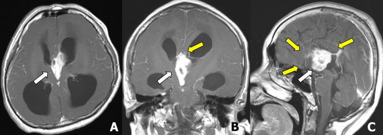
Figure 1. Initial brain magnetic resonance imaging.
Contrast-enhanced T1-weighted images show an intraventricular mass in the third ventricle to the anterior horn of the left lateral ventricle (A-C; white arrow) with hydrocephalus. Corpus callosum agenesis is noted in the coronal and sagittal images (B and C; yellow arrows).

