Abstract
The superantigen-encoding ypm gene and the pil gene cluster governing type IV pilus biogenesis have been laterally acquired by Yersinia pseudotuberculosis. PCR assays on 270 unrelated strains from various environmental and animal sources revealed a significant association of ypm and pil in isolates.
To date, Yersinia pseudotuberculosis is the only known gram-negative bacterium that synthesizes a superantigenic toxin (YPM), a protein which strongly stimulates the proliferation of polyclonal T lymphocytes (1). YPM is a distinct member of the bacterial superantigen family (5) in that (i) its molecular mass (14 kDa) is much lower than that of superantigens produced by Mycoplasma arthritidis and the gram-positive species Staphylococcus aureus and Streptococcus pyogenes (22 to 29 kDa) and (ii) it does not show significant amino acid similarity to other proteins. The three-dimensional structure of YPM has been recently established and the closest structural neighbors found, besides members of the tumor necrosis factor superfamily, were viral capsid proteins (10). Genes coding for bacterial superantigens are frequently located within mobile genetic elements in general and within bacteriophages in particular (16). Two features strongly suggest that Y. pseudotuberculosis horizontally acquired the superantigen-encoding gene: ypm is not distributed in all strains of the species and its G+C content is significantly lower than that of the genomic core (35 versus 47%) (5, 19). We have reported that ypm is present on the Y. pseudotuberculosis chromosome, 245 bp downstream of a 26-bp sequence (called yrs) which is homologous to dif (4), a site-specific recombination target used by filamentous bacteriophages for host chromosome integration (14). However, we failed to detect any phage remnants in the vicinity of this nucleotide motif (4). The yrs site is also present on the chromosome of Yersinia pestis, a ypm-negative species derived from Y. pseudotuberculosis (4), and strikingly, it is surrounded by filamentous phage-like (CUS-2) genes (4, 12). Therefore, one can reasonably speculate that a bacteriophage was involved in the incorporation of ypm into Y. pseudotuberculosis.
Fimbriae (and especially type IV pili) may serve as bacteriophage receptors at the bacterial cell surface (2, 3, 13, 15, 17, 18). We recently reported the presence in Y. pseudotuberculosis of an 11-kb, 11-gene pil locus (pilLMNOPQRSUVW) that encodes a type IV pilus (7). It is located in the 5′ part of a large (98-kb) pathogenicity island (PAI) called YAPI (6). Like ypm, the pil gene cluster is not present in all Y. pseudotuberculosis strains (7). The aim of the present work was to analyze ypm and pil association in a large collection of isolates, in order to determine the potential role of type IV pili in the emergence of ypm in Y. pseudotuberculosis.
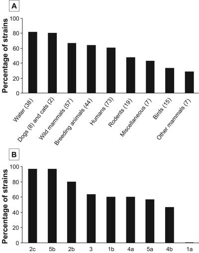
Thirty strains from each of the nine most frequent O serotypes (1a, 1b, 2b, 2c, 3, 4a, 4b, 5a, and 5b) were randomly chosen from a collection of 2,235 strains previously typed for presence of the ypm gene and the high-pathogenicity island (HPI) (11). The strains had been isolated from various environmental and animal sources (Fig. 1A). One hundred ninety-six of the 270 selected strains originated from Asia (mainly Japan with 188 strains, but also Korea and China). The remainder were collected from Europe (including Russia), America, and Oceania, with an unknown geographical origin for just two strains. Screening of pil segments was performed by PCR analysis as previously described, with primer sets 1 (forward, 5′ TATGTTGCTGGAGGCTCAG 3′; reverse, 5′ GCGAACTATCAGCTATACG 3′) and 2 (forward, 5′ GCAGGTTATTGTTGCTCCT 3′; reverse, 5′ GTCGTGGTATCACTGAAGC 3′) amplifying fragments of pilPQ (569 bp) and pilSUV (1223 bp), respectively (6). Amplimers were analyzed by agarose gel electrophoresis. One hundred sixty-eight strains (62.2% of the total) were found to be pil positive and all generated an amplification product of the expected size with each primer set. As shown in Fig. 1, pil genes were detected in strains from a broad natural reservoir, with a notably high frequency (≈80%) for those recovered from water. PCR analyses were negative for all O:1a strains (which are mainly isolated in European countries [11]), whereas the non-O:1a strains yielded amplimers with a frequency that varied according to the O serotype and ranged from 46.7% (O:4b) to 96.7% (O:2c and O:5b). Like ypm (19), pil genes were predominantly distributed in isolates from Asian rather than non-Asian countries (74.5 versus 29.2%, P < 10−7). However, in light of the geographical disparity of the O serotypes, this difference should be interpreted with caution. Indeed, O serotypes with the highest proportion of pil-positive strains (O:2c, O:5b, and to a lesser extent O:2b) were those with the highest percentages of Asian isolates (100, 100, and 82.8%, respectively). We next examined the distribution of pil and ypm genes in the Y. pseudotuberculosis collection (Table 1). Of the 168 pil-positive strains, 135 (80.4%) contained ypm, whereas the gene was detected in only 46 out of 102 (45.1%) pil-negative strains (odds ratio [OR], 4.98; 95% confidence interval [95% CI], 2.79 to 8.93; P < 10−7). Statistical tests also showed that pil genes were more often than not associated with ypm in both Asian and non-Asian isolates (OR, 2.52; 95% CI, 1.12 to 5.63; P = 0. 013; and OR, 4.33; 95% CI, 1.30 to 14.80; P = 0.005; respectively). In contrast, the HPI was present in 19 out of 168 (11.3%) pil-positive strains and in 46 out of 112 (41.1%) pil-negative strains (OR, 0.25; 95% CI, 0.16 to 0.40; P < 10−7). The lower frequency of the HPI in Pil+ Y. pseudotuberculosis strongly argues that these strains have no tendency to accumulate virulence genes nor to laterally acquire mobile genetic elements more readily.
FIG. 1.
pil distribution in Y. pseudotuberculosis strains in relation to their ecological niche (A) and O-antigen type (B). The number of strains in each class is given in parentheses. Water sources: mountain water (20), well water (5), river water (2), unknown origin (11). Wild mammal sources: raccoon dog (32), marten (7), hare (6), deer (5), reindeer (2), fox (2), mole (2), wild boar (1). Breeding animal sources: pig (29), rabbit (8), ruminant (7). Rodent sources: wild mouse (11), guinea pig (2), house rat (4), wild rat (1), unspecified (1). Miscellaneous sources: fish (2), plant (2), soil (1), unknown (2). Bird sources: pet birds (8), duck (2), pigeon (2), unspecified (3). Other mammal sources: monkey (3), goat (2), horse (2).
TABLE 1.
Distribution of pil and ypm genes in 270 Y. pseudotuberculosis strains
| Genotype | No. of strains (%)
|
|||
|---|---|---|---|---|
| Total | Asian origin | Non-Asian origin | Unknown area origin | |
| pil-positive ypm-positive | 135 (80.4) | 123 (84.2) | 12 (57.1) | |
| pil-positive ypm-negative | 33 (19.6) | 23 (15.8) | 9 (42.9) | 1 |
| pil-negative ypm-positive | 46 (45.1) | 34 (68.0) | 12 (23.5) | |
| pil-negative ypm-negative | 56 (54.9) | 16 (32.0) | 39 (76.5) | 1 |
The present work thus establishes that pil and ypm are specifically associated in Y. pseudotuberculosis. pil genes are present in both Y. pseudotuberculosis and Yersinia enterocolitica (8) and were probably acquired by the common Yersinia ancestor through PAI (YAPI) transfer (F. Collyn, C. A. Roten, L. Guy, M. Simonet, and M. Marceau, submitted for publication). In contrast, ypm is harbored by Y. pseudotuberculosis but not by Y. enterocolitica (5) and the gene may have arisen (most probably by transduction) after divergence from the Yersinia progenitor. It is therefore tempting to propose an evolutionary scenario for the origin of Y. pseudotuberculosis superantigen producers reminiscent of that suggested for the emergence of enterotoxinogenic Vibrio cholerae (9). Firstly, the Yersinia would have acquired the pil operon (the counterpart in V. cholerae is the PAI [VPI]-borne tcp operon) via YAPI transfer before it speciated, and secondly, some Pil+ strains would have been infected and lysogenized by an ypm-encoding prophage (the counterpart in V. cholerae is a filamentous temperate phage CTXφ encoding cholera toxin) using type IV pili as receptors. A previous in vitro study demonstrated that the PAI can be lost from Y. pseudotuberculosis (6), and nonproducers and superantigen producers lacking the pil operon would most probably result from spontaneous excision of YAPI. Characterization of the phage family that may have transferred ypm is a prerequisite step for validating the proposed evolutionary model. However, since there are no prophage remnants close to ypm (4), this identification cannot be driven using a helper bacteriophage. Comparative genometric analysis of the ypm locus with streptococcal and staphylococcal superantigen-encoding genes (especially those of phage origin) represents an alternative way of shedding light on this latter point.
Acknowledgments
F. Collyn received a doctoral studentship from the Ministère de l'Enseignement Supérieur, de la Recherche et de la Technologie and from the Fondation pour la Recherche Médicale. This work was supported in part by the European Regional Development Fund.
Editor: J. B. Bliska
REFERENCES
- 1.Abe, J., T. Takeda, Y. Watanabe, H. Nakao, N. Kobayashi, D. Y. Leung, and T. Kohsaka. 1993. Evidence for superantigen production by Yersinia pseudotuberculosis. J. Immunol. 151:4183-4188. [PubMed] [Google Scholar]
- 2.Bradley, D. E. 1973. A pilus-dependent Pseudomonas aeruginosa bacteriophage with a long noncontractile tail. Virology 51:489-492. [DOI] [PubMed] [Google Scholar]
- 3.Budzik, J. M., W. A. Rosche, A. Rietsch, and G. A. O'Toole. 2004. Isolation and characterization of a generalized transducing phage for Pseudomonas aeruginosa strains PAO1 and PA14. J. Bacteriol. 186:3270-3273. [DOI] [PMC free article] [PubMed] [Google Scholar]
- 4.Carnoy, C., S. Floquet, M. Marceau, F. Sebbane, S. Haentjens-Herwegh, A. Devalckenaere, and M. Simonet. 2002. The superantigen gene ypm is located in an unstable chromosomal locus of Yersinia pseudotuberculosis. J. Bacteriol. 184:4489-4499. [DOI] [PMC free article] [PubMed] [Google Scholar]
- 5.Carnoy, C., and M. Simonet. 1999. Yersinia pseudotuberculosis superantigenic toxins, p. 611-622. In J. E. Alouf and J. H. Freer (ed.), Bacterial protein toxins: a comprehensive sourcebook, 2nd ed. Academic Press, London, United Kingdom.
- 6.Collyn, F., A. Billault, C. Mullet, M. Simonet, and M. Marceau. 2004. YAPI, a new Yersinia pseudotuberculosis pathogenicity island. Infect. Immun. 72:4784-4790. [DOI] [PMC free article] [PubMed] [Google Scholar]
- 7.Collyn, F., M. A. Léty, S. Nair, V. Escuyer, A. Ben Younes, M. Simonet, and M. Marceau. 2002. Yersinia pseudotuberculosis harbors a type IV pilus gene cluster that contributes to pathogenicity. Infect. Immun. 70:6196-6205. [DOI] [PMC free article] [PubMed] [Google Scholar]
- 8.Collyn, F., M. Marceau, and M. Simonet. 2004. YAPI, a new pathogenicity island in enteropathogenic yersiniae, p. 307-317. In E. Carniel and B. J. Hinnebusch (ed.), Yersinia molecular and cellular biology. Horizon Biosciences, Wymondham, Norfolk, United Kingdom.
- 9.Davis, B. M., and M. K. Waldor. 2003. Filamentous phages linked to virulence of Vibrio cholerae. Curr. Opin. Microbiol. 6:35-42. [DOI] [PubMed] [Google Scholar]
- 10.Donadini, R., C. W. Liew, A. H. Kwan, J. P. Mackay, and B. A. Fields. 2004. Crystal and solution structures of a superantigen from Yersinia pseudotuberculosis reveal a jelly-roll fold. Structure (Cambridge) 12:145-156. [DOI] [PubMed] [Google Scholar]
- 11.Fukushima, H., Y. Matsuda, R. Seki, M. Tsubokura, N. Takeda, F. N. Shubin, I. K. Paik, and X. B. Zheng. 2001. Geographical heterogeneity between Far Eastern and Western countries in prevalence of the virulence plasmid, the superantigen Yersinia pseudotuberculosis-derived mitogen, and the high-pathogenicity island among Yersinia pseudotuberculosis strains. J. Clin. Microbiol. 39:3541-3547. [DOI] [PMC free article] [PubMed] [Google Scholar]
- 12.Gonzalez, M. D., C. A. Lichtensteiger, R. Caughlan, and E. R. Vimr. 2002. Conserved filamentous prophage in Escherichia coli O18:K1:H7 and Yersinia pestis biovar orientalis. J. Bacteriol. 184:6050-6055. [DOI] [PMC free article] [PubMed] [Google Scholar]
- 13.Horiuchi, T., and T. Komano. 1998. Mutational analysis of plasmid R64 thin pilus prepilin: the entire prepilin sequence is required for processing by type IV prepilin peptidase. J. Bacteriol. 180:4613-4620. [DOI] [PMC free article] [PubMed] [Google Scholar]
- 14.Huber, K. E., and M. K. Waldor. 2002. Filamentous phage integration requires the host recombinases XerC and XerD. Nature 417:656-659. [DOI] [PubMed] [Google Scholar]
- 15.Mindich, L., X. Qiao, J. Qiao, S. Onodera, M. Romantschuk, and D. Hoogstraten. 1999. Isolation of additional bacteriophages with genomes of segmented double-stranded RNA. J. Bacteriol. 181:4505-4508. [DOI] [PMC free article] [PubMed] [Google Scholar]
- 16.Muller-Alouf, H., C. Carnoy, M. Simonet, and J. E. Alouf. 2001. Superantigen bacterial toxins: state of the art. Toxicon 39:1691-1701. [DOI] [PubMed] [Google Scholar]
- 17.Roine, E., D. N. Nunn, L. Paulin, and M. Romantschuk. 1996. Characterization of genes required for pilus expression in Pseudomonas syringae pathovar phaseolicola. J. Bacteriol. 178:410-417. [DOI] [PMC free article] [PubMed] [Google Scholar]
- 18.Waldor, M. K., and J. J. Mekalanos. 1996. Lysogenic conversion by a filamentous phage encoding cholera toxin. Science 272:1910-1914. [DOI] [PubMed] [Google Scholar]
- 19.Yoshino, K., T. Ramamurthy, G. B. Nair, H. Fukushima, Y. Ohtomo, N. Takeda, S. Kaneko, and T. Takeda. 1995. Geographical heterogeneity between Far East and Europe in prevalence of ypm gene encoding the novel superantigen among Yersinia pseudotuberculosis strains. J. Clin. Microbiol. 33:3356-3358. [DOI] [PMC free article] [PubMed] [Google Scholar]


