Abstract
In our previous study, virus-binding proteins (VBPs) demonstrating the ability to strongly bind poliovirus type 1 (PV1) were recovered from a bacterial culture derived from activated sludge. The isolated VBPs would be useful as viral adsorbents for water and wastewater treatments. The VBP gene of activated sludge bacteria was isolated, and the cloning system of the VBP was established. The isolation of the VBP gene from DNA libraries for activated sludge bacteria was achieved with the colony hybridization technique. The sequence of the VBP gene consisted of 807 nucleotides encoding 268 amino acids. Fifteen amino acid sequences were retrieved from 2,137,877 sequences by a homology search using the BLAST server at the National Center for Biotechnology Information. The protein encoded in the isolated genome was considered to be a newly discovered protein from activated sludge culture, because any sequences in protein databases were not perfectly matched with the sequence of the VBP. It was confirmed that Escherichia coli BL21 transformed by pRSET carrying the isolated VBP gene could extensively produce the VBP clones. Enzyme-linked immunosorbent assay (ELISA) revealed that the VBP clone exhibited the binding ability with intact particles of PV1. The equilibrium binding constant between PV1 and VBP in the ELISA well was estimated to be 2.1 × 10⁷ (M−1), which also indicated that the VBP clones have a high affinity with the PV1 particle. The VBP cloning system developed in this study would make it possible to produce a mass volume of VBPs and to utilize them as a new material of the specific adsorbent in several technologies, including virus removal, concentration, and detection.
Waterborne infectious diseases caused by pathogenic viruses such as enteroviruses, adenoviruses, rotaviruses, and noroviruses have been frequently reported even in the developed countries, where hygiene conditions have been considerably improved (5-7, 12, 21, 26, 27). These pathogenic viruses can survive for a long period in water environments and transmit to humans (7, 10), so a public health hazard is posed by the spread of pathogenic viruses in water environments. Furthermore, the subsistence of these viruses in our society would raise concerns about viruses with virulent phenotypes by mutation or recombination, as has been reported with respect to vaccine-derived polioviruses (8, 15, 20) and the other several RNA viruses (22-24, 38). In order to prevent pathogenic viral contamination of the water environment, it would be significant to remove or inactivate pathogenic viruses in water and wastewater treatment processes. However, the conventional water and wastewater treatment systems have difficulty in removing or inactivating viruses. It is well known that the filtration process, the most fundamental technology in the water treatment system, is not operative in removing viruses (18). Several viruses have a higher resistance to disinfectants than indicator microorganisms (28). A new technology for virus removal needs to be developed in order to prevent the spread of pathogenic viruses in the water environment.
In our previous study, virus-binding proteins (VBPs) were successfully recovered from a bacterial culture derived from activated sludge by their affinity with the capsid peptide of poliovirus type 1 (PV1) (29). The isolated VBPs demonstrated the ability to strongly adsorb intact particles of PV1. Since the VBPs were expected to be stable under the conditions of water and wastewater treatments, it would be possible to utilize the VBP as a specific viral adsorbent in several technologies, including virus removal, concentration, and detection.
The objectives of this study were to isolate VBP genes from activated sludge bacteria and to construct the VBP cloning system in order to produce a mass volume of VBP clones. First, the DNA library for the activated sludge bacteria was constructed. Next, VBP genes were isolated from the constructed DNA library with DNA probes by the colony hybridization technique. The isolated gene of the VBP was introduced into a vector for protein cloning which was used for the transformation of Escherichia coli BL21. VBP clones were produced by the transformed E. coli BL21, and the ability of VBP clones to bind PV1 particles was confirmed. Finally, the equilibrium binding constant between PV1 and VBP was estimated, and the availability of the VBP as a specific viral adsorbent was discussed.
MATERIALS AND METHODS
Construction of the genomic DNA library for activated sludge bacteria.
Bacterial culture was developed from activated sludge as described previously (29). One hundred microliters of the bacterial culture derived from activated sludge was suspended in 2 ml of 50 mM EDTA in 50 mM Tris-HCl (pH 8.0). The suspension was frozen at −80°C for 10 min and thawed at 37°C. Two hundred microliters of 10 mg/ml of lysozyme (catalog no. 1.05281.0001; Merck, Darmstadt, Germany) in 0.25 M Tris-HCl (pH 8.0) was added to each tube, and cell suspensions were incubated at 37°C for 30 min. After the incubation, 400 μl of STEP solution (0.5% sodium dodecyl sulfate [SDS], 0.4 M EDTA, 1 mg/ml of proteinase K in 50 mM Tris-HCl [pH 7.5]) was added to each tube and stirred vigorously. The cell suspension was then incubated at 50°C for 60 min. After the incubation, 400 μl of TE solution (1 mM EDTA in 10 mM Tris-HCl [pH 8.0]) was added to each tube and mixed well. Three milliliters of a mixture of phenol and chloroform (phenol:chloroform, 1:1) were added and shaken by hand vigorously for 30 s. The cell suspension was then centrifuged at 10,000 × g at room temperature for 15 min with a centrifugal machine (catalog no. 6900; Kubota, Tokyo, Japan). The supernatant was transferred to other centrifugal tubes. The equivalent volume of 100% ethanol (chilled at −80°C) was added, mixed well, and centrifuged at 10,000 × g at 4°C for 20 min. The supernatant was decanted, and the pellet (extracted genomic DNA) was dissolved in 500 μl of TE solution. Next, 10 μl of 10 mg/ml of DNase-free RNase A (catalog no. 70194Z; Amersham Biosciences) was added to each tube, mixed well, and incubated at 37°C for 10 min. The equivalent volume of the mixture of phenol and chloroform (1:1) was added, mixed well, and centrifuged at 10,000 × g at room temperature for 15 min. The supernatant (400 μl) was transferred to a new tube. Forty microliters of 3 M sodium acetate (pH 5.2) and 880 μl of 100% ethanol (chilled at −80°C) were added, mixed well, and centrifuged at 10,000 × g at 4°C for 20 min. The supernatant was removed, and 400 μl of 80% ethanol (chilled at −80°C) was added. Samples were then mixed lightly and centrifuged at 10,000 × g at 4°C for 3 min. The supernatant was removed, and the pellet (extracted genomic DNA) was dissolved in 100 μl of TE solution and stored at −20°C.
The obtained genomic DNA mixture was fragmented with MboI and then separated with a 0.7% agarose gel (catalog no. 5033; TaKaRa, Shiga, Japan), followed by the extraction of DNA fragments of between 1 and 3 kbp in length by using a GENECLEAN II kit (catalog no. GL-1131-05; BIO 101 Systems, Qbiogene Inc., Irvine, CA). The fragments were ligated into the BamHI site of a plasmid vector, pUC118 (catalog no. 3321; TaKaRa, Shiga, Japan) by using a DNA Ligation kit, version 2 (catalog no. 6022; TaKaRa, Shiga, Japan). E. coli DH5α (catalog no. 9057; TaKaRa, Shiga, Japan) was transformed with the ligated plasmid, and transformants were plated onto Luria-Bertani (LB) medium containing 0.7% agar with X-Gal (5-bromo-4-chloro-3-indolyl-β-d-galactopyranoside) (catalog no. 027-07854; Wako, Tokyo, Japan).
Construction of the DNA probe for isolation of VBP genomes.
The DNA probe for isolation of VBP genomes was constructed based on the N-terminal sequences of VBP (29). The sequence of the constructed DNA probe is 5′-GAYATHCARAARAAYGAYTAYAARTGGTTYCARTTYAAY-3′. In the probe sequence, Y means thymine or cytosine, R means guanine or adenine, and H means adenine, thymine, or cytosine. This probe was modified by digoxigenin at the 5′ end.
Colony hybridization for isolation of VBP genomes.
Screening for the VBP gene was carried out by the colony hybridization technique. At first, colonies of E. coli DH5α transformed with the plasmid pUC118 containing genomic DNA fragments of activated sludge bacteria were transferred onto a nitrocellulose membrane filter (catalog no. 1 417 240; Roche Diagnostics GmbH, Mannheim, Germany). Plasmids in transformants were denaturated with 0.5 M NaOH including 1.5 M NaCl for 15 min and then neutralized with 1.5 M NaCl in 1 M Tris-HCl (pH 7.5) twice for 5 min each. After incubation in 2 mM Tris-HCl for 15 min at room temperature, DNA on the membrane filter was immobilized at 80°C for 2 h. The immobilized DNA was then hybridized in a solution of the digoxigenin-modified probe overnight at 40°C. The filter was washed twice at 40°C for 15 min in a 0.1× SSC (1× SSC is 0.15 M NaCl plus 0.015 M sodium citrate) and 0.1% SDS solution and colored with coloring reagent (catalog no. 1 093 657; Roche Diagnostics GmbH, Mannheim, Germany).
DNA sequencing of isolated genomes.
The sequences of the isolated genomes with the colony hybridization were analyzed by the direct sequencing of both strands of genomes with the ABI PRISM310 genetic analyzer (Applied Biosystems Japan Ltd., Tokyo, Japan) according to the BigDye Terminator cycle sequencing kit protocol. Plasmid in E. coli was extracted with Mini Prep (catalog no. 9085; TaKaRa, Shiga, Japan) and used for the template of the sequencing reaction, which was carried out according to the manufacturer's instructions. M13 primer M1 (5′-AGTCACGACGTTGTA-3′) and M13 primer RV (5′-CAGGAAACAGCTATGAC-3′) were used as the sequencing primers in the PCR cycle sequencing. Homology searches were conducted using the BLAST server at the National Center for Biotechnology Information (Bethesda, MD).
Construction of the cloning vector for the VBP.
The isolated gene was amplified by primers containing the EcoRI site at the 5′ end and the XhoI site at the 3′ end, respectively. PCR master (catalog no. 1 636 103; Roche Diagnostics GmbH, Mannheim, Germany) was used for the DNA amplification. The PCR profile was run with 1 cycle of 94°C for 10 min; 40 cycles of 94°C for 1 min, 50°C for 1 min, and 72°C for 1 min; 1 cycle of 72°C for 15 min; and 1 cycle of 20°C for 5 min.
The PCR product and a protein cloning vector, pRSET (Invitrogen Corporation, Carlsbad, CA), were simultaneously digested by EcoRI (catalog no. 1040A; TaKaRa, Shiga, Japan) and XhoI (catalog no. 1094A; TaKaRa, Shiga, Japan) and purified by phenol-chloroform (1:1). After precipitation with ethanol, the PCR product was processed for electrophoresis on a 0.7% agarose gel. The DNA fragment was extracted from the gel and used for the ligation. The digested vector was also precipitated with ethanol, dissolved in TE solution, and dephosphorylated with bacterial alkaline phosphatase (E. coli C75, catalog no. 2120B; TaKaRa, Shiga, Japan). The PCR product was then ligated into the dephosphorylated plasmid with DNA Ligation kit, version 2. The ligation product was used to transform the E. coli strain DH5α. DNA clones containing the isolated gene were identified by analyzing restriction enzyme (EcoRI) digestion after the extraction of the plasmid with Mini Prep. Furthermore, it was confirmed whether the isolated gene was appropriately conjugated with pRSET by sequencing the conjugation region using T7 primer (5′-TAATACGACTCACTATAGGG-3′) as described above.
Production and purification of the VBP.
The plasmid containing the isolated gene was used to transform E. coli strain BL21 (catalog no. NV470; TaKaRa, Shiga, Japan). The recombinant protein was expressed by E. coli BL21 as a fusion protein linked to a hexahistidine peptide at the N terminus. A preculture containing the expression plasmid was grown overnight at 37°C in LB medium with ampicillin (50 mg/ml) and used to inoculate (1/50) LB medium. The culture was grown for 4 h at 37°C, and protein expression was induced by the addition of isopropyl-β-d-thiogalactopyranoside (IPTG) (catalog no. 9030; TaKaRa, Shiga, Japan) to a final concentration of 1 mM. After 5-h growth at 37°C, the cells were immediately collected by centrifugation at 10,000 × g for 10 min at 4°C. The extraction of soluble proteins and the purification of the inclusion body were performed by BugBuster (catalog no. 70584-3; Novagen, Darmstadt, Germany). The insoluble proteins in the purified inclusion body were dissolved with a lysis buffer (6 M urea, 1 M NaCl in 20 mM phosphate buffer, pH 7.8). Extracted proteins were purified by using a HIS-BIND purification kit (catalog no. 70239-3; Novagen, Darmstadt, Germany) according to the manufacturer's instructions.
Protein assays.
Samples were mixed in an equal volume of the denaturation buffer (6 M urea, 2.5% SDS, 1% β-mercaptoethanol, 0.05 M Tris-HCl, pH 6.8, 0.01% bromophenol blue, 3 mM EDTA, and 10% glycerol) and heated for 5 min at 95°C. A 10% polyacrylamide gel was used to evaluate the level of protein expression and the relative molecular weight and to analyze the homogeneity of purified fractions. Protein bands were visualized by Coomassie brilliant blue (CBB) staining or silver staining.
Evaluation of virus-binding ability of the VBP with ELISA.
The virus-binding ability of the VBP candidate was assayed with enzyme-linked immunosorbent assay (ELISA). Prior to the assay, the solvent of the fusion protein was changed as described previously (29). The fusion protein in the pellet was dissolved in 50 mM carbonate buffer and used for the evaluation of virus-binding ability by ELISA using infectious particles of PV1 as described previously (29).
Estimation of the equilibrium binding constant between PV1 and VBP.
The equilibrium binding constant between PV1 and the immobilized VBP on ELISA wells was estimated as follows. Wells of an ELISA plate were covered with the VBP and blocked by bovine serum albumin (BSA). Unbound BSA was washed away, and PV1 in phosphate-buffered saline was inoculated into each well to adsorb to the immobilized VBP. Plates were incubated at room temperature for 60 min. After the incubation, the supernatant in each well was sampled, and the PFU of the supernatant was determined by the plaque method using the Buffalo green monkey (BGM) kidney cell line (36). The viral concentration in the sample before the inoculation was also obtained with the plaque method. The equilibrium binding constant between PV1 and VBP on the ELISA well was calculated by the following equation (equation 1):
 |
(1) |
where subscripts a and b represent before and after the incubation, respectively. The free-VBP stands for the VBP in the ELISA well which was not occupied by the PV1 particle. This concentration of free VBP was approximated by the total concentration of the immobilized VBP in the ELISA well, because the concentration of PV1 used in this test was much lower than that of the immobilized VBP. The concentration of the VBP was determined by the DC protein assay kit (catalog no. 500-0116; Bio-Rad, CA).
RESULTS
Construction of a DNA library for the activated sludge bacteria.
The transformation efficiency of E. coli DH5α with pUC118 carrying fractionated DNA of activated sludge bacteria was about 100 positive clones per microliter of ligation mixture as assayed on an LB agar plate. The constructed library consisted of 1,825 clones. In order to estimate the insert size, 10 clones were randomly selected and grown for plasmid preparation. Inserts were released from pUC118 by BamHI restriction and analyzed by 0.7% agarose gel electrophoresis. The average insert size was 2 kb with a range of 1 to 3 kb.
Isolation of the VBP genome.
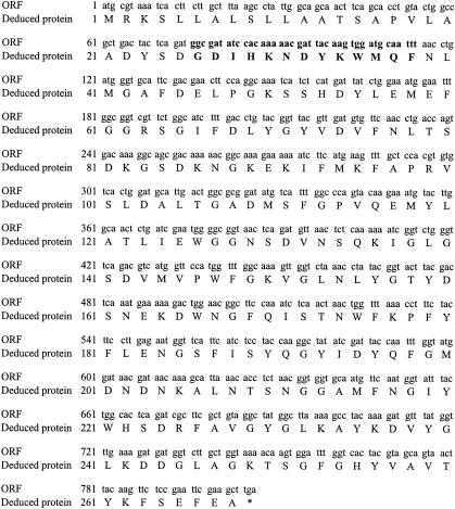
VBP genomes were searched from the genomic DNA library of activated sludge bacteria by the colony hybridization technique using the constructed DNA probe. As a result, one putative open reading frame (ORF) was obtained, which had a high identity with the amino acid sequence of the VBP obtained in our previous study (29). Figure 1 shows the sequence of the putative ORF and its deduced protein. This ORF consisted of 807 nucleotides encoding 268 amino acids. (Boldface type in Fig. 1 shows the region of the probe-like sequence in which 37 of 39 residues were identical to those in the probe.) Two mismatches between sequences of the ORF and the probe were located at Met36 (ATG) in the ORF. The counterpart sequence of the probe genome was TTT or TTC, which were the codon of Phe.
FIG. 1.
Putative ORF of the isolated gene by colony hybridization. Boldface type indicates the region of the probe-like sequence. The asterisk indicates the stop codon.
Figure 2 shows the comparison of the amino acid sequences between the deduced protein from the isolated gene and the VBP obtained in our previous study (29). Twenty of 22 residues were identical between the deduced protein and the VBP, although one space was necessary to obtain maximum matching alignment between Ser4 and Gly5 in the VBP.
FIG. 2.
Sequence alignment of the deduced protein from the isolated gene and VBP isolated in our previous study (29). Boldface type indicates the region of the probe-like sequence. The vertical solid bars indicate identical amino acids between the deduced protein and the VBP, and dashed bars indicate similar amino acids. The dash is a space introduced to maximize the alignment.
A homology search of the isolated ORF was conducted by the BLAST server against all nonredundant GenBank coding sequence translations, Protein Data Bank, SwissProt, Protein Identification Resource, and Protein Research Foundation. Fifteen amino acid sequences were retrieved from 2,137,877 sequences with high scores larger than 100 bits. The retrieved sequences included 14 bacterial proteins and one protein of Anopheles gambiae. Vibrio vulnificus OmpK (NP 760691.1) and Pseudomonas syringae Omp (ZP 00263676.1) were retrieved with the highest (79%) and the lowest (28%) identities, respectively. Disagreements in amino acid sequences between the isolated ORF and Vibrio vulnificus OmpK are observed especially in their midstreams. The first 20 amino acid residues of the deduced protein had characteristics common to bacterial signal peptide (1).
Production of the VBP candidate by E. coli BL21.
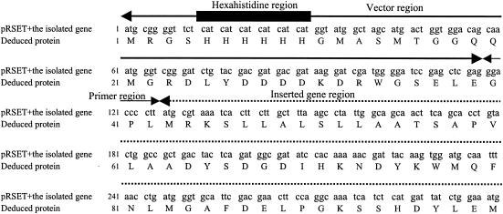
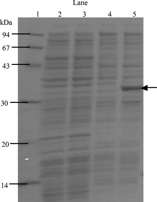
The isolated gene was amplified by the primers listed in Table 1 and inserted into the plasmid pRSET in order to construct the expression vector. The result of the DNA sequencing with T7 primer indicated that the isolated genome was successfully inserted into pRSET, as expected (Fig. 3). This constructed plasmid was used for the expression of the isolated gene. The recombinant VBP candidate was expressed in E. coli BL21 as a fusion protein linked to a hexahistidine peptide in the presence of 1 mM IPTG. Harvested cells were suspended in the denaturation buffer and processed for SDS-polyacrylamide gel electrophoresis (PAGE) (Fig. 4). One thick band between 30 and 43 kDa was observed in lane 4 (IPTG-induced cells transformed by pRSET carrying the isolated gene). This thick band was not obtained in lane 1 (noninduced cells transformed by pRSET), lane 2 (IPTG-induced cells transformed by pRSET), and lane 3 (noninduced cells transformed by pRSET carrying the isolated gene). These results indicate that both pRSET carrying the isolated gene and the IPTG induction were necessary to express the VBP candidate.
TABLE 1.
Primers used for amplification of the gene of the VBP candidate
| Primer code | Sequence |
|---|---|
| Upstream | 5′-TCAGTCAGCTCGAGGGACCCCTTATGCGTAAA-3′ |
| Downstream | 5′-CAGTCAGTGAATTCGGAGAACTTGTAAGTTAC-3′ |
FIG. 3.
Sequence of the N-terminal part of the VBP in the constructed expression vector. The thick line indicates the region originated in pRSET, and the thin line indicates the region derived from the primer. The dashed line indicates the region of the isolated gene.
FIG. 4.
SDS-PAGE analysis of proteins produced by E. coli BL21. A 15% polyacrylamide separating gel and a 5% stacking gel were used for SDS-PAGE. The cell pellet from 1 ml of harvested culture medium was suspended in 100 μl of MilliQ water. The sample buffer (50 mM Tris-HCl [pH 8.0], 2% SDS, 5% 2-mercaptoethanol, 10% glycerol, 0.02% bromophenol blue) was added, and the mixture was heated for 5 min at 95°C. VBPs were separated in an electrophoretic cell for 1.5 h in running buffer (25 mM Tris, 192 mM glycine, 0.1% SDS). The gel was stained by CBB. Lane 1, molecular mass marker; lane 2, proteins from noninduced E. coli cells transformed by pRSET; lane 3, proteins from IPTG (1 mM)-induced E. coli cells transformed by pRSET; lane 4, proteins from noninduced E. coli cells transformed by pRSET carrying the isolated gene; lane 5, proteins from IPTG (1 mM)-induced E. coli cells transformed by pRSET carrying the isolated gene. The arrow indicates the products of the isolated gene induced by IPTG.
Purification of the VBP candidate.
Figure 5 shows the PAGE profile of SDS-denatured soluble proteins and urea-soluble proteins from IPTG-induced E. coli BL21. It is apparent that the VBP candidate was involved in the urea-soluble proteins. Since the T7 promoter in pRSET is well known as the strong promoter, the VBP candidate was produced in the inclusion body form and cannot be obtained as a water-soluble protein.
FIG. 5.
SDS-PAGE analysis of proteins produced by E. coli BL21 transformed by pRSET carrying the isolated gene. A 15% polyacrylamide separating gel and a 5% stacking gel were used for SDS-PAGE. The cell pellet from 1 ml of harvested culture medium was suspended in 100 μl of MilliQ water. The sample buffer (50 mM Tris-HCl [ph 8.0], 2% SDS, 5% 2-mercaptoethanol, 10% glycerol, 0.02% bromophenol blue) was added, and the mixture was heated for 5 min at 95°C. VBPs were separated in an electrophoretic cell for 1.5 h in running buffer (25 mM Tris, 192 mM glycine, 0.1% SDS). The gel was stained by CBB. Lane 1, molecular mass marker; lane 2, proteins in supernatant; lane 3, proteins in purified inclusion body. The arrow indicates the product of the isolated gene induced by IPTG.
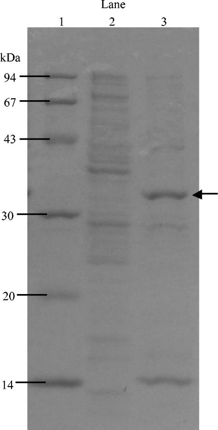
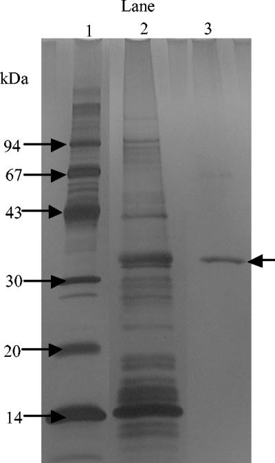
The VBP candidate was obtained in urea-soluble proteins and purified by nickel-charged gel that can capture the hexahistidine tag in the protein sequence. The homogeneity of the purified protein was evaluated by SDS-PAGE. As shown in Fig. 6, the eluate from the nickel-charged gel contains the VBP candidate alone, which means that the purification was successfully performed.
FIG. 6.
SDS-PAGE analysis of the fusion protein purified by the nickel chelate column. A 15% polyacrylamide separating gel and a 5% stacking gel were used for SDS-PAGE. The cell pellet from 1 ml of harvested culture medium was suspended in 100 μl of MilliQ water. The sample buffer (50 mM Tris-HCl [pH 8.0], 2% SDS, 5% 2-mercaptoethanol, 10% glycerol, 0.02% bromophenol blue) was added, and the mixture was heated for 5 min at 95°C. VBPs were separated in an electrophoretic cell for 1.5 h in running buffer (25 mM Tris, 192 mM glycine, 0.1% SDS). The gel was stained by silver. Lane 1, molecular mass marker; lane 2, proteins in purified inclusion body; lane 3, eluate from the nickel chelate column. The arrow indicates the position of the purified protein.
Evaluation of virus-binding ability of the VBP candidate.
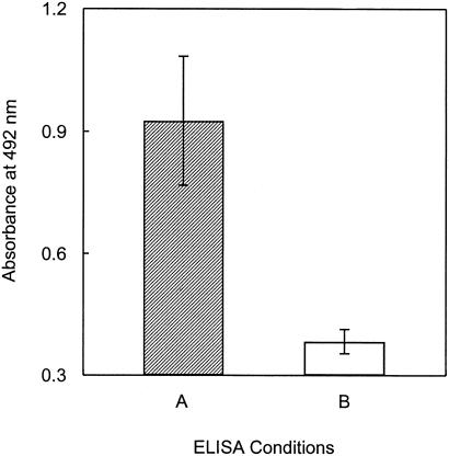
Figure 7 shows the result of ELISA for the evaluation of PV1-binding ability of the purified VBP candidate. The homoscedasticity between variances of the absorbance for conditions A and B was certified by the F test at a significance level of 0.01. The absorbance for the ELISA using the VBP candidate and PV1 (condition A) was apparently greater than that using BSA and PV1 (condition B). The significant difference between absorbances of conditions A and B was confirmed by the Student's t test at a significance level of 0.01. These results indicate that the fusion protein definitely has the ability to bind PV1 and can be regarded as poliovirus-binding protein.
FIG. 7.
Evaluation of the PV1-binding ability of the VBP with ELISA. The ELISA conditions are denoted A (ELISA with the VBP and PV1) and B (ELISA with BSA and PV1). The error bars indicate the standard errors for triplicate trials.
In order to quantitatively evaluate the binding ability of the VBP with PV1, the equilibrium binding constant between PV1 and VBP was estimated. VBP was immobilized on an ELISA plate as described above, and the amount of PV1 bound to the immobilized VBP was quantified by the plaque method. The estimated value of the equilibrium constant of the PV1 binding by the VBP was 2.1 × 10⁷ M−1 (Table 2). This value was equivalent to that of the affinity binding between protein A and antibodies (Table 3), although there is a difference in the procedures for estimating the equilibrium constant of protein binding. These results indicate that there is specific binding between the VBP and PV1.
TABLE 2.
Estimation of the binding constant between PV1 and VBP
| Concn of immobilized VBP in ELISA well (M) | Adsorption efficiency of PV1 to VBP in ELISA well (%) | Estimated value of binding constanta | Avg | SD |
|---|---|---|---|---|
| 8.2 × 10⁻⁷ | 88.5 | 9.33 × 10⁶ | ||
| 2.9 × 10⁻⁷ | 90.2 | 3.13 × 10⁷ | 2.1 × 10⁷ | 1.1 × 10⁷ |
| 3.7 × 10⁻⁷ | 89.8 | 2.36 × 10⁷ |
Binding constant was estimated with equation 1.
TABLE 3.
Binding constants for affinity interaction of protein
| Host | Guest | Kae (M−1) | Reference |
|---|---|---|---|
| VBP | PV1 | 2.14 × 10⁷ | |
| MabSelectb | IgGa | 4.76 × 10⁶ | 13 |
| rPr Sepharose FFc | IgG | 4.00 × 10⁶ | 13 |
| Prosep-rAd | IgG | 3.45 × 10⁶ | 13 |
| Carbohydrate-binding module of xylanase 10A from Thermotoga maritima | Cellopentaose | 2.26 × 10⁶ | 4 |
| Carbohydrate-binding module of xylanase 10A from Thermotoga maritima | Lactose | 1.30 × 10⁶ | 4 |
| Porcine pancreatic elastase | Turkey ovomucoid third domain | 4.98 × 10⁵ | 9 |
| Vancomycin | 9-fluorenylmethoxycarbonyl-amino acid-D-Ala-D-Ala | 1.75 × 10⁵ | 39 |
| Vancomycin | Carbonic anhidrase B | 9.9 × 10³ | 40 |
| β-Cyclodextrin | Methoxypoly(ethylene glycol)s | 4.38 × 10² | 16 |
| β-Cyclodextrin | Naproxen | 1.57 × 10² | 2 |
IgG, immunoglobulin G.
Catalog no. 288113, Amersham Bioscience.
rProtein A-Sepharose Fast Flow, catalog no. 256997, Amersham Bioscience(s).
Prosep-rA High Capacity, catalog no. 107009, Bioprocessing.
Ka, equilibrium binding constant.
DISCUSSION
Pathogenic viruses in water are more resistant to several disinfectants than indicator microorganisms. For example, the cycle threshold (CT value) for 3.78-log inactivation of hepatitis A virus by free chlorine was 200 mg/liter/min (19), which was much greater than the CT value for 3-log inactivation of E. coli (0.09 ± 0.003 mg/liter/min, strain C) (33). The U.S. Environmental Protection Agency reported that the average CT value for 3-log inactivation of viruses by ozone at 5°C was 0.6 mg/liter/min, which was about 30 times the CT value for E. coli (35). It was also reported that adenoviruses, one of the major leading causes of viral gastroenteritis among children, are UV light resistant (11, 34). Membrane technologies such as microfiltration and ultrafiltration are available to remove viruses (14, 25), but the operation of the membrane separation process still has some disadvantages, such as the large energy consumption and the nuisance of maintenance. The application of membrane technology to water and wastewater treatments would remain a matter of investigation. Under present conditions, the conventional water and wastewater treatment processes are not sufficient to prevent societies and water environments from viral contamination. In the near future, the spread of pathogenic viruses causing waterborne infectious diseases could be caused by population explosion, urban congestion, and water shortages, which will lead to the deterioration of sanitary conditions. It is important to develop a new scheme for virus inactivation or removal in water and wastewater treatment systems.
In our previous study, VBPs were successfully isolated from bacterial culture derived from activated sludge (29). Not hydrophobic and simple electrostatic interactions but the specific lock-and-key interaction was one of the persuasive explanations for the strong binding of PV1 capsid peptide to the isolated VBPs. The construction of the cloning system for the VBP is of significance to develop the VBP-based technology for virus removal, because a mass volume of the VBP is required to utilize it as a specific viral adsorbent. From this viewpoint, we searched for the VBP gene from the bacterial culture derived from activated sludge, and one putative ORF that has a similar sequence to the gene of VBP obtained in our previous study was successfully isolated (Fig. 1 and 2). Since the protein produced by the protein cloning technique with the isolated gene exhibited the ability to bind PV1 (Fig. 7), the obtained protein can be regarded as one of the VBPs for PV1.
The protein encoded in the isolated genome was considered to be a newly discovered protein from activated sludge culture, because any sequences in protein databases were not perfectly matched with the sequence of the VBP. V. vulnificus outer membrane protein OmpK (NP_760691.1) was retrieved with the highest identity with the deduced amino acid sequences of the isolated genome, but the identity was 79%. The result of the homology search may imply that the VBP is produced by bacteria in activated sludge culture as an outer membrane protein. Some researchers reported that no proteolysis was observed after intact bacteria were subjected to the protease digestion (30, 37), which means that some outer membrane proteins of bacteria are protease resistant and environmentally stable. VBPs which originated from the outer membrane protein of bacteria in activated sludge are expected to be stable under the conditions of water and wastewater treatments and useful as a viral adsorbent in the VBP-based technology.
The VBP has to be obtained as a soluble protein in order to utilize it as a viral adsorbent in the virus removal technology. The VBP in inclusion body form was dissolved in a lysis buffer including urea (Fig. 5). Since the VBP has a hexahistidine tag in its N terminus, it was easy to purify the VBP by one-step purification with nickel chelate gel (Fig. 6). The hexahistidine tag is useful for the simple purification of recombinant proteins produced by E. coli as reported previously (3, 17, 31). The facile purification of the VBP would make it possible to obtain a mass volume of the VBP in a financially feasible fashion.
We assumed that the binding between the VBP and PV1 can be Langmuir-type adsorption, but it was difficult to estimate the equilibrium binding constant by fitting data to the Langmuir equation because of the too-low concentration of PV1 compared with that of VBP. The estimated value of the equilibrium binding constant was obtained with equation 1. As a result, the equilibrium binding constant between PV1 and VBP in the ELISA well was estimated to be 2.1 × 10⁷ (M−1) (Table 2). This value was significantly larger than that of protein A, which is used as a ligand for purification of monoclonal and recombinant antibodies (13). Table 3 shows that the binding constants of affinity adsorption of protein were between 106 and 102 (M−1) (2, 4, 9, 16, 39, 40). Although there is the difference in the procedure for estimating the binding constant between these studies and our study, the large value of the estimated binding constant in this study would indicate that PV1 binds to the VBP with a high affinity.
The VBP cloning system developed in this study would make it possible to produce a mass volume of VBPs and to utilize them as a new material of the specific adsorbent in several technologies, including virus removal, concentration, and detection. In order to develop these VBP-based technologies, it is necessary to find VBPs for not only polioviruses but also other pathogenic viruses, including adenoviruses, rotaviruses, and noroviruses. It is difficult to find VBPs for all viruses in water, because more than 100 species of viruses are known as etiological agents of waterborne diseases (32). However, it would be practical to isolate VBPs for key viruses responsible for the outbreaks of waterborne infectious diseases. The isolation of VBPs for key viruses from the bacterial culture derived from activated sludge will be conducted in further studies.
Acknowledgments
This work has been funded by a Grant-in-Aid for the Development of Innovative Technology from the Ministry of Education, Culture, Sports, Science, and Technology of Japan.
REFERENCES
- 1.Arkowitz, R. A., and M. Bassilana. 1994. Protein translocation in Escherichia coli. Biochim. Biophys. Acta 1197:311-343. [DOI] [PubMed] [Google Scholar]
- 2.Bellini, M. S., Z. Deyl, G. Manetto, and M. Kohlickova. 2001. Determination of apparent binding constants of drugs by capillary electrophoresis using β-cyclodextrin as ligand and three different linear plotting methods. J. Chromatogr. A 924:438-491. [DOI] [PubMed] [Google Scholar]
- 3.Betemps, D., F. Mallet, V. Cheynet, and T. Baron. 1999. Overexpression and purification of an immunologically reactive His-BIV capsid fusion protein. Protein Expr. Purif. 15:258-264. [DOI] [PubMed] [Google Scholar]
- 4.Boraston, A. B., A. L. Creagh, M. M. Alam, J. M. Kormos, P. Tomme, C. A. Haynes, R. A. J. Warren, and D. G. Kilburn. 2001. Binding specificity and thermodynamics of a family 9 carbohydrate-binding module from Thermotoga maritima xylanase 10A. Biochemistry 40:6240-6247. [DOI] [PubMed] [Google Scholar]
- 5.Centers for Disease Control and Prevention. 2001. Echovirus type 13—United States, 2001. Morb. Mortal. Wkly. Rep. 50:777-780. [PubMed] [Google Scholar]
- 6.Centers for Disease Control and Prevention. 2000. Enterovirus surveillance. Morb. Mortal. Wkly. Rep. 49:913-916. [PubMed] [Google Scholar]
- 7.Centers for Disease Control and Prevention. 2000. Surveillance for waterborne-disease outbreaks—United States, 1997-1998. Morb. Mortal. Wkly. Rep. 49:1-36. [Google Scholar]
- 8.Cherkasova, E. A., E. A. Korotkova, M. L. Yakovenko, O. E. Ivanova, T. P. Eremeeva, K. M. Chumakov, and V. I. Agol. 2002. Long-term circulation of vaccine-derived poliovirus that causes paralytic disease. J. Virol. 76:6791-6799. [DOI] [PMC free article] [PubMed] [Google Scholar]
- 9.Edgcomb, S. P., B. M. Baker, and K. P. Murphy. 2000. The energetics of phosphate binding to a protein complex. Protein Sci. 9:927-933. [DOI] [PMC free article] [PubMed] [Google Scholar]
- 10.Enriquez, C. E., C. J. Hurst, and C. P. Gerba. 1995. Survival of the enteric adenoviruses 40 and 41 in tap, sea, and waste water. Water Res. 29:2548-2553. [Google Scholar]
- 11.Gerba, C. P., D. M. Gramos, and N. Nwachuku. 2002. Comparative inactivation of enteroviruses and adenovirus 2 by UV light. Appl. Environ. Microbiol. 68:5167-5169. [DOI] [PMC free article] [PubMed] [Google Scholar]
- 12.Glass, R. I., J. Noel, T. Ando, R. Fankhauser, G. Belliot, A. Mounts, U. D. Parashar, J. S. Bresee, and S. S. Monroe. 2000. The epidemiology of enteric caliciviruses from humans: a reassessment using new diagnostics. J. Infect. Dis. 181:S254-S261. [DOI] [PubMed] [Google Scholar]
- 13.Hahn, R., R. Schlegel, and A. Jungbauer. 2003. Comparison of protein A affinity sorbents. J. Chromatogr. B Analyt. Technol. Biomed. Life Sci. 790:35-51. [DOI] [PubMed] [Google Scholar]
- 14.Herath, G., K. Yamamoto, and T. Urase. 1999. Removal of viruses by microfiltration membranes at different solution environments. Water Sci. Technol. 40:331-338. [Google Scholar]
- 15.Horie, H., H. Yoshida, K. Matsuura, M. Miyazawa, Y. Ota, T. Nakayama, Y. Doi, and S. Hashizume. 2002. Neurovirulence of type 1 polioviruses isolated from sewage in Japan. Appl. Environ. Microbiol. 68:138-142. [DOI] [PMC free article] [PubMed] [Google Scholar]
- 16.Karakasyan, C., M. Taverna, and M.-C. Millot. 2004. Determination of binding constants of hydrophobically end-capped poly(ethylene glycol)s with β-cyclodextrin by affinity capillary electrophoresis. J. Chromatogr. A 1032:159-164. [DOI] [PubMed] [Google Scholar]
- 17.Laine, S., S. Salhi, and J.-M. Rossignol. 2002. Overexpression and purification of the hepatitis B e antigen precursor. J. Virol. Methods 103:67-74. [DOI] [PubMed] [Google Scholar]
- 18.Leong, L. Y. C. 1983. Removal and inactivation of viruses by treatment processes for potable water and wastewater—a review. Water Sci. Technol. 15:91-114. [Google Scholar]
- 19.Li, J. W., Z. T. Xin, X. W. Wang, J. L. Zheng, and F. H. Chao. 2002. Mechanisms of inactivation of hepatitis A virus by chlorine. Appl. Environ. Microbiol. 68:4951-4955. [DOI] [PMC free article] [PubMed] [Google Scholar]
- 20.Liu, H.-M., D.-P. Zheng, L.-B. Zhang, M. S. Oberste, O. M. Kew, and M. A. Pallansch. 2003. Serial recombination during circulation of type 1 wild-vaccine recombination polioviruses in China. J. Virol. 77:10994-11005. [DOI] [PMC free article] [PubMed] [Google Scholar]
- 21.Lopman, B. A., G. K. Adak, M. H. Reacher, and D. W. G. Brown. 2003. Two epidemiologic patterns of norovirus outbreaks: surveillance in England and Wales, 1992-2000. Emerg. Infect. Dis. 9:71-77. [DOI] [PMC free article] [PubMed] [Google Scholar]
- 22.Lukashev, A. N., V. A. Lashkevich, O. E. Ivanova, G. A. Koroleva, A. E. Hinkkanen, and J. Ilonen. 2003. Recombination in circulating enteroviruses. J. Virol. 77:10423-10431. [DOI] [PMC free article] [PubMed] [Google Scholar]
- 23.Oberste, M. S., K. Maher, and M. A. Pallansch. 2004. Evidence for frequent recombination within species Human Enterovirus B based on complete genomic sequences of all thirty-seven serotypes. J. Virol. 78:855-867. [DOI] [PMC free article] [PubMed] [Google Scholar]
- 24.Oberste, M. S., S. Peñaranda, and M. A. Pallansch. 2004. RNA recombination plays a major role in genomic change during circulation of coxsackie B viruses. J. Virol. 78:2948-2955. [DOI] [PMC free article] [PubMed] [Google Scholar]
- 25.Otaki, M., K. Yano, and S. Ohgaki. 1998. Virus removal in a membrane separation process. Water Sci. Technol. 37:107-116. [Google Scholar]
- 26.Parashar, U. D., E. G. Hummelman, J. S. Bresee, M. A. Miller, and R. I. Glass. 2003. Global illness and deaths caused by rotavirus disease in children. Emerg. Infect. Dis. 9:565-572. [DOI] [PMC free article] [PubMed] [Google Scholar]
- 27.Parshionikar, S. U., S. Willian-True, G. S. Fout, D. E. Robbins, S. A. Seys, J. D. Cassady, and R. Harris. 2003. Waterborne outbreaks of gastroenteritis associated with a norovirus. Appl. Environ. Microbiol. 69:5263-5268. [DOI] [PMC free article] [PubMed] [Google Scholar]
- 28.Payment, P. 1998. Waterborne viruses and parasites: resistance to treatment and disinfection. In Proceedings of the OECD Workshop on Molecular Methods for Safe Drinking Water, Interlaken, Switzerland. [Online.] http://www.eawag.ch/publications_e/proceedings/oecd/proceedings/Payment.pdf.
- 29.Sano, D., T. Matsuo, and T. Omura. 2004. Virus-binding proteins recovered from bacterial culture derived from activated sludge by affinity chromatography assay using a viral capsid peptide. Appl. Environ. Microbiol. 70:3434-3442. [DOI] [PMC free article] [PubMed] [Google Scholar]
- 30.Siehnel, R., N. L. Martin, and R. E. W. Hancock. 1990. Sequence and relatedness in other bacteria of the Pseudomonas aeruginosa oprP gene coding for the phosphate-specific porin P. Mol. Microbiol. 4:831-838. [DOI] [PubMed] [Google Scholar]
- 31.Tallet, B., T. Astier-Gin, M. Castroviejo, and X. Santarelli. 2001. One-step chromatographic purification procedure of a His-tag recombinant carboxyl half part of the HTLV-I surface envelope glycoprotein overexpressed in Escherichia coli as a secreted form. J. Chromatogr. B Biomed. Sci. Appl. 753:17-22. [DOI] [PubMed] [Google Scholar]
- 32.Taylor, F. B. 1974. Viruses—what is their significance in water supplies? JAWWA 66:306-311. [Google Scholar]
- 33.Taylor, R. H., J. O. Falkinham III, C. D. Norton, and M. W. Lechevallier. 2000. Chlorine, chloramine, chlorine dioxide, and ozone susceptibility of Mycobacterium avium. Appl. Environ. Microbiol. 66:1702-1705. [DOI] [PMC free article] [PubMed] [Google Scholar]
- 34.Thompson, S. S., J. L. Jackson, M. Suva-Castillo, W. A. Yanko, Z. E. Jack, J. Kuo, C. L. Chen, F. P. Williams, and D. P. Schnurr. 2003. Detection of infectious human adenoviruses in tertiary-treated and ultraviolet-disinfected wastewater. Water Environ. Res. 75:163-170. [DOI] [PubMed] [Google Scholar]
- 35.U.S. Environmental Protection Agency. 1989. Guidance manual for compliance with the filtration and disinfection requirements for public water systems using surface water sources. Publication no. EPA/570/9-89-018. U.S. Environmental Protection Agency, Washington, D.C.
- 36.U.S. Environmental Protection agency. 1999. Method for the recovery and assay of enteroviruses from sewage sludge, p. 117-145. Environmental regulations and technology. Control of pathogens and vector attraction in sewage sludge. Publication no. EPA/625/R-92/013. U.S. Environmental Protection Agency, Washington, D.C.
- 37.Worobec, E. A., N. L. Martin, W. D. McCubbin, C. M. Kay, G. D. Brayer, and R. E. W. Hancock. 1988. Large-scale purification and biochemical characterization of crystallization-grade porin protein P from Pseudomonas aeruginosa. Biochim. Biophys. Acta 939:366-374. [DOI] [PubMed] [Google Scholar]
- 38.Worobey, M., and E. C. Holmes. 1999. Evolutionary aspects of recombination in RNA viruses. J. Gen. Virol. 80:2535-2543. [DOI] [PubMed] [Google Scholar]
- 39.Zhang, Y., and F. A. Gomez. 2000. Multiple-step ligand injection affinity capillary electrophoresis for determining binding constants of ligands to receptors. J. Chromatogr. A 897:339-347. [DOI] [PubMed] [Google Scholar]
- 40.Zhang, Y., C. Kodama, C. Zurita, and F. A. Gomez. 2001. On-column ligand synthesis coupled to partial-filling affinity capillary electrophoresis to estimate binding constants of ligands to a receptor. J. Chromatogr. A 928:233-241. [DOI] [PubMed] [Google Scholar]


