Abstract
The cellular response to environmental signals is largely dependent upon the induction of responsive protein kinase signaling pathways. Within these pathways, distinct protein-protein interactions play a role in determining the specificity of the response through regulation of kinase function. The interferon-induced serine/threonine protein kinase, PKR, is activated in response to various environmental stimuli. Like many protein kinases, PKR is regulated through direct interactions with activator and inhibitory molecules, including P58IPK, a cellular PKR inhibitor. P58IPK functions to represses PKR-mediated phosphorylation of the eukaryotic initiation factor 2α subunit (eIF-2α) through a direct interaction, thereby relieving the PKR-imposed block on mRNA translation and cell growth. To further define the molecular mechanism underlying regulation of PKR, we have utilized an interaction cloning strategy to identify a novel cDNA encoding a P58IPK-interacting protein. This protein, designated P52rIPK, possesses limited homology to the charged domain of Hsp90 and is expressed in a wide range of cell lines. P52rIPK and P58IPK interacted in a yeast two-hybrid assay and were recovered as a complex from mammalian cell extracts. When coexpressed with PKR in yeast, P58IPK repressed PKR-mediated eIF-2α phosphorylation, inhibiting the normally toxic and growth-suppressive effects associated with PKR function. Conversely, introduction of P52rIPK into these strains resulted in restoration of both PKR activity and eIF-2α phosphorylation, concomitant with growth suppression due to inhibition of P58IPK function. Furthermore, P52rIPK inhibited P58IPK function in a reconstituted in vitro PKR-regulatory assay. Our results demonstrate that P58IPK is inhibited through a direct interaction with P52rIPK which, in turn, results in upregulation of PKR activity. Taken together, our data describe a novel protein kinase-regulatory system which encompasses an intersection of interferon-, stress-, and growth-regulatory pathways.
In eukaryotes, the cellular response to changing physiological and environmental conditions includes the induction of signaling and regulatory pathways which function to modulate gene expression and cell cycle control. The ability to rapidly respond to these changing conditions is often dependent upon cellular regulatory pathways defined by the many protein kinase signaling cascades which respond to environmental signals (70). Examples include specific stress-responsive protein kinases such as the mammalian Sek1 (62), Jnk/Sapk (77), and p38-Map (73) protein kinases. These enzymes and the components of their respective regulatory pathways provide rapid and sensitive mechanisms that allow the cell to initiate specific stress response programs. Similar to other stress-responsive kinases, the PKR protein kinase (59) is regulated in response to specific environmental stimuli (72). PKR is an interferon (IFN)-induced gene product which is activated by binding to double-stranded RNA or other polyanions (34, 45).
As a pivotal component of the IFN-induced cellular antiviral and antiproliferative states (78), PKR is the target of virally encoded inhibitors (reviewed in references 42 and 43), making it unique among protein kinases. In addition to its role in the IFN response, PKR is involved in cell growth control and has been identified as a tumor suppressor gene product (2, 48, 60). Activated PKR phosphorylates serine 51 within the alpha subunit of eukaryotic initiation factor 2 (eIF-2α), leading to inhibition of protein synthesis and concomitant growth suppression (reviewed in references 15 and 38). In addition to regulating protein synthesis, PKR has been identified as a pivotal component of double-stranded-RNA-mediated signaling events which lead to activation of nuclear factor kappa B (NF-κB) and IFN regulatory factor 1-dependent gene transcription (51, 90), and it is a mediator of stress-induced apoptosis (20, 91). Moreover, PKR is activated in response to such environmental challenges as viral infection (42), oxidative stresses, and possibly heat shock (7, 71). Each of these events lead to acute phosphorylation of eIF-2α and an immediate reduction in protein synthesis, thereby limiting potential proteotoxic and/or cytotoxic outcomes attributed to stress exposure. It is relevant that PKR is a member of a family of functionally related eIF-2α kinases, including reticulocyte-expressed HRI (12) and Saccharomyces cerevisiae GCN2 enzymes (23). Like PKR, GCN2 and HRI are regulated in response to cellular stress, where they play an important role in modulating gene expression in response to environmental stimuli (reviewed in reference 38).
The molecular mechanisms which regulate PKR function in normally dividing cells are largely unknown. However, we have previously identified P58IPK as a cellular inhibitor of PKR which is recruited by influenza virus to inhibit PKR function during viral infection (54–56). We hypothesized that P58IPK resides within the cell in specific complexes with its own inhibitory proteins. Formation of a P58IPK-PKR complex results in inhibition of kinase function and concomitant stimulation of mRNA translation (69, 86). In response to activating stimuli, which include viral infection, possibly other cellular stresses, and signal transduction processes, P58IPK is released from its inhibitor, enabling it to physically interact with PKR (31, 55). Overexpression of P58IPK in mammalian cells results in malignant transformation as a result of the ability of P58IPK to inhibit PKR (1). These studies indicate that P58IPK possesses growth-regulatory and oncogenic properties, possibly through inhibition of the PKR tumor suppressor phenotype. Containing nine tandemly arranged tetratricopeptide repeat (TPR) motifs (35) and a C-terminal DnaJ homology region (19), P58IPK has structural similarity to several members of the eukaryotic stress response protein family, including the heat shock protein (Hsp)-interacting TPR proteins Hip (39), Hop (81), FKBP52 (68), and CyP-40 (92) (reviewed in reference 28) as well as the eukaryotic DnaJ homologs Ydj-1 (10) and Hsp40 (64). The structural homology of P58IPK and stress response proteins putatively identifies P58IPK as a member of this protein family and suggests that P58IPK may play a role in the cellular response to stress. In support of this, we have recently identified Hsp40 as an inhibitor of P58IPK function (58).
In an effort to further delineate the regulatory mechanisms and upstream signals which function within the PKR-P58IPK pathway, we have undertaken interaction cloning to identify cellular proteins which bind to and regulate P58IPK function. Here we describe a novel P58IPK-regulatory protein, termed P52rIPK, which has homology to the charged domain of Hsp90. P52rIPK inhibits P58IPK through a direct protein-protein interaction. These events lead to an upregulation of PKR function, a resultant increase in eIF-2α phosphorylation, and cell growth suppression. Our results suggest that P52rIPK is a new member of the PKR-regulatory pathway. A model describing this increasingly complex protein kinase-regulatory pathway is presented.
MATERIALS AND METHODS
Yeast two-hybrid assays and library screen for P58IPK-interacting proteins.
The yeast two-hybrid system (5) was used to isolate the P52rIPK cDNA and to assess protein interactions in vivo. To screen for cDNAs encoding P58IPK-interacting proteins, S. cerevisiae Hf7c (MATa ura3-52 his3-200 lys2-801 ade2-101 trp1-901 leu2-3,112 gal4-542 gal80-538 LYS2::GAL1-HIS3 URA3 [GAL4 17-mers]3-CYC1-lacZ; Clontech) was transformed with the bait plasmid pBD-P58IPK, encoding P58IPK fused to the GAL4 DNA binding domain (BD) (5). After confirming expression of the BD-P58IPK fusion protein in transformed yeast, a library of HeLa cell cDNAs fused to the GAL4 activation domain (AD) in pGADGH (Clontech) was introduced by large-scale transformation, after which the yeast cells were plated onto selective medium lacking histidine. To increase the stringency of the two-hybrid library screen, we included 30 mM 3-aminotriazole, a competitive inhibitor of histidine biosynthesis (46), in the selective medium. Plates were incubated at 30°C for 8 days, and resultant yeast colonies were streaked onto medium lacking histidine in the presence of 30 mM 3-aminotriazole. To segregate the AD library plasmids from pBD-P58IPK, yeast two-hybrid library clones were cultured for 40 h in tryptophan-deficient liquid medium and replica plated onto medium lacking leucine or lacking both tryptophan and leucine. Segregated pGADGH library plasmids were isolated from yeast colonies which failed to grow in the absence of tryptophan, transformed into Escherichia coli, and purified. Specificity of the yeast two-hybrid protein interaction was determined by introducing the recovered library plasmids into S. cerevisiae Hf7c harboring pBD-P58IPK or control plasmids encoding the BD vector alone (pGBT9; Clontech), BD-simian virus 40 (SV40) T antigen (pTD1), or BD-lamin (pLAM5′) (31). BD-P58IPK interaction specificity was confirmed by introducing pBD-P58IPK into Hf7c harboring the AD plasmid pGAD425 or control plasmids encoding AD-PKR K296R (pAD-PKR K296R), AD-SV40 T antigen (pTD2) (31), or the AD library plasmid pGADGH. Only those library plasmids encoding proteins which exhibited specific interaction with BD-P58IPK were selected for further analyses. Of the 1.2 × 10⁶ CFU screened, we recovered two identical clones that met this criterion, each harboring the P52rIPK cDNA.
Yeast two-hybrid assays were conducted to test the specific induction of the individual His and LacZ reporter genes due to protein interaction in yeast strain Hf7c. Analysis of His reporter induction was conducted as described previously (31). Briefly, yeast strains cotransformed with plasmids encoding AD and BD fusion proteins were streaked onto histidine-containing medium. After growth on histidine-containing medium colonies were restreaked onto medium lacking histidine and incubated at 30°C for 3 to 5 days to allow depletion of residual histidine stores. Histidine-depleted colonies were streaked onto medium lacking histidine and assessed for growth after 3 to 5 days of incubation at 30°C. Strains that exhibited growth on medium lacking histidine were subsequently scored as positive for a two-hybrid protein interaction. For determination of lacZ induction, Hf7c cotransformants were plated as patches onto histidine-containing medium and incubated for 3 days at 30°C. Patches were then replica printed onto histidine-containing medium containing 70 mM phosphate buffer (pH 7.0) and 200 μg of the β-galactosidase substrate 5-bromo-4-chloro-3-indoyl-β-d-galactopyranoside per ml. After 3 to 5 days of growth at 30°C, patches were visually assessed for the development of blue color, which is indicative of LacZ induction and a two-hybrid protein interaction. In both the His and LacZ reporter assays, strains harboring pCL1 (27), encoding wild-type GAL4, were employed as a positive control.
Plasmid construction.
The yeast two-hybrid bait plasmid pBD-P58IPK encodes BD-fused full length human P58IPK (49), as described previously (31). Plasmid pAD-P52 harbors the P52rIPK cDNA in the yeast two-hybrid library plasmid pGADGH. To facilitate P52rIPK cDNA probe production and in vitro transcription reactions, the 1.5-kb P52rIPK cDNA insert was excised from pAD-P52 as an EcoRI fragment and cloned into the EcoRI site of pBSK (Stratagene) to yield pBSK-P52. pCDNA1neo-P58IPK and pGex2T-P58IPK encode full-length P58IPK alone or as a glutathione S-transferase (GST) fusion protein, respectively (54). pGST-P52 encodes full-length P52rIPK fused to the C-terminus of GST and was constructed by inserting the 1.5-kb EcoRI fragment from pAD-P52 into the EcoRI site of pGex4T-3 (Pharmacia). The yeast expression plasmid pYex-P58IPK was produced by inserting the EcoRV/XbaI fragment from pCDNA1neo-P58IPK into the galactose-inducible URA3 2μm yeast expression plasmid pEMBLYex4 (11). pYex-PKR Δ295–300 encodes a PKR deletion mutant lacking amino acids (aa) 295 to 300 cloned into pEMBLYex4 (74). For expression of P52rIPK in yeast, the P52rIPK coding region was amplified from pAD-P52 by PCR with the oligonucleotide primer pair 5′TTAGAATTCATGCCGAACTTCTGCGCTG (sense primer; the EcoRI site is underlined) and 5′GCGCCCGGGATTTCATTTTTAAGAACAACA (antisense primer; the SmaI site is underlined), encoding P52rIPK nucleotides (nt) 1 to 19 and 1479 to 1500, respectively. PCR products were cleaved with EcoRI and SmaI and cloned into the corresponding restriction sites of the galactose-inducible TRP1 2μm yeast expression plasmid pYX233 (Novagen) to yield pYX-P52.
Recombinant protein expression and generation of antiserum to P52rIPK.
For production of GST or GST fusion proteins, overnight 50-ml cultures of bacteria harboring pGex2T (80), pGex2T-P58IPK (54), or pGST-P52 were split to an optical density at 600 nm of 0.1 and grown for 1 h at 37°C. After the addition of 1 mM isopropylthiogalactopyranoside, cultures were grown for a further 3 h at 30°C and harvested by centrifugation. Production of protein extracts and purification of GST or GST fusion proteins were carried out essentially as described previously (80).
The generation of polyclonal antiserum to P52rIPK was carried out by immunizing New Zealand White rabbits with purified GST-P52rIPK as described previously (29). Rabbits were immunized once with 100 μg of GST-P52rIPK in 5.0 ml of incomplete Freund’s adjuvant containing 100 μg of N-acetylmuramyl-l-alanyl-d-isoglutamine. Subsequent boosts of 100 μg of GST-P52rIPK in incomplete Freund’s adjuvant were administered at monthly intervals. Anti-P52rIPK (α-P52rIPK) serum is specific to P52rIPK as determined by immunoblot and immunoprecipitation analyses.
Analysis of RNA.
For determination of mRNA expression, RNA was extracted by the guanidinium isothiocyanate method (13) from HeLa cells cultured overnight in the presence or absence of 1,000 U of human alpha IFN per ml. After poly(A) selection, RNA was fractionated by electrophoresis in agarose-formaldehyde gels and blotted to nylon membranes. 32P-labeled cDNA probe production and Northern (RNA) blot hybridizations were performed as described previously (30).
To determine if the P52rIPK cDNA isolated from our yeast two-hybrid library screen encoded the intact 5′ end of the corresponding mRNA, we carried out anchor PCR analysis of oligo(dT)-primed cDNA synthesized from subconfluent HeLa cells and containing the 30-nt 5′ anchor sequence 5′GATCCACTAGTTCTAGAGCGGCCGCCACCG3′. HeLa and control cDNAs were amplified by PCR with the Stratagene SK primer (5′CGCTCTAGAACTAGTGGATC3′; anchor primer) and the P52rIPK-specific primer 5′CTCACAGATCTCTAGCATCT3′ (52rIPK nt 725 to 744). PCR products from HeLa cDNA and pBSK-P52 (control) comigrated when resolved by agarose gel electrophoresis. By this method we determined that the P52rIPK cDNA clone isolated in our two-hybrid library screen encoded the intact 5′ end of the corresponding P52rIPK mRNA (data not shown) and that the size difference between the P52rIPK mRNA and cDNA clone was due to the presence of a 3′ untranslated region (UTR) within the mRNA not present in the cloned cDNA.
In vitro translation, protein labeling, and immunoprecipitation analysis.
P58IPK and P52rIPK were transcribed from the T7 promoter of pCDNA1neo-P58IPK and pBSK-P52, respectively, and translated in vitro by using the TNT reaction system (Promega). For the production of labeled proteins, translation reactions were carried out in the presence of [35S]methionine and radiolabel incorporation was quantitated by scintillation counting of the trichloroacetic acid-precipitable reaction products. For in vivo protein labeling, HeLa cells were incubated in methionine-deficient medium in the presence of [35S]methionine for 16 h and harvested for immunoprecipitation analysis as described previously (30). Immunoprecipitation reactions were carried out with a 1:100 dilution of rabbit preimmune or α-P52rIPK serum or a 1:500 dilution of α-P58IPK monoclonal antibody 9F10 ascites fluid (1). For immunoprecipitation of in vitro translation products, reticulocyte extracts containing 105 counts of [35S]methionine-labeled translation products were incubated with the indicated antibody in the presence of immunoprecipitation buffer I (50 mM KCl, 400 mM NaCl, 1 mM EDTA, 1 mM dithiothreitol [DTT], 20% glycerol, 1% Triton X-100, 100 U of aprotinin per ml, 0.2 mM phenylmethylsulfonyl fluoride [PMSF], 20 mM Tris, pH 7.5) as described previously (30). For coimmunoprecipitation analysis with α-P52rIPK serum, reticulocyte extracts containing 105 counts of labeled P58IPK were mixed with extracts containing unlabeled in vitro-translated P52rIPK. Similarly, for α-P58IPK coimmunoprecipitation analysis, reticulocyte extracts containing 105 counts of labeled P52rIPK were mixed with extracts containing unlabeled P58IPK. Mixtures were incubated for 1 h at 4°C, followed by the addition of the proper antibodies. For all immunoprecipitation reactions, immunocomplexes were recovered by centrifugation after the addition of protein G-agarose beads to the reaction mixture. Beads were washed extensively with ice-cold buffer I followed by three washes with ice-cold buffer II (100 mM KCl, 0.1 mM EDTA, 20% glycerol, 100 U of aprotinin per ml, 0.2 mM PMSF, 10 mM Tris, pH 7.5), resuspended in sodium dodecyl sulfate (SDS) sample buffer, and incubated at 100°C for 5 min. Immunoprecipitation products were resolved by electrophoresis on 12.5% acrylamide–SDS gels, followed by autoradiography of the dried gel.
Immunoblot analysis.
All immunoblot analyses were conducted essentially as described previously (30, 31). HeLa cell extracts were prepared by resuspending cells (2 × 10⁷/ml) in lysis buffer (50 mM KCl, 1 mM DTT, 2 mM MgCl, 1% Triton X-100, 10 mM Tris, pH 7.5) supplemented with 0.2 mM PMSF and 100 U of aprotinin per ml. Yeast cell extracts were prepared by collecting cells from 20-ml liquid cultures, washing once with water, and resuspending in lysis buffer. Yeast cells were lysed by the glass bead method with a bead homogenizer (21). All extracts were clarified by centrifugation at 12,000 × g, and supernatants were collected and stored frozen at −80°C until use. Cell extract protein concentrations were determined by using a protein assay reagent system as described by the manufacturer (Bio-Rad). Protein (25 to 50 μg) was separated by SDS-polyacrylamide gel electrophoresis (SDS-PAGE) and transferred to nitrocellulose membranes. After a 1-h blocking step, membranes were incubated with the indicated antibodies for an additional 1 h, washed, and probed with a horseradish peroxidase-conjugated secondary antibody. Bound antibodies were detected by enhanced chemiluminescence. P52rIPK, P58IPK, and PKR expression was determined with polyclonal α-P52rIPK serum or monoclonal 9F10 α-P58IPK (1) or α-PKR (53) primary antibodies, respectively. To control for potential errors in protein concentrations or gel loading, a monoclonal antibody to human actin was included as a probe on all blots.
In vitro assay for P52rIPK function.
Determination of P52rIPK function in vitro was carried out by using purified components essentially as described by Melville et al. (58). Increasing concentrations of purified GST or GST-P52rIPK were mixed with 2 pmol of GST-P58IPK and incubated at 30°C for 10 min. Following the incubation, PKR (2 pmol; purified from IFN-treated Daudi cells [30]), poly(I · C) (100-ng/ml final concentration), and 5 μCi of [γ-32P]ATP were added, and the mixture was incubated for a further 10 min at 30°C. Histone HIIA substrate (from calf thymus; Sigma) and 10 μCi of [γ-32P]ATP were added and left for an additional 20 min at 30°C, bringing the reaction volume to 30 μl in a final buffer of 17 mM Tris-HCl, 57 mM KCl, 40 mM NaCl, 2 mM MgCl2, 2 mM MnCl2, 1 mg of bovine serum albumin per ml, 1 mM DTT, 100 U of aprotinin per ml, 20 μM PMSF, 50 μM EDTA, 6.6% glycerol, 2 mM ATP, and 2 mM HEPES, pH 7.5. Reactions were terminated by the addition of 2× SDS-PAGE sample buffer, and the mixtures were incubated at 100°C for 5 min and subjected to SDS-PAGE analysis on 12.5% acrylamide gels. Histone substrate phosphorylation was visualized by autoradiography of the dried gel and quantitated by scanning laser densitometry. Previous studies from our laboratory have demonstrated a perfect correlation between the abilities of PKR to phosphorylate histone HIIA and eIF-2α (45).
For each assay, we carried out control experiments to eliminate the possibility that our recombinant protein preparations possessed P58IPK-specific protease or histone- and/or PKR-specific protein kinase activity (data not shown). Kinase assays in the presence of only GST-P52rIPK revealed that these recombinant protein preparations possessed no histone- or PKR-phosphorylating activities. Similarly, we conducted immunoblot analyses of the kinase assay reactants which were separated from 32P-labeled histones by SDS-PAGE. Using this approach, we determined that the input P58IPK remained intact and was not degraded during the assay.
Yeast growth assay for in vivo determination of P58IPK and P52rIPK function.
To examine the in vivo PKR-regulatory properties of P58IPK and P52rIPK, we utilized S. cerevisiae RY1-1 (MATa ura3-52 leu2-3 leu2-112 gcn2Δ trp1-Δ63 LEU2::[GAL-CYC1-PKR]2) (74) or the isogenic control strain J110 (MATa ura3-52 leu2-3 leu2-112 gcn2Δ trp1-Δ63 LEU2) (47). Both strains lack the gene encoding the yeast eIF-2α kinase GCN2 (23). However, RY1-1 carries two copies of human PKR integrated into the LEU2 locus, under control of the galactose-inducible GAL1-CYC1 promoter (74). Strains grown on dextrose will repress gene expression from the GAL1-CYC1 promoter, while conversely, high levels of GAL1-CYC1-dependent gene expression are achieved upon exposure of yeast strains to galactose. When grown on galactose medium, the RY1-1 strain exhibits a growth-suppressed phenotype due to the PKR-mediated phosphorylation of yeast eIF-2α (74). Strain J110 (a gift from Thomas Dever) is identical to RY1-1 except that it harbors a single LEU2-integrated expression vector devoid of insert DNA (47). For analysis of P58IPK function, strains were transformed with the galactose-inducible expression plasmid pEMBLYex4, pYex-P58IPK, or pYex-PKR Δ295-300 and streaked onto uracil-deficient dextrose medium (SD). After 3 days, individual colonies were replica streaked onto uracil-deficient SD medium or synthetic medium containing galactose as the sole carbon source (SGAL) and placed in a 30°C incubator. Strains were assessed for the ability to grow on SGAL medium, which is indicative of PKR repression by the plasmid-borne allele. For analysis of P52rIPK function, RY1-1 strains were cotransformed with the galactose-inducible expression plasmid combinations of pEMBLYex4-pYX233, pEMBLYex4–pYX-P52, pYex-P58IPK–pYX233, and pYex-P58IPK–pYX-P52 and plated onto SD medium lacking uracil and tryptophan. After colony growth (approximately 3 to 4 days), strains were replica streaked onto uracil- and tryptophan-deficient SD and SGAL media, incubated for 7 to 10 days at 30°C, and analyzed for growth. Protein expression from the transformation construct(s) was confirmed by immunoblot analysis of extracts prepared from yeast cells grown for 4 to 9 h in freshly diluted cultures of liquid SGAL selective medium. To ensure that all yeast expression plasmids had no growth-altering properties, either alone or in cotransfection combinations, all experiments included a parallel analysis of strain J110 transfected with the indicated plasmid(s). By this method we determined that both P58IPK and P52rIPK lacked growth-regulatory properties when expressed in the absence of PKR (data not shown).
eIF-2α phosphorylation analysis.
For isoelectric focusing of eIF-2α, yeast strains were grown for 16 h in selective SD medium, diluted to an optical density at 600 nm of 0.4 in selective SGAL medium, and grown for an additional 4 to 9 h at 30°C. Yeast extracts were prepared as described for immunoblot analysis, except that cells were lysed in buffer containing 50 mM PIPES [piperazine-N,N′-bis(2-ethanesulfonic acid)] (pH 6.2), 150 mM NaCl, 15 mM EDTA, 1 mM PMSF, 1 mM DTT, 50 mM NaF, 35 mM β-glycerolphosphate, and 10 mM 2-aminopurine. Proteins (16 μg) were separated by vertical isoelectric focusing (21, 23) and blotted to nitrocellulose membranes. eIF-2α was detected by immunoblot analysis with a rabbit polyclonal antiserum specific to yeast eIF-2α (a gift from Thomas Dever). In these experiments, an increase in the level of the less acidic, hypophosphorylated form of eIF-2α indicates a concomitant decrease in the level of hyperphosphorylated eIF-2α, which is phosphorylated by PKR on serine 51 (21, 22). The levels of hypophosphorylated eIF-2α relative to total eIF-2α were quantitated by scanning laser densitometry and are presented as a percentage of total eIF-2α for each sample.
DNA sequencing and computer analysis.
Both strands of the P52rIPK cDNA were sequenced by the dye termination method with an Applied Biosystems automated DNA sequencer and oligonucleotide primers designed to yield overlapping contigs of DNA sequence. Cloned PCR products were sequenced in their entirety to confirm that no mutations occurred during the amplification and cloning processes. Sequence contigs were assembled by using the Genetics Computer Group (GCG) Fragment Assembly System software package. Sequence alignments were conducted with the GCG Bestfit program. Searches of the nucleotide and protein databases were facilitated by using the BLAST program included in the GCG software package licensed to the University of Washington. We identified the Hsp90 homology domain of P52rIPK by preparing peptide sequences corresponding to contiguous 100-aa blocks of the deduced P52rIPK amino acid sequence. Each P52rIPK peptide sequence was used independently to search the protein databases for homologous sequences. By this method we identified a region of P52rIPK (aa 86 to 114) which has limited but significant amino acid sequence homology (P = 0.0005) to the charged domain of human Hsp90 (37).
Nucleotide sequence accession number.
The GenBank accession number for the P52rIPK cDNA is AF007393.
RESULTS
Identification and characterization of the P52rIPK cDNA.
Previous studies from our laboratory have determined that the PKR-inhibitory activity of P58IPK is regulated, at least in part, through direct P58IPK-protein interactions (31, 69). To determine the molecular mechanisms of P58IPK regulation and to identify P58IPK-dependent pathways which regulate PKR, we executed a search for cDNAs encoding regulators of P58IPK function. We used P58IPK fused to the GAL4 DNA BD (BD-P58IPK) as bait to screen a GAL4 transcription AD-fused HeLa cell cDNA library. To select for those BD-P58IPK–AD-protein interactions of the highest specificity, we increased the stringency of the two-hybrid library screen by the addition of 30 mM 3-aminotriazol, a competitive inhibitor of histidine biosynthesis (46), to the culture medium. Using this approach, we isolated two identical 1.5-kb cDNAs encoding a deduced protein of 492 aa (Fig. 1) which exhibited a strong and specific interaction with BD-P58IPK (see Fig. 3). Based upon the functional characterization described below, we have named this cDNA and its deduced 52-kDa protein P52rIPK (for 52-kDa repressor of the inhibitor of protein kinase).
FIG. 1.
P52rIPK possesses homology to the charged domain of Hsp90. (A) Nucleotide and deduced amino acid sequences of the 5′ UTR and open reading frame in the P52rIPK cDNA. Nucleotide and amino acid positions are indicated at the left. Position 1 denotes the initiator methionine codon (boxed); the asterisk denotes the site of translation termination. (B) Structural representation of P52rIPK and comparison with Hsp90. The Hsp90 homology domain of P52rIPK (aa 86 to 200) and the homologous charged domain of human Hsp90 (aa 170 to 300 [37]) are indicated in black (top) and are shown in an amino acid sequence alignment (bottom). Identical residues are indicated by a vertical line; double and single dots indicate conservative and less conservative amino acid replacements, respectively. Homology scores show 24% amino acid identity with 48% amino acid similarity over the region shown. (C) Hydropathy profile of the P52rIPK amino acid sequence. Positive (hydrophobic) amino acid sequences are represented by peaks extending above the neutral plane (dashed line). The bar indicates the Hsp90 homology domain.
FIG. 3.
P52rIPK interacts with P58IPK in vivo. (A) Histidine reporter assay of yeast two-hybrid strains. Hf7c yeast strains harboring GAL4 AD and DNA BD plasmids expressing AD-PKR and BD-P58IPK (controls; position 1), AD-vector (AD-V) (control) and BD-P58IPK (position 2), AD-P52rIPK and BD-P58IPK (position 3), AD-P52rIPK and BD-vector (control; position 4), AD-GAL4 wild type (control) and BD-vector (position 5), and AD-vector and BD-vector (control; position 6) were replica plated in the presence (+ His) or absence (−His) of histidine, incubated for 5 days, and scored for growth. Growth on medium lacking histidine is indicative of a two-hybrid protein interaction. (B) Analysis of β-galactosidase activity. Strain Hf7c was cotransformed with expression plasmids encoding the indicated combination of AD and BD fusion proteins and patch-plated onto medium containing histidine in the presence of β-galactosidase substrate. After 3 days of growth, patches were scored for color development. Induction of the LacZ reporter (dark patch) is indicative of β-galactosidase activity and a two-hybrid protein interaction. The top row shows strains expressing AD-P52rIPK and (from left to right) BD-vector, BD-SV40 T antigen (BD-T ag); BD-lamin, and BD-P58IPK. The bottom row, left, shows strains expressing BD-P58IPK with AD-vector or AD-SV40 T antigen. The bottom row, right, shows strain Hf7c expressing the wild-type (wt) GAL4 protein. In this analysis and that shown in panel A, AD and BD fusion protein expression was confirmed by immunoblot analysis (data not shown).
Examination of the P52rIPK cDNA revealed a potential translation start codon 17 nt from the 5′ terminus, followed by a translation termination codon at nt 1479 (Fig. 1A). Using anchor PCR to amplify the 5′ terminus of the P52rIPK mRNA from HeLa cells, we confirmed that the cloned cDNA included the intact 5′ end of the mRNA (data not shown). Thus, we identified the first ATG (Fig. 1A) as the translation start codon. However, this cDNA possessed a short 3′ UTR lacking a poly(A) tail (data not shown), indicating that this P52rIPK clone lacked the 3′ UTR of the native mRNA. A search of the nucleotide sequence database identified several expressed sequence tags having sequence identity with overlapping regions of the P52rIPK cDNA. However, no significant nucleotide sequence homologies to any known cDNAs were found. These results indicate that the P52rIPK clone represents a novel human cDNA. We similarly compared the deduced amino acid sequence of the P52rIPK open reading frame with known protein sequences in the GenBank database. We identified a region of 114 aa having limited homology (48% similarity, 24% identity) to the charged domain of mammalian isoforms of Hsp90 (human Hsp90 [37], aa 170 to 300) (Fig. 1B). As shown in Fig. 1C, this region of P52rIPK, termed the Hsp90 homology domain, is similarly rich with charged amino acid residues which would be expected to form a lobe structure protruding from an otherwise hydrophobic protein core (84). The homologous region of human Hsp90 has been implicated in mediating regulatory interactions with the glucocorticoid receptor (8, 9), suggesting that the P52rIPK Hsp90 homology domain may similarly mediate protein interactions. Taken together, these results show that the P52rIPK cDNA encodes a novel Hsp90-related protein having limited homology with this member of the stress response protein family.
Determination of P52rIPK mRNA and protein expression in human cells.
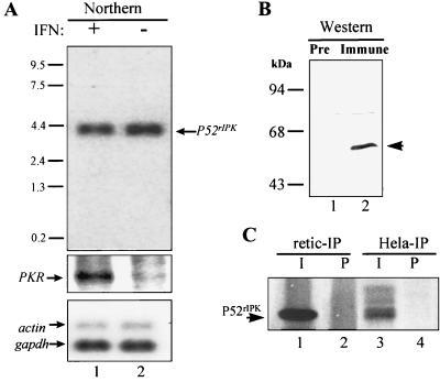
To determine the expression pattern(s) of P52rIPK mRNA, we carried out Northern blot analyses, using the P52rIPK cDNA as a probe. As shown in Fig. 2A, the P52rIPK mRNA was expressed as a single 4.2-kb species in HeLa cells, indicating that the P52rIPK cDNA clone, which possessed a complete open reading frame, lacks approximately 2.7 kb of the 3′ UTR. Exposure to IFN had no effect upon the steady-state level of P52rIPK mRNA (compare lanes 1 and 2 in Fig. 2A), indicating that P52rIPK is likely not an IFN-regulated gene. We similarly identified a single 4.2-kb P52rIPK transcript in various human cell lines, including 293 embryonal kidney cells, MT4 T cells, HL-60 myeloid cells, and Daudi B cells (data not shown). Thus, similar to P58IPK (49), the P52rIPK transcript is expressed in a range of cell types.
FIG. 2.
Expression of P52rIPK in mammalian cells. (A) Northern blot analysis of P52rIPK mRNA expression in HeLa cells cultured in the presence (lane 1) or absence (lane 2) of IFN. The 4.2-kb P52rIPK mRNA is indicated by the arrow at the right. Each lane contains 5 μg of poly(A)+ RNA. Gel loading was confirmed by sequentially hybridizing the same blot to probes specific for actin and glyceraldehyde dehydrogenase (gapdh) mRNAs. To confirm the induction of gene expression due to IFN treatment, the same blot was probed with 32P-labeled PKR cDNA. Positions of RNA size standards are indicated in kilobases. (B) Immunoblot analysis. Fifty micrograms of HeLa cell extract was separated by SDS-PAGE, blotted to membranes, and incubated with preimmune (Pre) rabbit serum (lane 1) or α-P52rIPK immune serum (lane 2). The arrow indicates the position of P52rIPK. The faint lower band in lane 2 corresponds to a background band recognized by rabbit serum. Positions of protein standards are shown. (C) Immunoprecipitation (IP) analysis. Reticulocyte lysate (retic) in vitro translation reaction mixtures containing [35S]methionine-labeled P52rIPK (lanes 1 and 2) or extracts from 106 HeLa cells metabolically labeled with [35S]-methionine (lanes 3 and 4) were immunoprecipitated with preimmune rabbit serum (P) (lanes 2 and 4) or α-P52rIPK immune serum (I) (lanes 1 and 3). Shown is an autoradiogram of immunoprecipitates separated by SDS-PAGE. The arrow indicates the position of P52rIPK.
To verify protein expression in mammalian cells and to facilitate additional biochemical studies, we prepared a rabbit polyclonal antiserum (α-P52) against recombinant P52rIPK expressed as a GST fusion protein. With α-P52 serum, immunoblot analysis revealed the expression of a 52-kDa protein in HeLa cells (Fig. 2B). Likewise, a 52-kDa protein was specifically isolated by α-P52 immunoprecipitation from HeLa cell extracts (Fig. 2C, lane 3). Interestingly, the HeLa α-P52 immunoprecipitation product showed the presence of a broad 52-kDa band possibly representing P52rIPK degradation products, coimmunoprecipitating proteins, or posttranslationally modified P52rIPK isoforms. We consider it unlikely that this broad band represents degraded P52rIPK, as the major product of this immunoprecipitation comigrated with full-length in vitro-translated P52rIPK immunoprecipitated from reticulocyte lysates (compare lanes 1 and 3 in Fig. 2C). The latter two possibilities are supported by the observation that P52rIPK and P58IPK form a stable complex in mammalian cell extracts (see Fig. 8) and by the identification of several potential protein kinase phosphorylation sites within the deduced P52rIPK amino acid sequence (data not shown), respectively. These results indicate that the P52rIPK cDNA encodes the entire native protein, which, importantly, is expressed and can be specifically detected with α-P52 serum.
FIG. 8.
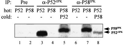
P58IPK and P52rIPK form a complex in mammalian cell extracts. P58IPK and P52rIPK were translated in vitro in the presence (hot) or absence (cold) of [35S]methionine by using a rabbit reticulocyte lysate translation system. Hot translation reactions were immunoprecipitated from reticulocyte extracts individually (lanes 1 to 4, 6, and 7) or as a mixture with the reciprocal cold translation extract (lanes 5 and 8), boiled in reducing sample buffer, and separated by SDS-PAGE. By this method, coimmunoprecipitating proteins will migrate through the gel independently based upon protein mass. The relative positions of labeled proteins after electrophoresis were determined by autoradiography of the dried gel. Lanes 1 and 2, preimmune serum (Pre) immunoprecipitations (IP) of hot P52rIPK and P58IPK, respectively; lanes 3 to 5, α-P52rIPK immunoprecipitations of hot P52rIPK (lane 3) and of hot P58IPK alone or in the presence of cold P52rIPK (lanes 4 and 5, respectively); lanes 6 to 8, α-P58IPK immunoprecipitations of hot P58IPK (lane 7) and of hot P52rIPK alone or mixed with lysates containing cold P58IPK (lanes 6 and 8, respectively). Arrows indicate the positions of P58IPK and P52rIPK.
Interaction of P58IPK with P52rIPK.
The P52rIPK cDNA was cloned based on the ability of the encoded protein to interact with P58IPK in a yeast two-hybrid system. To confirm and extend the results of our two-hybrid library screen, we conducted a more thorough analysis of P52rIPK-P58IPK interaction specificity. Using two independent reporter assays, we tested the ability of P52rIPK to interact with P58IPK or various control constructs in yeast strain Hf7c, employing the yeast two-hybrid assay. In this system, induction of the independent His and LacZ reporter genes is indicative of a two-hybrid protein interaction (5). As shown in Fig. 3A, coexpression of AD-P52rIPK and BD-P58IPK resulted in the specific induction of the His reporter, as determined by growth of this strain on histidine-deficient medium. Importantly, while all yeast two-hybrid strains grew on medium containing histidine, only those strains harboring wild-type GAL4 (GAL4/BD-V) or AD-PKR and BD-P58IPK (AD-PKR/BD-P58IPK) were capable of growth on medium lacking histidine, supporting our previous results (31). A similar examination of lacZ reporter gene expression independently verified the two-hybrid interaction between P52rIPK and P58IPK by the induction of β-galactosidase activity (Fig. 3B). Interaction specificity was demonstrated by the lack of detectable β-galactosidase activity in strains expressing AD-P52rIPK with BD-lamin, BD-P53, or the BD vector. With the yeast two-hybrid system, these results demonstrate that P52rIPK forms a specific complex with P58IPK in vivo.
Analysis of P52rIPK function in vitro.
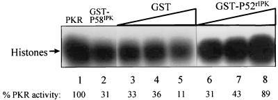
Previous results from our laboratory indicate that PKR function is inhibited through a direct interaction with P58IPK, which itself is regulated in large part through direct associations with specific regulatory proteins (31, 55, 69). We therefore reasoned that, through its ability to form a stable complex with P58IPK, P52rIPK may possess P58IPK-regulatory properties, thereby coupling P52rIPK to PKR regulation. To examine the function of P52rIPK, we expressed P52rIPK as a GST fusion protein (GST-P52rIPK) in E. coli and tested the ability of the purified recombinant protein to regulate P58IPK function and PKR activity in vitro. In these experiments, we utilized histone HIIA as a substrate, having previously demonstrated a perfect correlation between the phosphorylation of histones and eIF-2α by PKR (45). As determined previously (58) and seen in Fig. 4 (compare lanes 1 and 2), the high level of histone phosphorylation activity of PKR was repressed by GST-P58IPK, demonstrating the PKR-inhibitory activity of recombinant GST-P58IPK. Conversely, preincubation of GST-P58IPK with increasing concentrations of GST-P52rIPK resulted in a titratable inhibition of P58IPK function and concomitant retention of PKR activity (Fig. 4, lanes 6 to 8). We determined that the GST-P52rIPK-dependent restoration of kinase function was not due to the activity of contaminating kinase or P58IPK-specific protease activities (data not shown). Importantly, preincubation of PKR with P58IPK resulted in inhibition of PKR activity which could not be restored by the subsequent addition of P52rIPK to the reaction (data not shown). Preincubation of GST-P58IPK with low concentrations of GST had no effect on the ability of GST-P58IPK to repress PKR, while a high concentration of GST actually increased the level of PKR inhibition (Fig. 4, lanes 3 to 5). These results demonstrate that P52rIPK can specifically repress P58IPK function in vitro. Since inhibition of P58IPK function required preincubation with P52rIPK, P52rIPK may sequester P58IPK in an inactive complex rather than functioning to disrupt the PKR-P58IPK interaction.
FIG. 4.
P52rIPK inhibits P58IPK in vitro. Purified PKR (2 pmol) was mixed with buffer alone (lane 1) or buffer containing 2 pmol of GST-P58IPK either alone (lane 2) or preincubated with increasing amounts of GST (0.1, 2.0, or 5.0 pmol [lanes 3 to 5, respectively]) or GST-P52rIPK (0.1, 2.0, or 5.0 pmol [lanes 6 to 8, respectively]). To assess PKR activity, protein kinase assays were carried out in the presence of a histone HIIA substrate and [γ-32P]ATP. PKR-phosphorylated histones (arrow) were separated by SDS-PAGE and visualized by autoradiography. The level of PKR activity (histone phosphorylation) was quantitated by scanning laser densitometry and is presented below each lane as percent activity relative to that of the input control (lane 1).
Regulation of PKR function by P58IPK and P52rIPK in vivo.
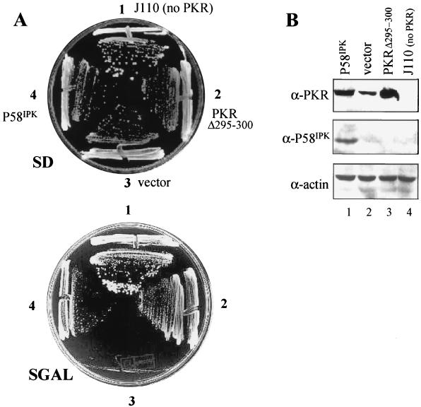
To determine the functional characteristics of P52rIPK in vivo and its relevance to the regulation of P58IPK and PKR, we took advantage of the growth-suppressive properties of PKR in S. cerevisiae (14, 74). It was first necessary to determine if P58IPK could inhibit PKR function when coexpressed in yeast. To facilitate these studies, we used the gcn2Δ S. cerevisiae strain RY1-1, which lacks GCN2 but harbors two copies of wild-type human PKR integrated in the LEU2 locus under control of a galactose-inducible promoter (74). When plated on galactose-containing medium, this strain expresses high levels of PKR, which phosphorylates the endogenous eIF-2α on serine 51 (22), leading to inhibition of protein synthesis and growth suppression. Coexpression of dominant-negative PKR mutants (74) or virally encoded PKR inhibitors (33, 47) in RY1-1 results in repression of PKR function and restoration of cell growth. We therefore transformed RY1-1 with the dominant-negative PKR mutant PKR Δ295-300 (74) (positive control), P58IPK, or the vector alone (negative control), each under control of a galactose-inducible promoter. Examination of the growth properties of the resulting strains revealed that while they grew on noninducing SD medium (Fig. 5A, upper plate), only those coexpressing P58IPK or the PKR Δ295–300 dominant-negative mutant with PKR retained the ability to grow on inducing SGAL medium (Fig. 5A, lower plate). Importantly, the isogenic gcn2Δ control strain, J110 (which lacks endogenous PKR) (47), grew equally well under both conditions, demonstrating that the galactose-inducible RY1-1 growth-suppressive phenotype was PKR specific. These results demonstrate that P58IPK can reverse the growth-suppressive effects of PKR in yeast. We verified that both the P58IPK and PKR Δ295-300 constructs were efficiently expressed yeast transformants (Fig. 5B). Previous studies have demonstrated that PKR autoregulates its own translation in mammalian cells and yeast by repressing de novo PKR synthesis (3, 47, 74). Importantly, RY1-1 strains coexpressing P58IPK with PKR exhibited an increase in PKR levels (Fig. 5B), consistent with a P58IPK-mediated inhibition of PKR autoregulation and stimulation of protein synthesis (86). Together with our previous results demonstrating in vivo complex formation between P58IPK and PKR (31), these results indicate that P58IPK, through a direct interaction with PKR, can repress kinase function and restore cell growth. Further, by reconstituting the PKR-regulatory pathway in yeast, we have established an in vivo assay for P58IPK function which is amenable to genetic analyses.
FIG. 5.
P58IPK represses PKR function in vivo. (A) Yeast growth analysis. Yeast strain RY1-1 was transformed with the 2μm expression plasmid pEMBLYex4 (vector; position 3), pYex-PKR Δ295–300 (PKR Δ295–300; position 2), or pYex-P58IPK (P58IPK; position 4) and streaked onto uracil-deficient medium containing 2% dextrose (SD) or 10% galactose (SGAL). To control for the specificity of growth effects due to PKR or P58IPK expression, the gcn2Δ isogenic control strain, J110, harboring vector alone (position 1) was included in all analyses. (B) Protein expression. Lanes 1 to 3 contain extracts from the RY1-1 strains shown in panel A harboring pYex-P58IPK (lane 1), pEMBLYex4 alone (lane 2), or pYex-PKR Δ295–300 (lane 3). An extract from the J110 parental control strain harboring pEMBLYex4 is shown in lane 4. Strains were grown for 5 h in galactose-containing liquid medium, and extracts were prepared as described in Materials and Methods. Proteins (25 μg) were separated by SDS-PAGE and subjected to immunoblot analysis. Panels show the same blot probed sequentially with antibodies specific to PKR, P58IPK, or actin.
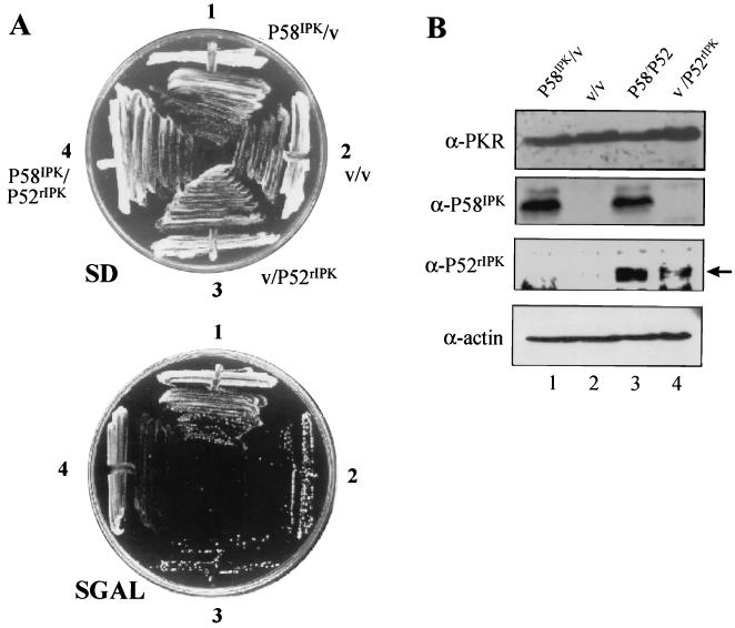
To determine the role of P52rIPK in this regulatory pathway, we introduced P52rIPK into RY1-1 harboring P58IPK. We predicted that in this strain P52rIPK, as an inhibitor of P58IPK, may itself possess growth-regulatory properties through restoration of PKR function. A comparison of the growth properties of the RY1-1 strains shown in Fig. 6A revealed that while all strains grew equally well on noninducing SD medium, only the strain coexpressing P58IPK alone with PKR (P58IPK/vector) exhibited growth on inducing SGAL medium, consistent with a P58IPK-mediated repression of PKR. Conversely, no growth was observed in RY1-1 coexpressing P52rIPK with P58IPK and PKR (P52rIPK/P58IPK) (Fig. 6A), indicating that P52rIPK could indeed block the PKR-inhibitory function of P58IPK. Importantly, we found that no toxic effects were associated with high levels of P52rIPK expression in the isogenic control strain J110 (data not shown). Similar to the RY1-1 strain expressing PKR alone (vector/vector) (Fig. 6A), those strains coexpressing PKR and P52rIPK in the absence of P58IPK (vector/P52rIPK) failed to grow on SGAL medium. Thus, both P58IPK and PKR are required for P52rIPK-mediated growth regulation in yeast. Collectively, these results confirm our in vitro data (Fig. 4) indicating that P52rIPK can repress the PKR-inhibitory function of P58IPK, resulting in restoration of kinase activity and suppression of cell growth. We found that coexpression of P52rIPK with P58IPK and PKR did not alter P58IPK levels in these strains (Fig. 6B). The differential effects on cell growth due to P58IPK and P52rIPK expression in cotransformed RY1-1 strains did not correlate with significant alterations in PKR levels (Fig. 6B), suggesting that PKR autoregulation may not be significantly affected in these strains. This seemingly disparate relationship between growth and PKR autoregulatory properties is likely due to the added complexity and competition for transcription and/or translation factors imposed by a dual plasmid system, or it may simply reflect limitations in our immunodetection methods.
FIG. 6.
P52rIPK inhibits P58IPK to restore PKR function in vivo. (A) Yeast growth analysis. RY1-1 harboring the indicated Ura and Trp 2μm expression plasmids was plated onto uracil- and tryptophan-deficient dextrose medium (SD) or inducing galactose medium (SGAL) and incubated at 30°C for 7 days. Plasmid combinations for each strain: pYex-P58IPK–pYX233 (P58IPK/v; position 1), pYex-pYX233 (v/v; position 2), pYex–pYX-P52 (v/P52rIPK; position 3), and pYex-P58IPK–pYX-P52 (P58IPK/P52rIPK; position 4). (B) Protein expression. Extracts were prepared from the yeast strains shown in panel A, which were grown for 7 h in galactose-containing liquid medium. Twenty-five micrograms of protein was separated by SDS-PAGE and subjected to immunoblot analysis. Shown is a single blot probed with antibodies specific to PKR, P58IPK, P52rIPK, or actin. Lanes represent extracts from RY1-1 harboring the plasmid combinations pYex-P58IPK–pYX233 (P58IPK/v; lane 1), pYex-pYX233 (v/v; lane 2), pYex-P58IPK–pYX-P52 (P58IPK/P52rIPK; lane 3), and pEMBLYex4–pYX-P52 (v/P52rIPK; lane 4). The arrow indicates the position of P52rIPK, which migrates above a background band found in all lanes.
To determine if P58IPK- and P52rIPK-mediated growth effects were due to alterations in eIF-2α phosphorylation, we used an isoelectric focusing technique to directly measure the phosphorylation state of endogenous eIF-2α in the yeast strains shown in Fig. 5 and 6. In this procedure, hyperphosphorylated eIF-2α, phosphorylated by PKR on serine 51, can be discriminated from the hypophosphorylated form of eIF-2α due to a phosphate-induced alteration in the protein isoelectric point (22, 23). This is demonstrated in Fig. 7B, in which all of the eIF-2α within the J110 control strain, which is devoid of PKR and serine 51 phosphorylation (47), is separated from the more acidic serine 51-phosphorylated form of eIF-2α found in RY1-1 expressing PKR (compare lanes 1 and 2). As seen in Fig. 7A, lane 1, virtually all of the eIF-2α in RY1-1 harboring the vector alone was phosphorylated on serine 51. In contrast, expression of P58IPK with PKR decreased the level of serine 51-phosphorylated eIF-2α, resulting in a 12-fold increase in the level of the hypophosphorylated form (Fig. 7A, lane 2). Similar results were obtained with an RY1-1 control strain expressing the dominant-negative PKR Δ295–300 mutant (Fig. 7A, compare lanes 2 and 3, respectively), consistent with inhibition of PKR function and restoration of cell growth (Fig. 5A) (74). Taken together, these results demonstrate that P58IPK can repress the function of PKR in vivo, resulting in a net decrease in the level of serine 51-phosphorylated eIF-2α and a concomitant restoration of cell growth.
FIG. 7.
P52rIPK and P58IPK regulate PKR-mediated eIF-2α phosphorylation. Protein extracts (16 μg) from yeast strains cultured in galactose-containing selective liquid medium for 9 h (A) or 5 h (B), were subjected to vertical gel isoelectric focusing and anti-eIF-2α immunoblot analysis. For each panel, the lower arrow indicates the position of hypophosphorylated eIF-2α. The upper arrow denotes the position of hyperphosphorylated eIF-2α, which is phosphorylated on serine 51 by PKR (22). The percentage of hypophosphorylated eIF-2α is presented beneath each lane. (A) Analysis of RY1-1 strains shown in Fig. 5, harboring pEMBLYex4 (vector; lane 1), pYex-P58IPK (lane 2), or pYex-PKR Δ295–300 (lane 3). (B) Analysis of the isogenic control strain J110 harboring pEMBLYex4 (lane 1) and the RY1-1 strains shown in Fig. 6, which harbor the plasmid combinations of pEMBLYex4-pYX233 (v/v; lane 2), pEMBLYex4–pYX-P52 (v/P52rIPK; lane 3), pYex-P58IPK–pYX233 (P58IPK/v; lane 4), and pYex-P58IPK–pYX-P52 (P58/P52; lane 5).
We then proceeded to examine eIF-2α phosphorylation within extracts prepared from the RY1-1 strains shown in Fig. 6. As seen in Fig. 7B, lane 2, expression of PKR in a strain cotransformed with vectors alone resulted in a high level of serine 51-phosphorylated eIF-2α, consistent with the growth-suppressed phenotype (Fig. 6). As demonstrated earlier, coexpression of PKR with P58IPK lowered the proportion of serine 51-phosphorylated eIF-2α and was reflected as a significant increase in the level of the hypophosphorylated species (Fig. 7B, lane 4). This P58IPK-mediated decrease in serine 51 eIF-2α phosphorylation was blocked by P52rIPK, as coexpression of P52rIPK with P58IPK and PKR (Fig. 7B, lane 5) restored the level of serine 51-phosphorylated eIF-2α to one comparable to that of the control strain expressing PKR alone (compare lanes 2, 4, and 5 in Fig. 7B). Coexpression of P52rIPK with PKR (Fig. 7B, lane 3) had no significant effect on eIF-2α phosphorylation levels, which again were similar to those of RY1-1 expressing PKR alone. Taken together with the growth data presented in Fig. 6, these results indicate that P52rIPK regulates PKR activity through inhibition of P58IPK function. P52rIPK inhibition of P58rIPK results in restoration of PKR-mediated eIF-2α phosphorylation and concomitant cell growth suppression. Moreover, and in support of previous studies (33, 47), our data demonstrates that small alterations in the levels of serine 51-phosphorylated eIF-2α can have dramatic effects on cell growth.
P52rIPK/P58IPK interaction in mammalian cell extracts.
P52rIPK specifically interacted with P58IPK in a yeast two-hybrid system (Fig. 3), demonstrating that the two proteins interact in vivo. However, it was essential to verify that P52rIPK could similarly bind to P58IPK within mammalian cell extracts. We therefore carried out a detailed immunoprecipitation analysis, assaying for P52rIPK-P58IPK complexes expressed within reticulocyte lysates. By using a rabbit reticulocyte translation system, cDNAs encoding wild-type P52rIPK and P58IPK were independently translated, and extracts were mixed together and subjected to immunoprecipitation analysis with P58IPK- or P52rIPK-specific antibodies. As shown in Fig. 8, analysis of immunoprecipitations from mixed translation reactions revealed that P52rIPK and P58IPK formed a complex in vitro; each was recovered in an immunoprecipitation with the reciprocal antibody. Importantly, coimmunoprecipitation of P58IPK with α-P52 serum was dependent on the presence of P52rIPK in the reaction mixture (Fig. 8, compare lanes 4 and 5). Likewise, recovery of P52rIPK from α-P58IPK immunoprecipitations was similarly dependent upon the presence of P58IPK in the reaction mixture (Fig. 8, compare lanes 6 and 8).
DISCUSSION
P52rIPK defines a novel PKR-regulatory pathway.
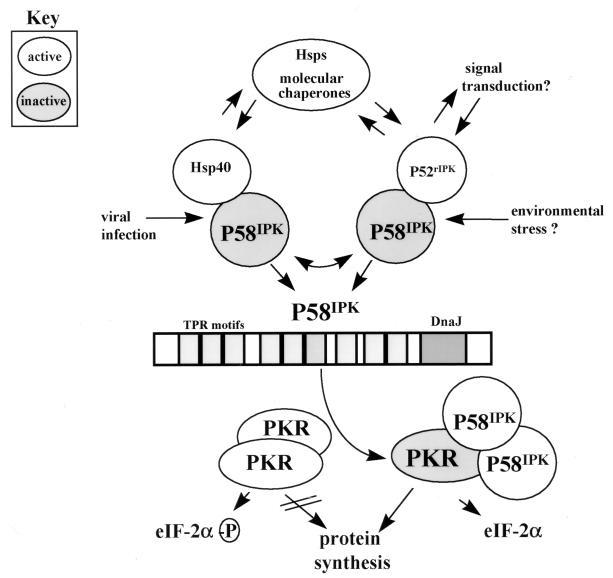
Our current model of the PKR-regulatory pathway is shown in Fig. 9. Serine 51 of eIF-2α is phosphorylated by PKR (22), the activity of which may be dependent on the formation of PKR dimers (Fig. 9, bottom) (67, 74). PKR-mediated phosphorylation of eIF-2α results in a block in protein synthesis initiation and concomitant growth suppression (14, 22, 74). P58IPK, which binds to PKR (31, 69), represses kinase function by disrupting active PKR dimers (85), resulting in a decrease in the level of serine 51-phosphorylated eIF-2α (1), stimulation of protein synthesis (86), and restoration of cell growth. In this model, P58IPK functions as a central element in the control of PKR, undergoing regulatory interactions with a complex set of proteins which define specific pathways for the control of PKR function. Earlier work from our laboratory has identified Hsp40 as a regulator of P58IPK (58). In the current study, we have identified P52rIPK as an additional and independent (Fig. 4) regulator of P58IPK and an upstream regulator of PKR. After either virus infection (44) or other stimuli, Hsp40 or P52rIPK dissociates from P58IPK. P58IPK then binds to PKR, resulting in repression of kinase function (31, 69). We have not yet identified the signal(s) which induces dissociation of the P52rIPK-P58IPK inhibitory complex. However, in an analogous system in mast cells, signaling processes lead to dissociation of the c-Cbl proto-oncogene product from an inactive protein complex, whereby it binds to and represses the function of the Syk protein kinase (65). The P52rIPK-dependent signal transduction process may similarly lead to activation of P58IPK and inhibition of PKR function. Further, Shim et al. (79) have determined that the p21WAF1/CIP1/Sdi1 DNA damage-inducible cell cycle inhibitor can bind to and repress the activity of the stress-activated protein kinase group of Map kinases. Thus, the p21WAF1/CIP1/Sdi1 and P58IPK protein kinase inhibitors define specific intersections between cellular stress response pathways and cell growth control. The PKR-dependent cellular IFN response defines yet a third intersecting pathway regulated in part by P58IPK. It is tempting to speculate that in addition to being involved in PKR regulation, P52rIPK, Hsp40, and P58IPK may also participate in PKR-independent regulatory events in which P58IPK regulation may lead to modulation of P52rIPK and/or Hsp40 function (Fig. 9, top). Such a pathway could have profound effects on molecular chaperone activities and signal transduction cascades which require Hsp40 (17, 61) or P52rIPK. Determination of the upstream signals which regulate P52rIPK function will allow us to more clearly define the extent of the biological role(s) played by this protein and its contributions within cellular regulatory processes.
FIG. 9.
Regulation of PKR. Two distinct cellular pathways of PKR regulation are defined by P52rIPK and Hsp40, both of which converge upon P58IPK to modulate PKR function. P58IPK is regulated through the formation of independent inhibitory complexes with P52rIPK and Hsp40 (58). Cellular stress, including virus infection (55, 56), may induce dissociation of the P58IPK-Hsp40 inhibitory complex. Possibly representing a protein signaling motif, the Hsp90 homology domain of P52rIPK may participate in signal transduction processes induced by exposure to environmental stress or mediated through specific signaling cascades which result in dissociation of the P58IPK-P52rIPK complex. Once released from its inhibitor, P58IPK forms a dimer or higher-ordered homotypic complex (31) competent to bind and disrupt active PKR dimers (lower) (85). Inhibition of PKR results in a block in PKR-mediated eIF-2α phosphorylation, stimulation of protein synthesis, and concomitant repression of PKR tumor suppressor function (lower middle and lower right) (1, 74, 86). P58IPK-mediated inhibition of PKR may also confer alterations in PKR-mediated signal transduction processes (50, 51, 89). Finally, P52rIPK and Hsp40 may interact with P58IPK as regulatory components of PKR-independent processes which lead to alterations in Hsp40 and/or P52rIPK function (upper). Other stress response proteins which regulate Hsp40, including heat shock proteins and molecular chaperones (19), could participate in both PKR-dependent and -independent regulatory pathways.
Stress response, molecular chaperones, and protein kinase regulation.
Interaction with elements of the stress response protein family and molecular chaperones is becoming a widely recognized mechanism of protein kinase regulation. In many cases, molecular chaperones have been identified as critical components of the maturation, subcellular localization, and subunit interactions of a wide variety of protein kinases (reviewed by Hunter [40]). A relevant example is the interaction between Hsp90 and the reticulocyte-expressed eIF-2α kinase HRI (57). Uma et al. (87) have recently demonstrated that HRI is conformationally maintained in an inactive, heme-responsive state by interaction with Hsp90 and that disruption of the HRI-Hsp90 complex results in loss of heme-regulated kinase function. Similarly, the interaction between PKR and P58IPK may induce conformational changes in the kinase which could lead to inhibition of catalytic function. Furthermore, the structural features of TPR protein P58IPK are consistent with the functions of related members within the stress response protein family, which participate in protein folding and signaling reactions. For example, the TPR proteins Hip and Hop interact with Hsc70 as cofactors in protein-folding, maturation, and signaling processes (28). Moreover, we have identified P58IPK TPR domain 6 as the PKR binding motif (31), and recent results indicate that interaction with P52rIPK is mediated by P58IPK TPR domain 7 (32). The C-terminal DnaJ domain of P58IPK may impart DnaJ-related chaperone and signaling functions (19) to P58IPK, possibly mediated through interaction with Hsp40 (58). It is relevant that the DnaJ region is essential for P58IPK function in vivo (86). Thus, through TPR motif- and DnaJ-dependent events, P58IPK participates in a protein kinase regulation, effecting PKR function through specific protein interactions.
The deduced structure of P52rIPK revealed that aa 86 to 200 have limited but significant homology to the charged domain, aa 170 to 300, of Hsp90 (Fig. 1) (37). Based on the three-dimensional structure of an Hsp90-geldanamycin complex (84), this domain is expected to be exposed to solvent and thereby available for molecular interactions. Previous studies have determined that the Hsp90 charged domain was necessary for steroid hormone signal transduction (8), where it may mediate a direct interaction with the glucocorticoid receptor (9). Recently, a genetic analysis of the Drosophila Hsp90 homolog, Hsp83, and its association with the Raf protein kinase has identified independent mutations which cluster near the charged domain of the Hsp83 gene product, resulting in reduced Raf binding and a decrease in Raf activity (88). Cutforth and Rubin (18) previously determined that a subset of these Hsp83 mutations abrogated signal transduction through the sevenless receptor tyrosine kinase in Drosophila. Together these results suggest that the charged domain of the Drosophila Hsp90 homolog may participate in protein kinase regulation and signal transduction processes. The limited homology between P52rIPK and Hsp90 indicates that P52rIPK is unlikely to be a functional Hsp90 homolog but rather suggests that the Hsp90 homology domain may similarly mediate heterotypic protein interactions and signal transduction processes. Indeed, preliminary results from our laboratory suggest that the Hsp90 homology region of P52rIPK may mediate interaction with P58IPK and is required for PKR regulation (32). Moreover, Owens-Grillo et al. (66) have demonstrated that Hsp90 binds to several different TPR proteins in a manner indicative of a universal TPR domain binding region within Hsp90. Thus, the Hsp90 homology region may function as a TPR domain-interactive site, mediating interaction with one or more TPR domains of P58IPK or other, yet-unidentified TPR proteins. Consistent with its role in Hsp90-dependent glucocorticoid and protein kinase signaling, we propose that the Hsp90 homology domain may represent a novel signaling motif, functioning through direct protein interactions to mediate signal transduction processes, including P52rIPK-dependent regulation of the PKR protein kinase.
PKR regulation, translational control, and cell growth.
The control of mRNA translation, through PKR-dependent and -independent processes, may provide checkpoint mechanisms regulating the expression of genes which are critical to cellular growth control (41, 82). Our results support the growing field of evidence that regulation of PKR can have profound effects on cell growth. Abrogation of eIF-2α function through the altered expression of PKR-regulatory components (1, 6, 48) or through eIF-2α mutation (24) leads to malignant transformation of mammalian cells. Several studies have demonstrated that in a pathway independent of PKR, deregulated eIF-4E expression can induce malignant transformation in murine fibroblasts (reviewed by Sonenberg [83]). Moreover, mutations in the S. cerevisiae TOR locus which abrogate eIF-4E function result in a block in mRNA translation initiation and a G1 cell cycle arrest, linking the mRNA translation initiation process to cell cycle control (4). In support of this, recent studies have identified translational control as a regulatory component of specific gene products involved in cell cycle regulation (reviewed in reference 82). These include, for example, the cyclin-dependent kinase inhibitor p27Kip1 (36), cyclin D1 (75, 76), the p53 tumor suppressor (25), and CDK4 (26). As a candidate tumor suppressor itself, PKR and its upstream regulatory components may play a role in the translational regulation of these gene products and possibly gene products involved in cell growth control. Indeed, both the oncogenic potential of P58IPK (1) and the tumor suppressor properties of PKR (16) are potential targets for regulation by P52rIPK. By functioning as a positive regulator of PKR, P52rIPK may itself represent a candidate tumor suppressor gene product. Loss of P52rIPK function could conceivably unmask the oncogenic potential of P58IPK, while hyperactive P52rIPK may potentiate PKR function. We have preliminarily mapped the P52rIPK locus to q13.5 of human chromosome 11 (32), a known tumor suppressor region which is deleted in various malignancies (52, 63). A clear definition of the roles played by P52rIPK and the PKR-regulatory pathway in cell cycle progression and cell growth control will allow us to determine the potential roles and contributions of this pathway in human malignancies.
ACKNOWLEDGMENTS
We thank Pat Mcgiffort and University of Washington photography for data photos. We are grateful to Thomas Dever for advice on isoelectric focusing analyses, as well as for providing the J110 yeast strain and antibody to yeast eIF-2α. We also thank Marcus Korth and Gary Geiss for critical review of the manuscript. We are grateful to Seng-Lai Tan for sharing results prior to publication.
This work was supported by National Institutes of Health grants AI22646 and RR00166 to M.G.K. C.M.B. was supported in part by an undergraduate research fellowship award from the Howard Hughes Medical Institute and by a University of Washington Mary Gates research fellowship. M.W.M. was supported by a Public Health Service grant, National Research Service Award 32 GM07270, from the National Institute of General Medical Sciences. M.G. was supported by a fellowship from the Helen Hay Whitney Foundation.
REFERENCES
- 1.Barber G N, Thompson S, Lee T G, Strom T, Jagus R, Darveau A, Katze M G. The 58-kilodalton inhibitor of the interferon-induced double-stranded RNA-activated protein kinase is a tetratricopeptide repeat protein with oncogenic properties. Proc Natl Acad Sci USA. 1994;91:4278–4282. doi: 10.1073/pnas.91.10.4278. [DOI] [PMC free article] [PubMed] [Google Scholar]
- 2.Barber G N, Wambach M, Thompson S, Jagus R, Katze M G. Mutants of the RNA-dependent protein kinase (PKR) lacking double-stranded RNA binding domain I can act as transdominant inhibitors and induce malignant transformation. Mol Cell Biol. 1995;15:3138–3146. doi: 10.1128/mcb.15.6.3138. [DOI] [PMC free article] [PubMed] [Google Scholar]
- 3.Barber G N, Wambach M, Wong M-L, Dever T E, Hinnebusch A G, Katze M G. Translational regulation by the interferon-induced dsRNA activated 68,000 Mr protein kinase. Proc Natl Acad Sci USA. 1993;90:4621–4625. doi: 10.1073/pnas.90.10.4621. [DOI] [PMC free article] [PubMed] [Google Scholar]
- 4.Barbet N C, Schneider U, Helliwell S B, Stansfield I, Tuite M F, Hall M N. TOR controls translation initiation and early G1 progression in yeast. Mol Biol Cell. 1996;7:25–42. doi: 10.1091/mbc.7.1.25. [DOI] [PMC free article] [PubMed] [Google Scholar]
- 5.Bartel P L, Fields S. Analyzing protein-protein interactions using the two-hybrid system. Methods Enzymol. 1995;254:241–263. doi: 10.1016/0076-6879(95)54018-0. [DOI] [PubMed] [Google Scholar]
- 6.Benkirane M, Neuveut C, Chun R F, Smith S M, Samuel C E, Gatignol A, Jeang K-T. Oncogenic potential of TAR RNA binding protein TRBP and its regulatory interaction with RNA-dependent protein kinase PKR. EMBO J. 1997;16:611–624. doi: 10.1093/emboj/16.3.611. [DOI] [PMC free article] [PubMed] [Google Scholar]
- 7.Brostrom C O, Prostko C R, Kaufman R J, Brostrom M A. Inhibition of translational initiation by activators of the glucose-regulated stress protein and heat shock protein stress response systems. J Biol Chem. 1996;271:24995–25002. doi: 10.1074/jbc.271.40.24995. [DOI] [PubMed] [Google Scholar]
- 8.Cadepond F, Binart N, Chambraud B, Jibard N, Schweizer-Groyer G, Segard-Maurel I, Baulieu E E. Interaction of glucocorticosteroid receptor and wild-type or mutated 90-kDa heat shock protein coexpressed in baculovirus-infected Sf9 cells. Proc Natl Acad Sci USA. 1993;90:10434–10438. doi: 10.1073/pnas.90.22.10434. [DOI] [PMC free article] [PubMed] [Google Scholar]
- 9.Cadepond F, Jibard N, Binart N, Schweizer-Groyer G, Segard-Maurel I, Baulieu E E. Selective deletions in the 90 kDa heat shock protein (hsp90) impede hetero-ligomeric complex formation with the glucocorticoid receptor (GR) and hormone binding by GR. Steroid Biochem Mol Biol. 1994;48:361–367. doi: 10.1016/0960-0760(94)90076-0. [DOI] [PubMed] [Google Scholar]
- 10.Caplan A J, Douglas M G. Characterization of YDJ1: a yeast homologue of the bacterial dnaJ protein. J Cell Biol. 1991;114:609–621. doi: 10.1083/jcb.114.4.609. [DOI] [PMC free article] [PubMed] [Google Scholar]
- 11.Cesareni G, Murray J A H. Plasmid vectors carrying the replication origin of filamentous single-stranded phages. In: Setlow J K, Hollaender A, editors. Genetic engineering: principles and methods. New York, N.Y: Plenum Press; 1987. pp. 135–154. [Google Scholar]
- 12.Chen J, Pal J, Throop M S, Gehrke L, Kuo I, Pal J K, Brodsky M, London I M. Cloning of the cDNA of the heme-regulated eukaryotic initiation factor 2a (eIF-2a) kinase of rabbit reticulocytes: homology to yeast GCN2 protein kinase and human double-stranded-RNA-dependent eIF-2a kinase. Proc Natl Acad Sci USA. 1991;88:7729–7733. doi: 10.1073/pnas.88.17.7729. [DOI] [PMC free article] [PubMed] [Google Scholar]
- 13.Chomczynski P, Sacchi N. Single-step method of RNA isolation by guanidium thiocyanate-phenol-chloroform extraction. Anal Biochem. 1987;162:156–159. doi: 10.1006/abio.1987.9999. [DOI] [PubMed] [Google Scholar]
- 14.Chong K L, Schappert K, Meurs E, Feng F, Donahue T F, Friesen J D, Hovanessian A G, Williams B R G. Human P68 kinase exhibits growth suppression in yeast and homology to the translational regulator GCN2. EMBO J. 1992;11:1553–1562. doi: 10.1002/j.1460-2075.1992.tb05200.x. [DOI] [PMC free article] [PubMed] [Google Scholar]
- 15.Clemens M J. Protein kinases that phosphorylate eIF-2 and eIF-2B, and their role in eukaryotic cell translational control. In: Hershey J, Mathews M, Sonenberg N, editors. Translational control. Cold Spring Harbor, N.Y: Cold Spring Harbor Laboratory Press; 1996. pp. 139–172. [Google Scholar]
- 16.Clemens M J, Elia A. The double-stranded RNA-dependent protein kinase PKR - structure and function. J Interferon Cytokine Res. 1997;17:503–524. doi: 10.1089/jir.1997.17.503. [DOI] [PubMed] [Google Scholar]
- 17.Craig E A, Gambill B D, Nelson R J. Heat shock proteins: molecular chaperones of protein biogenesis. Microbiol Rev. 1993;57:402–414. doi: 10.1128/mr.57.2.402-414.1993. [DOI] [PMC free article] [PubMed] [Google Scholar]
- 18.Cutforth T, Rubin G M. Mutations in Hsp83 and cdc37 impair signaling by the sevenless receptor tyrosine kinase in Drosophila. Cell. 1994;77:1027–1036. doi: 10.1016/0092-8674(94)90442-1. [DOI] [PubMed] [Google Scholar]
- 19.Cyr D M, Langer T, Douglas M G. DnaJ-like proteins: molecular chaperones and specific regulators of Hsp70. Trends Biochem Sci. 1994;19:176–181. doi: 10.1016/0968-0004(94)90281-x. [DOI] [PubMed] [Google Scholar]
- 20.Der S D, Yang Y L, Weissman C, Williams B R. A double-stranded RNA-activated protein kinase-dependent pathway mediating stress-induced apoptosis. Proc Natl Acad Sci USA. 1997;94:3279–3283. doi: 10.1073/pnas.94.7.3279. [DOI] [PMC free article] [PubMed] [Google Scholar]
- 21.Dever T E. Using GCN4 as a reporter of eIF-2α phosphorylation and translational regulation in yeast. Methods Companion Methods Enzymol. 1997;11:403–417. doi: 10.1006/meth.1996.0437. [DOI] [PubMed] [Google Scholar]
- 22.Dever T E, Chen J-J, Barber G N, Cigan A M, Feng L, Donahue T F, London I, Katze M G, Hinnebusch A G. Mammalian eukaryotic initiation factor eIF2a kinases functionally substitute for GCN2 protein kinase in the GCN4 translational control mechanism in yeast. Proc Natl Acad Sci USA. 1994;90:4616–4620. doi: 10.1073/pnas.90.10.4616. [DOI] [PMC free article] [PubMed] [Google Scholar]
- 23.Dever T E, Feng L, Wek R C, Cigan A M, Donahue T F, Hinnebusch A G. Phosphorylation of initiation factor 2 alpha by protein kinase GCN2 mediates gene specific translational control of GCN4 in yeast. Cell. 1992;68:585–596. doi: 10.1016/0092-8674(92)90193-g. [DOI] [PubMed] [Google Scholar]
- 24.Donze O, Jagus R, Koromilas A E, Hershey J W B, Sonneberg N. Abrogation of translation initiation factor eIF-2 phosphorylation causes malignant transformation of NIH 3T3 cells. EMBO J. 1995;14:3828–3834. doi: 10.1002/j.1460-2075.1995.tb00052.x. [DOI] [PMC free article] [PubMed] [Google Scholar]
- 25.Ewen M E, Miller S J. p53 and translational control. Biochim Biophys Acta. 1996;1242:181–184. doi: 10.1016/0304-419x(95)00010-d. [DOI] [PubMed] [Google Scholar]
- 26.Ewen M E, Oliver C J, Sluss H K, Miller S J, Peeper D S. p53-dependent repression of CDK4 translation in TGF-beta-induced G1 cell cycle arrest. Genes Dev. 1997;9:204–217. doi: 10.1101/gad.9.2.204. [DOI] [PubMed] [Google Scholar]
- 27.Fields S, Song O. A novel genetic system to detect protein-protein interactions. Nature. 1989;340:245–246. doi: 10.1038/340245a0. [DOI] [PubMed] [Google Scholar]
- 28.Frydman J, Hohfeld J. Chaperones get in touch: the Hip-Hop connection. Trends Biochem Sci. 1997;22:87–92. doi: 10.1016/s0968-0004(97)01005-0. [DOI] [PubMed] [Google Scholar]
- 29.Gale M, Jr, Carter V, Parsons M. Translational control mediates the developmental regulation of the Trypanosoma brucei Nrk protein kinase. J Biol Chem. 1994;269:31659–31665. [PubMed] [Google Scholar]
- 30.Gale M, Jr, Katze M G. What happens inside lentivirus or influenza virus infected cells: insights into the regulation of cellular and viral protein synthesis. Methods Companion Methods Enzymol. 1997;11:383–401. doi: 10.1006/meth.1996.0436. [DOI] [PubMed] [Google Scholar]
- 31.Gale M J, Jr, Tan S-L, Wambach M, Katze M G. Interaction of the interferon-induced PKR protein kinase with inhibitory proteins P58IPK and vaccinia virus K3L is mediated by unique domains: implications for kinase regulation. Mol Cell Biol. 1996;16:4172–4181. doi: 10.1128/mcb.16.8.4172. [DOI] [PMC free article] [PubMed] [Google Scholar]
- 32.Gale, M. J., Jr., C. M. Blakely, and M. G. Katze. Unpublished observations.
- 33.Gale M J, Jr, Korth M J, Tang N M, Tan S L, Hopkins D A, Dever T E, Polyak S J, Gretch D R, Katze M G. Evidence that hepatitis C virus resistance to interferon is mediated through repression of the PKR protein kinase by the nonstructural 5A protein. Virology. 1997;230:217–227. doi: 10.1006/viro.1997.8493. [DOI] [PubMed] [Google Scholar]
- 34.George C X, Thomis D C, McCormack S J, Svahn C M, Samuel C E. Characterization of the heparin-mediated activation of PKR, the interferon-inducible RNA-dependent protein kinase. Virology. 1996;221:180–188. doi: 10.1006/viro.1996.0364. [DOI] [PubMed] [Google Scholar]
- 35.Goebl M, Yanagida M. The TPR snap helix: a novel protein repeat motif from mitosis to transcription. Trends Biochem Sci. 1991;16:173–177. doi: 10.1016/0968-0004(91)90070-c. [DOI] [PubMed] [Google Scholar]
- 36.Hengst L, Reed S I. Translational control of p27Kip1 accumulation during the cell cycle. Science. 1997;271:1861–1864. doi: 10.1126/science.271.5257.1861. [DOI] [PubMed] [Google Scholar]
- 37.Hickey E, Brandon S E, Smale G, Lloyd D, Weber L A. Sequence and regulation of a gene encoding a human 89-kilodalton heat shock protein. Mol Cell Biol. 1989;9:2615–2626. doi: 10.1128/mcb.9.6.2615. [DOI] [PMC free article] [PubMed] [Google Scholar]
- 38.Hinnebusch A G. The eIF-2α kinases: regulators of protein synthesis in starvation and stress. Semin Cell Biol. 1994;5:417–426. doi: 10.1006/scel.1994.1049. [DOI] [PubMed] [Google Scholar]
- 39.Höhfeld J, Minami Y, Hartl F U. Hip, a novel cochaperone involved in the eukaryotic Hsc70/Hsp40 reaction cycle. Cell. 1995;83:589–598. doi: 10.1016/0092-8674(95)90099-3. [DOI] [PubMed] [Google Scholar]
- 40.Hunter T. Cdc37: a protein kinase chaperone? Trends Cell Biol. 1997;7:157–161. doi: 10.1016/S0962-8924(97)01027-1. [DOI] [PubMed] [Google Scholar]
- 41.Hunter T. Oncoprotein networks. Cell. 1997;88:333–346. doi: 10.1016/s0092-8674(00)81872-3. [DOI] [PubMed] [Google Scholar]
- 42.Katze M G. The regulation of the interferon-induced dsRNA activated protein kinase in virus-infected cells by viral and cellular gene products. Semin Virol. 1993;508:1–5. [Google Scholar]
- 43.Katze M G. Regulation of the interferon-induced PKR: can viruses cope? Trends Microbiol. 1995;3:75–78. doi: 10.1016/s0966-842x(00)88880-0. [DOI] [PubMed] [Google Scholar]
- 44.Katze M G. Translational control in cells infected with influenza virus and reovirus. In: Hershey J W B, Mathews M B, Sonenberg N, editors. Translational control. Cold Spring Harbor, N.Y: Cold Spring Harbor Laboratory Press; 1996. pp. 607–630. [Google Scholar]
- 45.Katze M G, Wambach M, Wong M-L, Garfinkel M S, Meurs E, Chong K L, Williams B R G, Hovanessian A G, Barber G N. Functional expression of interferon-induced, double-stranded-RNA-activated 68,000-Mr protein kinase in a cell-free system. Mol Cell Biol. 1991;11:5497–5505. doi: 10.1128/mcb.11.11.5497. [DOI] [PMC free article] [PubMed] [Google Scholar]
- 46.Kishore G M, Shaw D M. Amino acid biosynthesis inhibitors as herbicides. Annu Rev Biochem. 1988;57:627–663. doi: 10.1146/annurev.bi.57.070188.003211. [DOI] [PubMed] [Google Scholar]
- 47.Kobayashi M, Locke E, Silverman J, Ung T, Dever T E. Regulation of the protein kinase PKR by the vaccinia virus pseudosubstrate inhibitor K3L is dependent on residues conserved between the K3L protein and the PKR substrate eIF-2α. Mol Cell Biol. 1997;17:4146–4158. doi: 10.1128/mcb.17.7.4146. [DOI] [PMC free article] [PubMed] [Google Scholar]
- 48.Koromilas A E, Roy S, Barber G N, Katze M G, Sonneberg N. Malignant transformation by a mutant of the interferon-inducible dsRNA dependent protein kinase. Science. 1992;257:1685–1689. doi: 10.1126/science.1382315. [DOI] [PubMed] [Google Scholar]
- 49.Korth M J, Lyons C N, Wambach M, Katze M G. Cloning, expression, and cellular localization of the oncogenic 58-kDa inhibitor of the RNA-activated human and mouse protein kinase. Gene. 1996;170:181–188. doi: 10.1016/0378-1119(95)00883-7. [DOI] [PubMed] [Google Scholar]
- 50.Kumar A, Haque J, Lacoste J, Hiscott J, Williams B R G. The dsRNA-dependent protein kinase, PKR, activates transcription factor NF-kB by phosphorylating IkB. Proc Natl Acad Sci USA. 1994;91:6288–6292. doi: 10.1073/pnas.91.14.6288. [DOI] [PMC free article] [PubMed] [Google Scholar]
- 51.Kumar A, Yang Y, Flati V, Der S, Kadereit S, Deb A, Haque J, Reis L, Weissmann C, Williams B R G. Deficient cytokine signaling in mouse embryo fibroblasts with a targeted deletion in the PKR gene: role of IRF-1 and NF-kB. EMBO J. 1997;16:406–413. doi: 10.1093/emboj/16.2.406. [DOI] [PMC free article] [PubMed] [Google Scholar]
- 52.Lammie G A, Peters G. Chromosome 11q13 abnormalities in human cancer. Cancer Cells. 1997;3:413–420. [PubMed] [Google Scholar]
- 53.Laurent A G, Krust B, Galabru J, Svab J, Hovanessian A G. Monoclonal antibodies to interferon induced 68,000 Mr protein and their use for the detection of double-stranded RNA dependent protein kinase in human cells. Proc Natl Acad Sci USA. 1985;82:4341–4345. doi: 10.1073/pnas.82.13.4341. [DOI] [PMC free article] [PubMed] [Google Scholar]
- 54.Lee T G, Tang N, Thompson S, Miller J, Katze M G. The 58,000-dalton cellular inhibitor of the interferon-induced double-stranded RNA-activated protein kinase (PKR) is a member of the tetratricopeptide repeat family of proteins. Mol Cell Biol. 1994;14:2331–2342. doi: 10.1128/mcb.14.4.2331. [DOI] [PMC free article] [PubMed] [Google Scholar]
- 55.Lee T G, Tomita J, Hovanessian A G, Katze M G. Characterization and regulation of the 58,000-dalton cellular inhibitor of the interferon-induced, dsRNA-activated protein kinase. J Biol Chem. 1992;267:14238–14243. [PubMed] [Google Scholar]
- 56.Lee T G, Tomita J, Hovanessian A G, Katze M G. Purification and partial characterization of a cellular inhibitor of the interferon-induced 68,000 Mr protein kinase from influenza virus-infected cells. Proc Natl Acad Sci USA. 1990;87:6208–6212. doi: 10.1073/pnas.87.16.6208. [DOI] [PMC free article] [PubMed] [Google Scholar]
- 57.Matts R L, Xu Z, Pal J K, Chen J-J. Interactions of the heme-regulated eIF-2 alpha kinase with heat shock proteins in rabbit reticulocyte lysates. J Biol Chem. 1992;267:18160–18167. [PubMed] [Google Scholar]
- 58.Melville M W, Hansen W J, Freeman B C, Welch W J, Katze M G. The molecular chaperone, hsp40, regulates the activity of P58IPK, the cellular inhibitor of PKR. Proc Natl Acad Sci USA. 1997;94:97–102. doi: 10.1073/pnas.94.1.97. [DOI] [PMC free article] [PubMed] [Google Scholar]
- 59.Meurs E, Chong K L, Galabru J, Thomas N, Kerr I, Williams B R G, Hovanessian A G. Molecular cloning and characterization of the human double-stranded RNA-activated protein kinase induced by interferon. Cell. 1990;62:379–390. doi: 10.1016/0092-8674(90)90374-n. [DOI] [PubMed] [Google Scholar]
- 60.Meurs E, Galabru J, Barber G N, Katze M G, Hovanessian A G. Tumour suppressor function of the interferon-induced double-stranded RNA-activated 68,000-Mr protein kinase. Proc Natl Acad Sci USA. 1993;90:232–236. doi: 10.1073/pnas.90.1.232. [DOI] [PMC free article] [PubMed] [Google Scholar]
- 61.Morimoto R I, Tissieres A, Georgopoulos C. Progress and perspectives on the biology of heat shock proteins and molecular chaperones. In: Morimoto R I, Tissieres A, Georgopoulos C, editors. The biology of heat shock proteins and molecular chaperones. Cold Spring Harbor, N.Y: Cold Spring Harbor Laboratory Press; 1994. pp. 1–30. [Google Scholar]
- 62.Nishina H, Fiscer K D, Radvanyi L, Shahinian A, Hakem R, Rubie E A, Bernstein A, Mak T W, Woodgett J R, Penninger J M. Stress-signalling kinase Sek1 protects thymocytes from apoptosis mediated by CD95 and CD3. Nature. 1997;385:350–353. doi: 10.1038/385350a0. [DOI] [PubMed] [Google Scholar]
- 63.Nowak N J, Shows T B. Genetics of chromosome 11: loci for pediatric and adult malignancies, developmental disorders, and other diseases. Cancer Inv. 1995;13:646–659. doi: 10.3109/07357909509024936. [DOI] [PubMed] [Google Scholar]
- 64.Ohtsuka K. Cloning of a cDNA for heat-shock protein hsp40, a human homologue of bacterial DnaJ. Biochem Biophys Res Commun. 1993;197:235–240. doi: 10.1006/bbrc.1993.2466. [DOI] [PubMed] [Google Scholar]
- 65.Ota Y, Samelson L E. The product of the proto-oncogene c-cbl: a negative regulator of the Syk tyrosine kinase. Science. 1997;276:418–420. doi: 10.1126/science.276.5311.418. [DOI] [PubMed] [Google Scholar]
- 66.Owens-Grillo J K, Czar M J, Hutchinson K A, Hoffman K, Perdew G H, Pratt W B. A model of protein targeting mediated by immunophilins and other proteins that bind to hsp90 via tetratricopeptide repeat domains. J Biol Chem. 1996;271:13468–13475. doi: 10.1074/jbc.271.23.13468. [DOI] [PubMed] [Google Scholar]
- 67.Patel R C, Stanton P, McMillan N M J, Williams B R G, Sen G C. The interferon-inducible double-stranded RNA-activated protein kinase self-associates in vitro and in vivo. Proc Natl Acad Sci USA. 1995;92:8283–8287. doi: 10.1073/pnas.92.18.8283. [DOI] [PMC free article] [PubMed] [Google Scholar]
- 68.Peattie D A, Harding M W, Fleming M A, DeCenzo M T, Lippke J A, Livingston D J, Benasutti M. Expression and characterization of human FKBP52, an immunophilin that associates with the 90-kDa heat shock protein and is a component of steroid receptor complexes. Proc Natl Acad Sci USA. 1997;89:10974–10978. doi: 10.1073/pnas.89.22.10974. [DOI] [PMC free article] [PubMed] [Google Scholar]
- 69.Polyak S J, Tang N, Wambach M, Barber G N, Katze M G. The p58 cellular inhibitor complexes with the interferon-induced, double-stranded RNA-dependent protein kinase, PKR, to regulate its autophosphorylation and activity. J Biol Chem. 1996;271:1702–1707. doi: 10.1074/jbc.271.3.1702. [DOI] [PubMed] [Google Scholar]
- 70.Poussegur J, Seuwen K. Transmembrane receptors and intracellular pathways that control cell proliferation. Annu Rev Physiol. 1992;54:195–210. doi: 10.1146/annurev.ph.54.030192.001211. [DOI] [PubMed] [Google Scholar]
- 71.Prostko C R, Dholakia J N, Brostrom M A, Brostrom C O. Activation of the double-stranded RNA-regulated protein kinase by depletion of endoplasmic reticular calcium stores. J Biol Chem. 1995;270:6211–6215. doi: 10.1074/jbc.270.11.6211. [DOI] [PubMed] [Google Scholar]
- 72.Proud C G. PKR: a new name and new roles. Trends Biochem Sci. 1995;20:241–246. doi: 10.1016/s0968-0004(00)89025-8. [DOI] [PubMed] [Google Scholar]
- 73.Raingeaud J, Gupta S, Rodgers J S, Dickens M, Han J, Ulevitch R J, Davis R J. Pro-inflammatory cytokines and environmental stress cause p38 mitogen-activated protein kinase activation by dual phosphorylation on tyrosine and threonine. J Biol Chem. 1997;270:7420–7426. doi: 10.1074/jbc.270.13.7420. [DOI] [PubMed] [Google Scholar]
- 74.Romano P R, Green S R, Barber G N, Mathews M B, Hinnebusch A G. Structural requirements for double-stranded RNA binding, dimerization, and activation of the human eIF-2α kinase DAI in yeast. Mol Cell Biol. 1995;15:365–378. doi: 10.1128/mcb.15.1.365. [DOI] [PMC free article] [PubMed] [Google Scholar]
- 75.Rosenwald I B, Kaspar R, Rousseau D, Gehrke L, Leboulch P, Chen J J, Schmidt E V, Sonenberg N, London I M. Eukaryotic translation initiation factor 4E regulates expression of cyclin D1 at transcriptional and post-transcriptional levels. J Biol Chem. 1995;270:21176–21180. doi: 10.1074/jbc.270.36.21176. [DOI] [PubMed] [Google Scholar]
- 76.Rosenwald I B, Lazaris-Karatzas A, Sonenberg N, Schmidt E V. Elevated levels of cyclin D1 protein in response to increased expression of eukaryotic initiation factor 4E. Mol Cell Biol. 1993;13:7358–7363. doi: 10.1128/mcb.13.12.7358. [DOI] [PMC free article] [PubMed] [Google Scholar]
- 77.Rosette C, Karin M. Ultraviolet light and osmotic stress: activation of the JNK cascade through multiple growth factor and cytokine receptors. Science. 1996;274:1194–1197. doi: 10.1126/science.274.5290.1194. [DOI] [PubMed] [Google Scholar]
- 78.Sen G C, Ransohoff R M. Interferon-induced antiviral actions and their regulation. Adv Virus Res. 1993;42:57. doi: 10.1016/s0065-3527(08)60083-4. [DOI] [PubMed] [Google Scholar]
- 79.Shim J, Lee H, Park J, Kim H, Choi E-J. A non-enzymatic p21 protein inhibitor of stress-activated protein kinase. Nature. 1997;381:804–807. doi: 10.1038/381804a0. [DOI] [PubMed] [Google Scholar]
- 80.Smith D B, Johnson K S. Single-step purification of polypeptides expressed in Escherichia coli as fusions with glutathione-S-transferase. Gene. 1988;67:31–40. doi: 10.1016/0378-1119(88)90005-4. [DOI] [PubMed] [Google Scholar]
- 81.Smith D F, Sullivan W P, Marion T N, Zaitsu K, Madden B, McCormick D J, Toft D O. Identification of a 60-kilodalton stress-related protein, p60, which interacts with hsp90 and hsp70. Mol Cell Biol. 1993;13:869–876. doi: 10.1128/mcb.13.2.869. [DOI] [PMC free article] [PubMed] [Google Scholar]
- 82.Sonenberg N. Translation factors as effectors of cell growth and tumorigenesis. Curr Opin Cell Biol. 1993;5:955–960. doi: 10.1016/0955-0674(93)90076-3. [DOI] [PubMed] [Google Scholar]
- 83.Sonenberg N. mRNA 5′ cap-binding protein eIF4E and control of cell growth. In: Hershey J, Mathews M, Sonenberg N, editors. Translational control. Cold Spring Harbor, N.Y: Cold Spring Harbor Laboratory Press; 1996. pp. 245–270. [Google Scholar]
- 84.Stebbins C E, Russo A A, Schneider C, Rosen N, Hartl F U, Pavletich N P. Crystal structure of an hsp90-geldanamycin complex: targeting of a protein chaperone by an antitumor agent. Cell. 1997;89:239–250. doi: 10.1016/s0092-8674(00)80203-2. [DOI] [PubMed] [Google Scholar]
- 85.Tan, S.-L., M. G. Gale, Jr., and M. G. Katze. Interaction with P58IPK prevents dimerization of the interferon-induced, dsRNA-activated PKR protein kinase: implications for kinase modulation and DnaJ domain function. Submitted for publication.
- 86.Tang N M, Ho C Y, Katze M G. The 58 kDa cellular inhibitor of the dsRNA-dependent protein kinase (PKR) requires the TPR 6 and DnaJ motifs to stimulate protein synthesis in vivo. J Biol Chem. 1996;271:28660–28666. doi: 10.1074/jbc.271.45.28660. [DOI] [PubMed] [Google Scholar]
- 87.Uma S, Hartson S D, Chen J J, Matts R L. Hsp90 is obligatory for the heme-regulated eIF-2 alpha kinase to acquire and maintain an activable conformation. J Biol Chem. 1997;272:11648–11656. doi: 10.1074/jbc.272.17.11648. [DOI] [PubMed] [Google Scholar]
- 88.van der Straten A, Rommel C, Dickson B, Hafen E. The heat shock protein 83 (Hsp83) is required for Raf-mediated signalling in Drosophila. EMBO J. 1997;16:1961–1969. doi: 10.1093/emboj/16.8.1961. [DOI] [PMC free article] [PubMed] [Google Scholar]
- 89.Wong A H-T, Tam N W N, Yang Y-L, Cuddihy A R, Li S, Kirchoff S, Hauser H, Decker T, Koromilas A. Physical interaction between STAT1 and the interferon-induced protein kinase PKR and implications for interferon and double-stranded RNA signaling pathways. EMBO J. 1997;16:1291–1304. doi: 10.1093/emboj/16.6.1291. [DOI] [PMC free article] [PubMed] [Google Scholar]
- 90.Yang Y-L, Reis L F L, Pavlovic J, Aguzzi A, Schafer R, Kumar A, Williams B R G, Ague M, Weissmann C. Deficient signalling in mice devoid of double-stranded RNA-dependent protein kinase, PKR. EMBO J. 1995;14:6095–6106. doi: 10.1002/j.1460-2075.1995.tb00300.x. [DOI] [PMC free article] [PubMed] [Google Scholar]
- 91.Yeung M C, Liu J, Lau A S. An essential role for the interferon-inducible, double-stranded RNA-activated protein kinase PKR in the tumor necrosis factor-induced apoptosis in U937 cells. Proc Natl Acad Sci USA. 1996;93:12451–12455. doi: 10.1073/pnas.93.22.12451. [DOI] [PMC free article] [PubMed] [Google Scholar]
- 92.Yokoi H, Kondo H, Furuya A, Hanai N, Ikeda J E, Anazawa H. Characterization of cyclophilin 40: a highly conserved protein that directly associates with Hsp90. Biol Pharm Bull. 1997;19:506–511. doi: 10.1248/bpb.19.506. [DOI] [PubMed] [Google Scholar]


