Image of the month
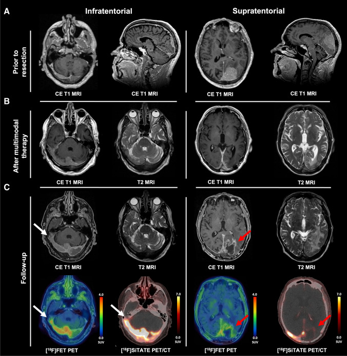
An 84-year-old male presented with transitional meningioma WHO° 1 (Figure part A, prior to resection). After completed therapy (resection, cyberknife radiosurgery, fractionated radiation), he showed right-sided residues infiltrating the transverse and sigmoid sinuses (Figure part B, after multimodal therapy). At follow-up 12 months later, MRI showed a new heterogeneous contrast enhancement in the left occipital resection cavity, suggestive of tumor progression (Figure part C, follow-up) [1]. Due to limited availability of somatostatin receptor (SSTR) PET imaging, [18F]FET PET was performed as an alternative method. The left occipital lesion showed minor radionuclide uptake on [18F]FET PET (TBRmax: 2.4, TBRmean: 1.2; red arrow), whereas the right-sided meningioma showed intense uptake (TBRmax: 4.6; white arrow). Analysis of [18F]FET uptake dynamics revealed decreasing time–activity curves (TTPmin:12.5 min) in the right-sided meningioma and increasing curves in the left occipital lesion. Three weeks later, we performed SSTR imaging using [18F]SiTATE, showing typical SSTR expression of the right-sided meningioma (SUVmax: 17.1; white arrow), but no typical SSTR expression in the left occipital lesion (SUVmax: 1.9; red arrow) [2]. Together with the moderate [18F]FET uptake, these findings were interpreted as pseudoprogression, confirmed by further follow-up.
The incidence of posttherapeutic pseudoprogression in meningioma is still unknown but considered rare [3]. With an increasing range of treatment options, diagnostic strategies are required to distinguish tumor recurrence more accurately from pseudoprogression [3]. However, when rapid clinical access to SSTR imaging is limited, this may delay diagnosis [4, 5]. To our knowledge, this is the first case demonstrating the value of dual tracer PET imaging in the detection of pseudoprogression in meningioma.
Acknowledgements
We thank the patient for granting permission to publish this article.
Author contribution
K. J. M.: writing original draft, visualization. A. B.: writing-review and editing. C. S.: writing-review and editing. L. B.: writing-review and editing. N. L. A.: supervision, visualization, writing-review and editing.
Funding
Open Access funding enabled and organized by Projekt DEAL.
Declarations
Ethical approval
Written informed consent in respect of this case report was obtained in accordance with the Declaration of Helsinki. No ethics board approval was required for this case report. The article was written according to the CARE reporting guidelines.
Conflict of interest
The authors declare no competing interests.
Footnotes
Publisher's Note
Springer Nature remains neutral with regard to jurisdictional claims in published maps and institutional affiliations.
References
- 1.Rogers L, Barani I, Chamberlain M, et al. Meningiomas: knowledge base, treatment outcomes, and uncertainties. A RANO review. JNS. 2015;122(1):4–23. doi: 10.3171/2014.7.JNS131644. [DOI] [PMC free article] [PubMed] [Google Scholar]
- 2.Ilhan H, Lindner S, Todica A, et al. Biodistribution and first clinical results of 18F-SiFAlin-TATE PET: a novel 18F-labeled somatostatin analog for imaging of neuroendocrine tumors. Eur J Nucl Med Mol Imaging. 2020;47(4):870–880. doi: 10.1007/s00259-019-04501-6. [DOI] [PubMed] [Google Scholar]
- 3.Wirsching HG, Steiner L, Becker D, et al. Increase in contrast-enhancing volume of irradiated meningiomas reflects tumor progression and not pseudoprogression. Neuro Oncol. 2021;23(9):1612–1613. doi: 10.1093/neuonc/noab119. [DOI] [PMC free article] [PubMed] [Google Scholar]
- 4.Hennrich U, Benešová M. [68Ga]Ga-DOTA-TOC: the first FDA-approved 68Ga-radiopharmaceutical for PET imaging. Pharmaceuticals. 2020;13(3):38. doi: 10.3390/ph13030038. [DOI] [PMC free article] [PubMed] [Google Scholar]
- 5.Unterrainer M, Kunte SC, Unterrainer LM, et al. Next-generation PET/CT imaging in meningioma—first clinical experiences using the novel SSTR-targeting peptide [18F]SiTATE. Eur J Nucl Med Mol Imaging. 2023;50:3390–3399. doi: 10.1007/s00259-023-06315-z. [DOI] [PMC free article] [PubMed] [Google Scholar]


