Abstract
The Srb10-Srb11 protein kinase of Saccharomyces cerevisiae is a cyclin-dependent kinase (cdk)-cyclin pair which has been found associated with the carboxy-terminal domain (CTD) of RNA polymerase II holoenzyme forms. Previous genetic findings implicated the Srb10-Srb11 kinase in transcriptional repression. Here we use synthetic promoters and LexA fusion proteins to test the requirement for Srb10-Srb11 in repression by Ssn6-Tup1, a global corepressor. We show that srb10Δ and srb11Δ mutations reduce repression by DNA-bound LexA-Ssn6 and LexA-Tup1. A point mutation in a conserved subdomain of the kinase similarly reduced repression, indicating that the catalytic activity is required. These findings establish a functional link between Ssn6-Tup1 and the Srb10-Srb11 kinase in vivo. We also explored the relationship between Srb10-Srb11 and CTD kinase I (CTDK-I), another member of the cdk-cyclin family that has been implicated in CTD phosphorylation. We show that mutation of CTK1, encoding the cdk subunit, causes defects in transcriptional repression by LexA-Tup1 and in transcriptional activation. Analysis of the mutant phenotypes and the genetic interactions of srb10Δ and ctk1Δ suggests that the two kinases have related but distinct roles in transcriptional control. These genetic findings, together with previous biochemical evidence, suggest that one mechanism of repression by Ssn6-Tup1 involves functional interaction with RNA polymerase II holoenzyme.
RNA polymerase II holoenzyme forms purified from the yeast Saccharomyces cerevisiae contain a mediator complex, which functions in transcriptional activation (3, 12, 18, 22, 23, 53). Components of mediator/holoenzyme forms include Srb proteins, Gal11, Sin4, Rgr1, Rox3, and general transcription factors (16, 18, 22, 23, 29, 30). Genetic evidence suggests that the mediator/holoenzyme plays a role not only in transcriptional activation but also in repression (for a review, see reference 4). Mutations in the genes encoding Srb8 to Srb11, Gal11, Sin4, Rgr1, and Rox3 appear to relieve negative regulation of diversely regulated genes (6, 9, 13, 20, 25, 39, 44, 49, 52, 60, 61).
Two of these proteins, Srb10 and Srb11, constitute a cyclin-dependent kinase (cdk)-cyclin pair (25, 30). The connection to RNA polymerase II was first established by the isolation of srb10 and srb11 alleles as suppressors of truncations in the carboxy-terminal repeat domain (CTD) of the largest subunit of polymerase (30). The Srb10-Srb11 kinase was found associated with an RNA polymerase II holoenzyme form and was shown to affect phosphorylation of the CTD in vitro (18, 30). Mutations in SRB10 and SRB11 also reduced the activation of GAL promoters (25, 30). Mutations in both genes had previously been isolated in genetic selections for specific effects on gene regulation. Alleles called ssn3 and ssn8 were identified as suppressors of a defect in the Snf1 protein kinase and were shown to affect glucose repression of the SUC2 gene (5, 25, 60). A related selection for suppressors affecting the Snf1-dependent expression of gluconeogenic genes yielded the gig mutations (2). A search for mutations that allow the expression of meiotic genes in vegetatively growing cells yielded ume3 and ume5 (8, 51, 52). The kinase subunit was also identified by mutations (are1) that impair α2 repression, the repression of a-specific genes in MATα cells (61). Both glucose repression of SUC2 and α2 repression depend on the Ssn6(Cyc8)-Tup1 complex, a global corepressor (21, 33, 46, 47, 56, 62). Thus, these genetic findings implicate the Srb10-Srb11 kinase in transcriptional repression and also raise the possibility of a direct role in the response to Ssn6-Tup1.
The Ssn6-Tup1 corepressor is recruited to many different promoters by specific DNA-binding regulatory proteins (1, 21, 24, 32, 34, 48, 55, 59, 65). Tup1 plays a primary role in repression (58), while Ssn6 mediates most, although not all, contacts with DNA-binding proteins (24, 48, 59). The mechanisms responsible for repression are not yet understood. Several lines of evidence indicate that Ssn6-Tup1 is required for the formation of chromatin structures that may be inhibitory to the binding of transcription factors (reviewed in reference 40). Positioned nucleosomes are observed at Ssn6-Tup1-repressed promoters in the wild type but not in ssn6 or tup1 mutants (7). In addition, Tup1 interacts in vitro with histones H3 and H4, and mutations in the histones that reduce this interaction also reduce repression by Ssn6-Tup1 in vivo (11). These findings suggest that Ssn6-Tup1 functions by establishing or maintaining repressive chromatin. However, other evidence suggests that the corepressor directly contacts components of the transcription machinery; Ssn6-Tup1-dependent repression was reconstituted in an in vitro transcription system without chromatin assembly (19, 36), and α2 repression can be achieved in the apparent absence of positioned nucleosomes (37).
In this study, we present evidence that the Srb10-Srb11 kinase plays a role in repression by Ssn6-Tup1 in vivo. We have used simple synthetic reporters and LexA fusion proteins to test the requirement for Srb10-Srb11 in repression by Ssn6-Tup1. Previous studies showed that LexA-Ssn6 and LexA-Tup1 fusion proteins, when bound to a promoter via LexA operators, repress transcription; LexA-Ssn6 requires Tup1 for repression (21), whereas LexA-Tup1 functions independently of Ssn6 (58). We show that mutations in the Srb10-Srb11 kinase substantially impair transcriptional repression by DNA-bound LexA-Ssn6 and LexA-Tup1.
The srb10Δ and srb11Δ mutations cause modest defects in the repression of natural promoters, indicating that the repression mechanism involving the Srb10-Srb11 kinase is only one of the mechanisms that contribute to repression. Because Srb10-Srb11 has been implicated in CTD phosphorylation, we have explored the functional relationship between Srb10-Srb11 and CTD kinase I (CTDK-I), also a member of the cdk-cyclin family. CTDK-I exhibits CTD kinase activity in vitro and affects CTD phosphorylation in vivo (26, 50). In this study, we determined the effects of mutation of CTK1, encoding the cdk subunit, on transcriptional repression and activation and examined the genetic interactions of srb10Δ and ctk1Δ mutations. The genetic evidence suggests that the two kinases play related but distinct roles in transcriptional control.
MATERIALS AND METHODS
Strains and genetic methods.
The S. cerevisiae strains used in this work are listed in Table 1. The Escherichia coli strains used for propagation of plasmid DNA were XL1-Blue or DH5α. Genetic methods were as described previously (38), except that yeast extract-peptone (YEP), or rich, medium required supplemental tryptophan (40 μg/ml) to allow germination of ctk1Δ trp1 spores and was used for all rich medium cultures. To introduce the ctk1ΔE::URA3 allele into the S288C background, a 2.4-kb ClaI fragment from plasmid pSZ17 (26) was used to replace (42) one of two allelic copies of CTK1 in diploid FY251 × FY86 cells, followed by sporulation and tetrad analysis. To generate strain MCY3664 (ctk1ΔE::ura3::LEU2), the HindIII fragment from plasmid pWJ460 (41) carrying a ura3::LEU2 allele was used to transform strain MCY3663.
TABLE 1.
Strains of S. cerevisiae used in this study
| Straina | Genotypeb | Source |
|---|---|---|
| FY86 | MATα ura3-52 leu2-Δ1 his3-Δ200 | F. Winston |
| FY250 | MATα ura3-52 leu2-Δ1 his3-Δ200 trp1-Δ63 | F. Winston |
| FY251 | MATa ura3-52 leu2-Δ1 his3-Δ200 trp1-Δ63 | F. Winston |
| MCY3639 | MATα ura3-52 leu2-3,112 his3-Δ200 lys2-801 mig1Δ2::LEU2 | This laboratory |
| MCY3644 | Identical to MCY3647, except srb11Δ::LEU2 | This laboratory |
| MCY3647 | MATα ura3-52 leu2-3,112 his3-Δ200 lys2-801 | This laboratory |
| MCY3655 | Identical to FY250, except srb11Δ::LEU2 | This work |
| MCY3657 | MATα ura3-52 leu2-Δ1 his3-Δ200 trp1-Δ63 srb10Δ::HIS3 | This work |
| MCY3658 | MATa ura3-52 leu2-Δ1 his3-Δ200 ctk1ΔE::URA3 | This work |
| MCY3659 | MATa ura3-52 leu2-3,112 or -Δ1 his3-Δ200 lys2-801 | This work |
| MCY3660 | MATα ura3-52 leu2-3,112 or -Δ1 his3-Δ200 mig1Δ2::LEU2 ctk1ΔE::URA3 | This work |
| MCY3661 | MATa ura3-52 leu2-Δ1 his3-Δ200 trp1-Δ63 | This work |
| MCY3662 | MATa ura3-52 leu2-Δ1 his3-Δ200 trp1-Δ63 srb10Δ::HIS3 | This work |
| MCY3663 | MATα ura3-52 leu2-Δ1 his3-Δ200 trp1-Δ63 ctk1ΔE::URA3 | This work |
| MCY3664 | Identical to MCY3663, except ctk1ΔE::ura3::LEU2 | This work |
| MCY3667 | MATα ura3-52 leu2-Δ1 his3-Δ200 srb10Δ::HIS3 ctk1ΔE::URA3 | This work |
| MCY3668 | Identical to MCY3912, except srb10Δ::HIS3 | This work |
| MCY3694 | Identical to FY250, except srb10-D290A | This work |
| MCY3912 | MATa ura3-52 leu2-3,112 his3-Δ200 lys2-801 trp1-Δ1 ade2-101 | This laboratory |
| CTY10-5d | MATa ade2-101 leu2-Δ1 his3-Δ200 trp1-Δ901 gal4 gal80 URA3::lexAop-lacZ | R. Sternglanz |
All strains except CTY10-5d are derived from S288C.
srb10Δ::HIS3 and srb11Δ::LEU2 are the same as ssn3Δ1::HIS3 and ssn8Δ1::LEU2, respectively (25).
Construction of the srb10-D290A mutant.
A DNA fragment encoding the C-terminal part of Srb10-D290A was generated by PCR with Vent DNA polymerase (New England Biolabs), plasmid pPY24 (25) as a template, and primers K-24 (5′-GGATGTGTTAAAATTGGaGcTcTAGGTTTGGCCAGAAA-3′) and K-17 (5′-GACGGATCCTGAATGTTGCAGACTTGC-3′). K-24 incorporates mismatching nucleotides (lowercase type) that encode the Asp290-to-Ala substitution and create a silent diagnostic SacI site (underlined); K-17 is complementary to a region 3′ to the SRB10 gene and contains a BamHI site (underlined). The fragment was gel purified and used as a primer in the second round of PCR with the second primer K-16 (5′-CGGGATCCTAATGTATAATGGCAAGGATAGAGC-3′) and pPY24 as a template; K-16 is at the 5′ end of the gene and contains a BamHI site (underlined). The resulting mutant fragment was digested with BamHI and cloned into the vector pBTM116 (a gift of Stan Fields, University of Washington, Seattle, Wash.), which expresses LexA from a weak version of the ADH1 promoter, to generate pSK74. The LexA-Srb10-D290A protein expressed from pSK74 interacts with GAD-Srb11 in the two-hybrid system but, in contrast to the wild-type LexA-Srb10, confers a dominant flocculent phenotype. The BamHI fragment from pSK74 was cloned into the integrating URA3 vector pRS306, yielding pSK90. pSK90 was digested with HpaI and used to transform strain FY250. Because the mutant fragment contains no yeast promoter sequence and the unique HpaI site is downstream of the mutated site, only the wild-type copy of SRB10 was expressed from the resulting gene duplication, and all of the transformants were nonflocculent. Three transformants were subjected to selection against URA3 on plates containing 5-fluoroorotic acid. Two independent flocculent Ura− isolates were colony purified. To confirm the presence of the srb10-D290A allele at the correct chromosomal location, genomic DNA was used as a template in PCRs directed by primers K-41 (5′-AGGCCGCCTAGTTTTGAC-3′) and K-42 (5′-GGGCTGTAATCCTATCAG-3′). K-41 anneals to the chromosome 5′ to the PCR-amplified region; K-42 anneals within the amplified region 3′ to the mutation. The fragments resulting from the mutants were the same size as those from a wild-type control and contained the diagnostic SacI site.
Plasmid construction.
Plasmid pBM2762 (35) was modified to carry one LexA operator 5′ to UASLEU2, yielding reporter plasmid pMT27 (54). pSK101 was constructed by inserting the BamHI-SalI MIG1 fragment from pLexA-MIG1 (55) between the BamHI and SalI sites of the vector pBTM116. The CTK1 and CTK2 coding regions were amplified from genomic DNA of FY250 by PCR. The synthetic primers were as follows: for CTK1, 5′-GCGCGGATCCTAATGTCCTACAATAATGGC-3′ and 5′-GCGCGGATCCTTATTTATCATCATC-3′; for CTK2, 5′-GCGCGGATCCTAATGCCTAGCACGTTTGAATC-3′ and 5′-GCGCGGATCCTATGCATGTCTTGTAGAAC-3′ (BamHI sites and ATG are underlined). To generate fusions to the Gal4 activation domain (GAD) and the DNA-binding domain (amino acids 1 to 87) of LexA (LexA87), the PCR fragments were digested with BamHI and cloned into the BamHI site of pSH2-1 (17) and pACTII (28), respectively. The resulting plasmids, pSK63, pSK64, and pSK65, express LexA87-Ctk2, GAD-Ctk2, and LexA87-Ctk1, respectively. Expression of LexA87-Ctk1 and LexA87-Ctk2 was confirmed by immunoblot analysis; LexA87-Ctk1 complements ctk1Δ for the slow-growth and cold-sensitive phenotypes and interacts with GAD-Ctk2 in a two-hybrid assay. LexA87-Srb10, GAD-Srb10, and GAD-Srb11 were expressed from pSK39, pSK40, and pSK36, respectively (25). pSK45 is pSK40 in which the sequences encoding GAD are deleted.
Enzyme assays.
For reporter repression assays, cells were grown to mid-log phase in selective synthetic complete (SC) medium containing 2% glucose. Because the mutant strains used here are flocculent, the density of cell cultures was determined after the addition of EDTA to 5 or 10 mM to disperse cell clumps. β-Galactosidase activity was assayed in permeabilized cells or in protein extracts (38) and expressed in Miller units or in arbitrary units (1 unit = 1,000 × optical density at 420 nm [OD420] per min per mg of protein), respectively. Glucose-repressed cells were obtained by growth to mid-log phase in selective synthetic complete (SC) or rich medium containing 2% glucose; for derepression, repressed cells were shifted for 3 h to medium containing 0.05% glucose. Invertase activity was assayed in whole cells (60). For two-hybrid interaction assays, filter lift assays of β-galactosidase activity in transformants were performed (63) after growth on selective SC plates containing 2% glucose.
Immunoblot analysis.
Cells were grown to the mid-log phase in selective SC medium containing 2% glucose. Protein extracts were prepared as described previously (63). Alternatively, cell pellets were directly resuspended in sample buffer (1×) containing 2% sodium dodecyl sulfate and 2% β-mercaptoethanol (0.1 ml of sample buffer per equivalent of 1 ml of culture at an OD600 of 1.0), boiled for 5 min, and cleared by centrifugation at 12,000 × g for 1 min. Proteins were separated on sodium dodecyl sulfate–8% polyacrylamide gels and analyzed by immunoblotting with rabbit polyclonal LexA antibodies (gift of C. Denis, University of New Hampshire) and enhanced chemiluminescence (ECL reagents; Amersham).
RESULTS
Transcriptional repression by LexA-Tup1 requires the Srb10-Srb11 kinase.
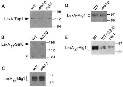
We first tested whether transcriptional repression by LexA-Tup1 requires the Srb10-Srb11 kinase. Wild-type and isogenic srb10Δ and srb11Δ mutants were cotransformed with a plasmid carrying genes expressing LexA-Tup1 (58) and CYC1-lacZ reporters (15, 21) containing no or one LexA operator located 5′ to the CYC1 upstream activation sequence (UAS) (Fig. 1). Transformants were assayed for β-galactosidase activity after growth in glucose. In wild-type cells, LexA-Tup1 repressed transcription 14-fold, as calculated by comparing the expression of the reporter containing a LexA operator to that of the reporter lacking an operator (Table 2). In the srb10Δ and srb11Δ mutants, however, LexA-Tup1 repressed transcription only 1.7- and 1.3-fold, which was not significantly different from the results with the LexA controls. Thus, repression was reduced 8- to 10-fold in the mutants. Immunoblot analysis indicated that LexA-Tup1 was expressed at the same level in the mutants and wild type (Fig. 2A and data not shown). The use of a reporter with a larger number of LexA operators (four) (pJK1621; Fig. 1) did not improve repression or relieve the dependence on Srb10-Srb11 (data not shown).
FIG. 1.
Reporter plasmids used in repression assays. The CYC1-lacZ reporter plasmid pLGΔ312S (15) has no LexA operator, and its derivatives pCK26 and pCK30 (21) carry one LexA operator located 5′ or 3′ to UASCYC1, respectively; pJK1621 carries four LexA operators 5′ to UASCYC1. The UASLEU2-HIS3-lacZ reporter plasmid pBM2762 (35) has no operator, and its derivative pMT27 carries one LexA operator 5′ to UASLEU2. Representations are not to scale.
TABLE 2.
Mutations in SRB10, SRB11, and CTK1 affect repression of CYC1-lacZ by LexA-Tup1a
| Protein | Genotype | β-Gal activityb
|
Fold repressionc | |
|---|---|---|---|---|
| −LexAop | +LexAop | |||
| 5′ to UAS | ||||
| LexA | WT | 162 | 95 | 1.7 |
| srb10Δ | 277 | 207 | 1.3 | |
| srb11Δ | 270 | 200 | 1.4 | |
| srb10-D290A | 281 | 129 | 2.2 | |
| ctk1Δ | 247 | 157 | 1.6 | |
| LexA-Tup1 | WT | 102 | 7.5 | 14 |
| srb10Δ | 182 | 105 | 1.7 | |
| srb11Δ | 152 | 115 | 1.3 | |
| srb10-D290A | 240 | 87 | 2.8 | |
| ctk1Δ | 60 | 18 | 3.3 | |
| 3′ to UAS | ||||
| LexA | WT | 167 | 36 | 4.6 |
| srb10Δ | 321 | 94 | 3.4 | |
| LexA-Tup1 | WT | 124 | 2.1 | 59 |
| srb10Δ | 261 | 42 | 6.2 | |
Isogenic wild-type (WT) and mutant strains were cotransformed with plasmid YCp91 or pLexA-Tup1 (58) expressing LexA and LexA-Tup1, respectively, and CYC1-lacZ reporter plasmids (Fig. 1) with no LexA operator (−LexAop) or one LexA operator (+LexAop) located 5′ or 3′ to UASCYC1, as indicated.
β-Galactosidase (β-Gal) activity was assayed in protein extracts, and values are averages for 4 to 10 independent transformants. Standard errors were typically 10 to 15%.
Fold repression was calculated as the ratio of the values obtained for reporters with and without a LexA operator. The host strains were FY250, MCY3655, MCY3661, MCY3662, MCY3664, and MCY3694.
FIG. 2.
Immunoblot analysis of LexA fusion proteins. Protein extracts (5 μg [A] or 20 μg [B and C]) or boiled cells (D and E) [equivalent of 0.2 ml of culture at an OD600 of 1.0, except lane WT(0.3×)] were analyzed by immunoblotting with LexA antibodies. (A) LexA-Tup1 protein in wild-type (WT) and srb10Δ transformants carrying pLexA-Tup1 and pCK26. These transformants were assayed in Table 2. Two srb11Δ transformants were also analyzed and expressed LexA-Tup1 at levels equal to those of the wild type (data not shown). (B) LexA87-Ssn6 protein in wild-type and srb10Δ transformants carrying pCK23 and pMT27. These transformants were assayed, and the results are shown in Table 3. The asterisk marks a degradation product. (C) LexA87-Mig1 protein in wild-type and srb11Δ transformants carrying pLexA-Mig1 (Table 4, experiment A). (D) LexA-Mig1 protein in wild-type and srb10Δ transformants carrying pSK101 and pCK26. These transformants were assayed, and the results are shown in Table 4 (experiment B). (E) LexA87-Mig1 protein in wild-type and ctk1Δ transformants carrying pLexA-Mig1 and pCK26. These transformants were assayed, and the results are shown in Table 4 (experiment C). The lane labeled WT(0.3×) was loaded with threefold less protein than the other lanes. In panels C to E, the multiple bands correspond to different phosphorylation states (55).
To demonstrate that efficient repression requires the catalytic activity of Srb10, we constructed a mutation that causes Asp290 in subdomain VII to be replaced with Ala, here designated srb10-D290A. This mutation encodes the same amino acid substitution as the allele srb10-3, which was reported by Liao et al. (30) to inactivate the kinase without affecting its incorporation into the RNA polymerase II holoenzyme. In the srb10-D290A mutant, LexA-Tup1 repressed transcription of the reporter with one LexA operator only 2.8-fold, which is comparable to the LexA control value, 2.2-fold (Table 2). Immunoblot analysis detected the same levels of LexA-Tup1 in the mutant and wild type (data not shown).
We also performed repression assays by using a CYC1-lacZ reporter with a LexA operator inserted between the UAS and TATA sequence (pCK30; Fig. 1). In this case, LexA-Tup1 repressed transcription 59-fold (Table 2). Repression by LexA-Tup1 in an isogenic srb10Δ mutant was 6.2-fold, which is only slightly higher than that of the LexA control (3.4-fold).
Thus, repression by LexA-Tup1 requires the catalytic activity of the Srb10-Srb11 kinase. Deletion of either subunit of the kinase reduces repression 8- to 10-fold, which is close to the limit of sensitivity in this assay. Moreover, repression is dependent on Srb10-Srb11 when LexA-Tup1 is tethered to sites located either 5′ or 3′ to the UAS.
Repression of a UASLEU2-HIS3-lacZ reporter by LexA-Ssn6 requires Srb10.
To show that this Srb10-Srb11 dependence is not specific to the CYC1-lacZ reporter, we next examined the repression of a reporter with different UAS and TATA elements. We used a pair of reporters in which UASLEU2 drives the expression of a HIS3-lacZ fusion from the TATAHIS3 sequence (35), with no or one LexA operator inserted 5′ to the UAS (Fig. 1). In addition, to confirm that Srb10-Srb11 dependence is not specific to the LexA-Tup1 fusion protein, we used LexA87-Ssn6 (21) as the DNA-bound protein, which represses by recruiting the native Tup1. Repression was assayed in wild-type and isogenic srb10Δ mutant cells. In the wild type, DNA-bound LexA87-Ssn6 caused 6.7-fold repression of the reporter containing a LexA operator relative to that of the reporter lacking an operator (Table 3). In the srb10Δ mutant, repression was reduced to levels comparable to those for the LexA control (1.4-fold). Immunoblot analysis confirmed that the repression defect is not caused by lower levels of LexA87-Ssn6 (Fig. 2B).
TABLE 3.
Effect of srb10Δ on repression of UASLEU2-HIS3-lacZ by LexA87-Ssn6a
| Protein | Genotype | β-Gal activityb
|
Fold repression | |
|---|---|---|---|---|
| −LexAop | +LexAop | |||
| LexA | WT | 292 | 232 | 1.3 |
| srb10Δ | 330 | 232 | 1.4 | |
| LexA87-Ssn6 | WT | 370 | 55 | 6.7 |
| srb10Δ | 436 | 253 | 1.7 | |
Isogenic strains MCY3912 (wild type [WT]) and MCY3668 (srb10Δ) were cotransformed with plasmid pBTM116 or pCK23 (21) expressing LexA and LexA87-Ssn6, respectively, and UASLEU2-HIS3-lacZ reporter plasmids with no or one LexA operator 5′ to UASLEU2 (Fig. 1).
β-Galactosidase (β-Gal) activity was assayed in permeabilized cells. Values are averages for three to five independent transformants. Standard errors were <17%.
Repression of CYC1-lacZ by LexA-Mig1 is partially dependent on Srb10 and Srb11.
We next examined repression by LexA87-Mig1. Mig1 is one of two DNA-binding proteins that function with Ssn6-Tup1 to repress SUC2 (21, 32, 34, 55, 59, 62), and this LexA87-Mig1 fusion protein is an Ssn6-Tup1-dependent repressor (55). LexA87-Mig1 repressed CYC1-lacZ expression 16-fold in the wild type but only 5.9-fold in an srb11Δ mutant (Table 4, experiment A). The latter value, however, is still significantly greater than that for the LexA87 control (1.3-fold). LexA87-Mig1 protein levels in the mutant and wild type were comparable; moreover, the repression defect in the srb11Δ mutant was not associated with a major change in phosphorylation of LexA87-Mig1 (Fig. 2C) (55).
TABLE 4.
Effects of srb10Δ, srb11Δ, and ctk1Δ on repression of CYC1-lacZ by LexA-Mig1a
| Protein | Genotype | β-Gal activity
|
Fold repression
|
|||
|---|---|---|---|---|---|---|
| −LexAop | +1 LexAop | +4 LexAop | 1op | 4op | ||
| Expt Ab | ||||||
| LexA87 | WT | 76 | 78 | ND | 1.0 | ND |
| srb11Δ | 199 | 148 | ND | 1.3 | ND | |
| LexA87-Mig1 | WT | 106 | 6.6 | ND | 16 | ND |
| srb11Δ | 124 | 21 | ND | 5.9 | ND | |
| Expt Bc | ||||||
| LexA | WT | 91 | 66 | 63 | 1.4 | 1.4 |
| srb10Δ | 105 | 74 | 73 | 1.4 | 1.4 | |
| LexA-Mig1 | WT | 111 | 19 | 16 | 5.8 | 6.9 |
| srb10Δ | 86 | 35 | 21 | 2.5 | 4.1 | |
| Expt Cd | ||||||
| LexA87 | WT | 77 | 51 | 46 | 1.5 | 1.7 |
| ctk1Δ | 106 | 72 | 58 | 1.5 | 1.8 | |
| LexA87-Mig1 | WT | 99 | 6.3 | 2.8 | 16 | 35 |
| ctk1Δ | 144 | 26 | 14 | 5.5 | 10 | |
Pairs of isogenic strains were cotransformed with plasmids expressing fusion proteins and CYC1-lacZ reporter plasmids carrying zero, one, or four LexA operators 5′ to the UAS (Fig. 1). Fold repression for both one-operator (1op) and four-operator (4op) reporters is relative to the operator-less reporter. ND, not determined.
The strains were MCY3647 and MCY3644, and the plasmids were pSH2-1 or pLexA-Mig1. β-Galactosidase (β-Gal) activity was assayed in permeabilized cells. Values are averages for three independent transformants. Standard errors were <20%.
The strains were MCY3661 and MCY3662, and the plasmids were pBTM116 and pSK101. β-Galactosidase activity was assayed in protein extracts. Values are averages for five transformants. Standard errors were <12%.
Same as described for experiment B, except that the strains were MCY3661 and MCY3664 and the plasmids were pSH2-1 or pLexA-Mig1. Standard errors were <14%.
The fact that this dependence was only partial could result from the massive overexpression of LexA87-Mig1 from the strong ADH1 promoter of the vector pSH2-1. To address this possibility, we tested a different LexA-Mig1 fusion protein, which was expressed at a much lower level (data not shown) from a shorter version of the ADH1 promoter present in the vector pBTM116. Again, the Srb10 dependence was partial (Table 4, experiment B). The LexA-Mig1 protein levels were comparable in the wild type and srb10Δ mutant (Fig. 2D).
CTDK-I contributes to repression of SUC2.
For all natural promoters tested, the repression defects caused by srb10 and srb11 mutations are modest. Thus, the repression mechanism involving the Srb10-Srb11 kinase is only one of the mechanisms that contribute to repression. We considered the possibility that another, related kinase functions redundantly, or partly so, with Srb10-Srb11. CTDK-I is similar to Srb10-Srb11 in that two of the subunits are cdk and cyclin homologs (Ctk1 and Ctk2, respectively) and CTDK-I exhibits CTD kinase activity (26, 50). In addition, ctk1 and ctk2 mutants apparently resembled srb10 and srb11 mutants with respect to their flocculent, cold-sensitive, and slow-growth phenotypes and their failure or reduced ability to sporulate, although the characterized mutants were in different genetic backgrounds (25, 26, 30, 50, 52). Thus, CTDK-I seemed a likely candidate.
To test whether CTDK-I is functionally related to Srb10-Srb11, we first introduced a ctk1Δ allele (26) into the S288C genetic background, which we have used for previous studies of srb10Δ (25) (see Materials and Methods). The resulting mutants exhibited slow growth, cold sensitivity, and inability of homozygous diploids to sporulate, as reported previously (26, 50); however, in the S288C background, ctk1Δ mutants were not flocculent, although microscopic examination revealed clustered cells. We also found that these mutants were impaired in growth on galactose. The srb10Δ mutants in the S288C background are similar with respect to slow growth and impaired utilization of galactose, but they are highly flocculent, less cold sensitive, and able to sporulate (25).
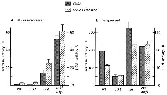
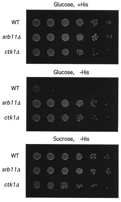
We next examined the ctk1Δ mutants for repression of the SUC2 (invertase) gene in response to glucose, which requires Ssn6-Tup1 (5, 46, 56). The ctk1Δ mutation did not substantially relieve repression; however, ctk1Δ impaired positive regulation of SUC2 a fewfold, which could mask an effect on repression (Fig. 3). We therefore turned to a more sensitive assay to detect relief of glucose repression, based on a SUC2-HIS3 fusion in which SUC2 regulatory sequences mediate glucose repression of HIS3 expression (57). When cells are grown on glucose, the SUC2 promoter is repressed and HIS3 is not expressed; however, when cells are grown on sucrose or when cells are defective in glucose repression, the fusion confers a His+ phenotype. A CEN-TRP1 plasmid carrying this reporter was introduced into isogenic wild-type, ctk1Δ, and srb11Δ strains, all with the chromosomal HIS3 locus deleted. Both ctk1Δ and srb11Δ strains grew on glucose in the absence of histidine, whereas the wild type did not (Fig. 4). In the control experiment, all the strains grew on sucrose without histidine. Thus, ctk1Δ and srb11Δ relieve glucose repression of the SUC2 promoter in this assay.
FIG. 3.
Combined effect of ctk1Δ and mig1Δ on the regulation of SUC2 and SUC2-LEU2-lacZ. Invertase assays were performed in glucose-repressed and derepressed segregants of the MCY3639 × MCY3658 cross. Segregants bearing SUC2-LEU2-lacZ were derived from the cross of MCY3660 (ctk1Δ mig1Δ) with a transformant of MCY3659 (wild type [WT]) carrying integrated pLS11 (45). β-Galactosidase (β-Gal) activity was assayed in permeabilized cells. Values are averages for at least three segregants. (A) Glucose-repressed cultures. (B) Derepressed cultures.
FIG. 4.
ctk1Δ relieves glucose repression of a SUC2-HIS3 reporter. The trp1Δ his3Δ strains FY250 (WT), MCY3663 (ctk1Δ), and MCY3655 (srb11Δ) were transformed with the CEN-TRP1 plasmid pYSH (57). Serial sixfold dilutions of cell suspensions (diluted in 10 mM Tris-HCl [pH 7.0] containing 10 mM EDTA to disperse flocculated cells) were spotted on SC-Trp or SC-Trp-His containing 2% glucose or sucrose, as indicated. The plates were photographed after 4 days at 30°C. For either mutant, the number of colonies on glucose-His was approximately equal to that in the corresponding dilution on sucrose-His, suggesting that possible cross-feeding and stochastic effects did not occur.
Both srb10Δ and srb11Δ synergize strongly with the mig1Δ mutation to relieve repression of SUC2 (60). In a mig1Δ mutant, the recruitment of Ssn6-Tup1 to the SUC2 promoter is impaired but the Mig2 protein partially substitutes for Mig1 (32); however, the resulting repression is highly dependent on Srb10-Srb11. We therefore tested ctk1Δ for synergy with mig1Δ. In ctk1Δ mig1Δ double mutants, constructed by genetic crossing, the repressed invertase activity was about fourfold higher than in mig1Δ single-mutant segregants (Fig. 3). We also examined the regulation of an integrated SUC2-LEU2-lacZ fusion, under control of the SUC2 regulatory region and the LEU2 promoter (45), and the β-galactosidase activity correlated well with invertase activity (Fig. 3). Although the synergistic effect of srb10Δ is more pronounced (about 10-fold [60]), the effect of ctk1Δ on repression may be underestimated due to the defect in SUC2 activation. Together, these results strongly suggest that CTDK-I contributes to the transcriptional repression of SUC2.
Effect of ctk1Δ on repression by LexA-Tup1.
To test whether CTDK-I contributes to SUC2 repression via the Ssn6-Tup1 corepressor, we examined the effect of ctk1Δ on the ability of LexA-Tup1 to repress transcription of a lexAop-CYC1-lacZ reporter (pCK26; Fig. 1). LexA-Tup1 repressed transcription only 3.3-fold in the mutant, compared to 14-fold in the isogenic wild type (Table 2). LexA-Tup1 expression in the mutant was confirmed by immunoblot analysis (Fig. 2A).
We also tested the effect of ctk1Δ on repression by LexA-Mig1. Repression by LexA-Mig1 in the mutant was reduced to the LexA control levels, but we could not detect the fusion protein (data not shown). In the case of LexA87-Mig1, expressed from a stronger version of the ADH1 promoter, repression was threefold lower in the mutant (Table 4, experiment C) but the LexA87-Mig1 protein level was also at least threefold lower than in the wild type (Fig. 2E). Therefore, it is not clear that ctk1Δ has any effect on repression by LexA-Mig1.
Ctk1 and Srb10 are not functionally interchangeable.
The similarities between the ctk1Δ and srb10Δ mutant phenotypes suggested that Ctk1 and Srb10 are functionally related and raised the possibility that they are interchangeable cdk subunits; the differences in phenotype could conceivably result from different levels of expression of the two proteins. To address this issue, we first tested whether overexpression of Srb10 can suppress ctk1Δ for the SUC2 repression defect. We transformed a mig1Δ ctk1Δ double mutant (MCY3660) with a multicopy plasmid expressing a functional LexA87-Srb10 protein from the strong ADH1 promoter (pSK39) (25). Invertase activity in glucose-repressed transformants was not significantly different from that in transformants expressing LexA (average values, 81 and 87 U, respectively). In another experiment, overexpression of Srb10 from the ADH1 promoter (pSK45) in a ctk1Δ mutant did not improve growth on galactose relative to that of vector controls. Thus, these assays provide no evidence that elevated levels of Srb10 can compensate for the absence of Ctk1.
Taking a different approach, we next used the two-hybrid system to test for heterotypic interaction between the kinase and cyclin subunits of the two complexes, using the lexAop-GAL1-lacZ reporter in strain CTY10-5d. No interaction was detected between LexA87-Ctk1 and GAD-Srb11 or between LexA87-Ctk2 and GAD-Srb10; in control experiments, LexA87-Srb10 interacted with GAD-Srb11 and LexA87-Ctk1 interacted with GAD-Ctk2 (see Materials and Methods). This lack of heterotypic interaction is consistent with the sequence divergence between Srb10 and Ctk1; for example, the sequence corresponding to the PSTAIRE motif, which is required for specific interaction with cyclins, is SQSACRE in Srb10 and PITSIRE in Ctk1.
These two-hybrid experiments also revealed another difference between the subunits of the two kinases. In contrast to LexA87-Srb10 and LexA87-Srb11 (25), LexA87 fusions to Ctk1 and Ctk2 showed no ability to activate transcription of reporters (data not shown).
Evidence for independent function of the Srb10-Srb11 and CTDK-I kinases.
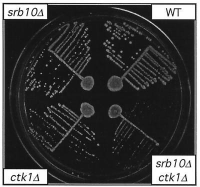
To explore the functional relationship between the CTDK-I and Srb10-Srb11 kinases, we examined the genetic interactions between the ctk1Δ and srb10Δ mutations. If the two kinases function independently, the double mutant should exhibit a more pronounced phenotypic defect than either single mutant. We constructed such double mutants by genetic crossing, and we found that the srb10Δ ctk1Δ segregants were viable but grew more slowly than the single mutants (Fig. 5). The double mutants exhibited a substantially longer doubling time in rich medium (6.1 h) than did either single mutant (3.4 h for ctk1Δ; 2.4 h for srb10Δ) or the wild type (1.8 h). These findings indicate that CTDK-I and Srb10-Srb11 have independent inputs into some cellular processes. Moreover, the viability of the double mutants indicates that the two kinases cannot together be responsible for an essential function.
FIG. 5.
Growth of srb10Δ ctk1Δ double mutants. A diploid heterozygous for srb10Δ::HIS3 and ctk1ΔE::URA3 (MCY3657 × MCY3658) was sporulated and subjected to tetrad analysis. Double-mutant segregants were identified by their His+ Ura+ phenotype and corresponded to slowly growing spore clones (data not shown). Representative segregants were streaked on rich medium containing 2% glucose. The plate was photographed after 2 days at 30°C. The single-mutant colonies are smaller than those of the wild type (WT) during the early stages of growth.
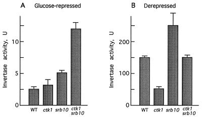
We next tested whether the SUC2 repression defect of the srb10Δ ctk1Δ double mutant exceeds the defect of either single mutant. Under glucose-repressing conditions, invertase activity in the srb10Δ ctk1Δ double mutant was 2.4- and 3.8-fold higher than in srb10Δ and ctk1Δ single mutants, respectively (Fig. 6A). However, the fold regulation of SUC2 was not substantially different in the double mutant than in the ctk1Δ mutant, which is defective in derepression (Fig. 6B). Derepressed invertase activity was closer to the wild-type level in the double mutant.
FIG. 6.
Combined effect of srb10Δ and ctk1Δ on SUC2 expression. Segregants from the cross described in the legend to Fig. 5 were assayed for invertase activity after growth under glucose-repressing conditions (A) or after a shift to derepressing conditions (B). WT, wild type.
The Srb10-Srb11 kinase has been implicated not only in repression but also in transcriptional activation, and both srb10Δ and srb11Δ mutants are defective in induction of GAL genes (25, 30). The growth defect of ctk1Δ mutants on galactose suggested that CTDK-I plays a similar role, so we tested whether ctk1Δ similarly impairs the induction of an integrated GAL1-lacZ fusion (64). Wild-type and mutant strains were assayed for β-galactosidase activity after growth in galactose (Table 5). Activity in the srb10Δ and ctk1Δ mutants was 2.5- and 8.0-fold lower, respectively, than in the wild type. To test whether Srb10-Srb11 and CTDK-I contribute independently to the induction of GAL1-lacZ, we examined the srb10Δ ctk1Δ double mutants. The defect was more pronounced in the double mutants, and activity was decreased 18-fold relative to that of the wild type, suggesting that the two kinases function independently.
TABLE 5.
Effects of ctk1Δ and srb10Δ on GAL1-lacZ expressiona
| Relevant genotype | β-Gal activityb | Fold induction defect |
|---|---|---|
| Wild type | 2,210 | 1.0 |
| srb10Δ | 871 | 2.5 |
| ctk1Δ | 275 | 8.0 |
| srb10Δ ctk1Δ | 122 | 18 |
Strains carrying an integrated GAL1-lacZ fusion gene were derived from the cross of MCY3667 (srb10Δ ctk1Δ) with MCY3659 (wild type), which had been transformed with plasmid pRY171 (64). Cells were grown in rich medium with 2% galactose (inducing conditions).
β-Galactosidase (β-Gal) activity was assayed in permeabilized cells. Values are averages for at least three segregants of each genotype. Standard errors were <15%.
DISCUSSION
In this study, we have examined the role of the Srb10-Srb11 kinase in transcriptional repression by the Ssn6-Tup1 corepressor. We show that srb10Δ and srb11Δ mutations reduce the repression of synthetic reporters by DNA-bound LexA-Ssn6 and LexA-Tup1. Repression was reduced 8- to 10-fold when LexA-Tup1 was tethered to sites either 5′ or 3′ to the UAS of the reporter. Moreover, using a point mutation that inactivates the kinase, we showed that repression requires the catalytic activity of Srb10. The Srb10-Srb11 kinase has been found associated with an RNA polymerase II holoenzyme form (18, 30). Together with biochemical evidence that Ssn6-Tup1-dependent repression acts on the transcription apparatus (19, 36), these genetic data suggest that one mechanism of repression involves functional interaction between Ssn6-Tup1 and RNA polymerase II holoenzyme.
Repression of the native SUC2 promoter by Ssn6-Tup1 is only partially dependent on the Srb10-Srb11 kinase. Mutation of SSN6 or TUP1 abolishes the repression of SUC2, yielding a greater than 100-fold increase in expression relative to the level of expression of the wild type. In contrast, mutation of SRB10 or SRB11 causes a modest effect (a fewfold alone and 10-fold when combined with mig1, which partially impairs the recruitment of Ssn6-Tup1 [25, 60]). We show here that the absence of a stronger phenotype in these mutants cannot be attributed to a redundant function provided by CTDK-I, a related cdk-cyclin which has been implicated in CTD phosphorylation. Mutation of CTK1 causes a similarly modest effect on repression, and the combination of srb10Δ and ctk1Δ does not reproduce the effect of ssn6Δ or tup1Δ. Mutations in various RNA polymerase II holoenzyme components also cause a partial loss of repression at Ssn6-Tup1-dependent promoters (6, 44, 49, 60, 61). These results are consistent with models in which Ssn6-Tup1 effects repression by at least two mechanisms, one involving functional interaction with RNA polymerase II holoenzyme and another most probably involving chromatin (7, 11, 40). In the assays presented here, repression by LexA-Mig1 was less dependent on Srb10-Srb11 than was repression by LexA-Tup1 and LexA-Ssn6, suggesting that the relative contributions of Srb10-Srb11-dependent and -independent repression mechanisms vary with the particulars of the recruitment of Ssn6-Tup1. The spatial relationship of the Ssn6-Tup1 corepressor to chromatin, RNA polymerase II holoenzyme, and other transcription factors should more closely approximate the natural configuration when Ssn6-Tup1 is recruited by LexA-Mig1 than when the corepressor is tethered by direct fusion to LexA.
We have explored the relationship of the CTDK-I kinase to Srb10-Srb11, and we present genetic evidence that these two kinases have related but distinct roles in transcriptional control. First, we compared the effects of mutations in the cognate genes in the S288C genetic background and showed that ctk1Δ and srb10Δ cause similar but not identical phenotypes. Both mutants exhibit slow growth and impaired utilization of galactose. However, the mutant phenotypes differ with respect to flocculence, degree of cold sensitivity, and sporulation proficiency. We also examined the ctk1Δ mutant for defects in transcriptional regulation of SUC2. Like srb10Δ, the ctk1Δ mutation alone causes mild defects in glucose repression of SUC2 but acts synergistically with mig1Δ to relieve repression. We present evidence that repression by LexA-Tup1 is reduced in the ctk1Δ mutant. A difference between the two mutants is that the ctk1Δ mutant exhibits a significant defect in derepression of SUC2 whereas the srb10Δ mutant derepresses invertase activity to somewhat higher levels than does the wild type. Finally, we showed that ctk1Δ impairs induction of the GAL1 promoter, as previously shown for srb10Δ (25, 30). The many shared mutant phenotypes indicate that the Ctk1 and Srb10 proteins function in some of the same cellular processes, including both transcriptional repression and activation. The mutant phenotypes are not identical, however, strongly suggesting that CTDK-I and Srb10-Srb11 have related but distinct functions.
Genetic studies further support the view that Ctk1 and Srb10 are not functionally interchangeable. First, overexpression of Srb10 did not suppress defects caused by the loss of Ctk1. Second, in two-hybrid assays, Ctk1 did not interact with the Srb11 cyclin and Srb10 did not interact with the Ctk2 cyclin. These studies also showed that Srb10 and Srb11 differ from Ctk1 and Ctk2 with respect to the ability of the corresponding LexA fusions to activate transcription of reporters.
Evidence that the CTDK-I and Srb10-Srb11 kinases contribute separately to transcriptional control was provided by analysis of ctk1Δ srb10Δ double mutants. These double mutants are viable, indicating that the two kinases are not together responsible for any essential function. Moreover, the double mutants exhibited more severe phenotypes than the single mutants with respect to slow growth (on glucose) and induction of GAL1. Thus, strains lacking both kinases are more defective for certain transcriptional regulatory responses than are cells lacking only one of the kinases. In contrast, derepressed invertase activity was closer to the wild-type level in the double mutant than in either single mutant. These findings support the view that the two kinases have some related, overlapping roles but are in other respects functionally distinct.
Biochemical studies have also revealed both similarities and differences between the two kinases. The Srb10-Srb11 kinase is associated with the CTD in some RNA polymerase II holoenzyme forms (18, 30), whereas CTDK-I has not been detected in purified holoenzyme (50). However, CTDK-I may interact transiently or may be a component of an alternative form of the holoenzyme that has not yet been characterized. Both kinases have been implicated in phosphorylation of the CTD. Phosphorylation of the CTD accompanies the transition from initiation to elongation (reviewed in reference 10) and therefore represents a potentially important step for transcriptional control. Although the general transcription factor TFIIH is responsible for the major phosphorylation of the CTD (14), CTDK-I phosphorylates the CTD in vitro and phosphorylation is reduced in ctk1Δ mutants in vivo (26, 50). Recent work suggests that CTDK-I stimulates the elongation efficiency of RNA polymerase II (27). Srb10-Srb11 also phosphorylates the CTD, or modulates the activity of another CTD kinase, since phosphorylation of the CTD was 10-fold reduced in assays of holoenzyme purified from an srb10-3 mutant (30).
The evidence that Srb10-Srb11 and CTDK-I affect repression by Ssn6-Tup1 establishes a functional connection between the corepressor and RNA polymerase II holoenzyme. A simple model is that Ssn6-Tup1 affects CTD phosphorylation by these kinases. However, it also remains possible that Ssn6-Tup1 affects the activity of these kinases in phosphorylation of other proteins, such as general transcription factors or components of the mediator/holoenzyme, and thereby represses transcription. Alternatively, Srb10-Srb11 and CTDK-I may modulate transcriptional repression and activation by phosphorylating repressors and activators. The activator Gal4 is known to be phosphorylated as a consequence of transcriptional activation (31, 43). Both Ssn6 and Tup1 are phosphorylated in vivo (36, 47), and it is possible that the Srb10-Srb11 and CTDK-I kinases potentiate transcriptional repression by phosphorylating the Ssn6-Tup1 corepressor.
ACKNOWLEDGMENTS
We thank K. Struhl, A. Johnson, M. Treitel, M. Johnston, D. Sterner, A. Greenleaf, and S. Fields for generously providing plasmids.
This work was supported by grants GM34095 and GM47259 from the NIH.
REFERENCES
- 1.Balasubramanian B, Lowry C V, Zitomer R S. The Rox1 repressor of the Saccharomyces cerevisiae hypoxic gene is a specific DNA-binding protein with a high-mobility-group motif. Mol Cell Biol. 1993;13:6071–6078. doi: 10.1128/mcb.13.10.6071. [DOI] [PMC free article] [PubMed] [Google Scholar]
- 2.Balciunas D, Ronne H. Three subunits of the RNA polymerase II mediator complex are involved in glucose repression. Nucleic Acids Res. 1995;23:4421–4425. doi: 10.1093/nar/23.21.4421. [DOI] [PMC free article] [PubMed] [Google Scholar]
- 3.Barberis A, Pearlberg J, Simkovich N, Farrell S, Reinagel P, Bamdad C, Sigal G, Ptashne M. Contact with a component of the polymerase II holoenzyme suffices for gene activation. Cell. 1995;81:359–368. doi: 10.1016/0092-8674(95)90389-5. [DOI] [PubMed] [Google Scholar]
- 4.Carlson M. Genetics of transcriptional regulation in yeast: connections to the RNA polymerase II CTD. Annu Rev Cell Dev Biol. 1997;13:1–23. doi: 10.1146/annurev.cellbio.13.1.1. [DOI] [PubMed] [Google Scholar]
- 5.Carlson M, Osmond B C, Neigeborn L, Botstein D. A suppressor of snf1 mutations causes constitutive high-level invertase synthesis in yeast. Genetics. 1984;107:19–32. doi: 10.1093/genetics/107.1.19. [DOI] [PMC free article] [PubMed] [Google Scholar]
- 6.Chen S, West R W, Johnson S L, Gans H, Kruger B, Ma J. TSF3, a global regulatory protein that silences transcription of yeast GAL genes, also mediates repression by α2 repressor and is identical to SIN4. Mol Cell Biol. 1993;13:831–840. doi: 10.1128/mcb.13.2.831. [DOI] [PMC free article] [PubMed] [Google Scholar]
- 7.Cooper J P, Roth S Y, Simpson R T. The global transcriptional regulators, SSN6 and TUP1, play distinct roles in the establishment of a repressive chromatin structure. Genes Dev. 1994;8:1400–1410. doi: 10.1101/gad.8.12.1400. [DOI] [PubMed] [Google Scholar]
- 8.Cooper K F, Mallory M J, Smith J B, Strich R. Stress and developmental regulation of the yeast C-type cyclin Ume3p (Srb11p/Ssn8p) EMBO J. 1997;16:4665–4675. doi: 10.1093/emboj/16.15.4665. [DOI] [PMC free article] [PubMed] [Google Scholar]
- 9.Covitz P A, Song W, Mitchell A P. Requirement for RGR1 and SIN4 in RME1-dependent repression in Saccharomyces cerevisiae. Genetics. 1994;138:577–586. doi: 10.1093/genetics/138.3.577. [DOI] [PMC free article] [PubMed] [Google Scholar]
- 10.Dahmus M E. Phosphorylation of the C-terminal domain of RNA polymerase II. Biochim Biophys Acta. 1995;1261:171–182. doi: 10.1016/0167-4781(94)00233-s. [DOI] [PubMed] [Google Scholar]
- 11.Edmondson D G, Smith M M, Roth S Y. Repression domain of the yeast global repressor Tup1 interacts directly with histones H3 and H4. Genes Dev. 1996;10:1247–1259. doi: 10.1101/gad.10.10.1247. [DOI] [PubMed] [Google Scholar]
- 12.Farrell S, Simkovich N, Wu Y, Barberis A, Ptashne M. Gene activation by recruitment of the RNA polymerase II holoenzyme. Genes Dev. 1996;10:2359–2367. doi: 10.1101/gad.10.18.2359. [DOI] [PubMed] [Google Scholar]
- 13.Fassler J S, Winston F. The Saccharomyces cerevisiae SPT13/GAL11 gene has both positive and negative regulatory roles in transcription. Mol Cell Biol. 1989;9:5602–5609. doi: 10.1128/mcb.9.12.5602. [DOI] [PMC free article] [PubMed] [Google Scholar]
- 14.Feaver W J, Svejstrup J Q, Henry N L, Kornberg R D. Relationship of CDK-activating kinase and RNA polymerase II CTD kinase TFIIH/TFIIK. Cell. 1994;79:1103–1109. doi: 10.1016/0092-8674(94)90040-x. [DOI] [PubMed] [Google Scholar]
- 15.Guarente L, Hoar E. Upstream activation sites of the CYC1 gene of Saccharomyces cerevisiae are active when inverted but not when placed downstream of the “TATA box.”. Proc Natl Acad Sci USA. 1984;81:7860–7864. doi: 10.1073/pnas.81.24.7860. [DOI] [PMC free article] [PubMed] [Google Scholar]
- 16.Gustafsson C M, Myers L C, Yang L, Redd M J, Lui M, Erdjument-Bromage H, Tempst P, Kornberg R D. Identification of Rox3 as a component of mediator and RNA polymerase II holoenzyme. J Biol Chem. 1997;272:48–50. doi: 10.1074/jbc.272.1.48. [DOI] [PubMed] [Google Scholar]
- 17.Hanes S D, Brent R. DNA specificity of the bicoid activator protein is determined by homeodomain recognition helix residue 9. Cell. 1989;57:1275–1283. doi: 10.1016/0092-8674(89)90063-9. [DOI] [PubMed] [Google Scholar]
- 18.Hengartner C J, Thompson C M, Zhang J, Chao D M, Liao S-M, Koleske A J, Okamura S, Young R A. Association of an activator with an RNA polymerase II holoenzyme. Genes Dev. 1995;9:897–910. doi: 10.1101/gad.9.8.897. [DOI] [PubMed] [Google Scholar]
- 19.Herschbach B M, Arnaud M B, Johnson A D. Transcriptional repression directed by the yeast α2 protein in vitro. Nature. 1994;370:309–311. doi: 10.1038/370309a0. [DOI] [PubMed] [Google Scholar]
- 20.Jiang Y W, Stillman D J. Involvement of the SIN4 global transcriptional regulator in the chromatin structure of Saccharomyces cerevisiae. Mol Cell Biol. 1992;12:4503–4514. doi: 10.1128/mcb.12.10.4503. [DOI] [PMC free article] [PubMed] [Google Scholar]
- 21.Keleher C A, Redd M J, Schultz J, Carlson M, Johnson A D. Ssn6-Tup1 is a general repressor of transcription in yeast. Cell. 1992;68:709–719. doi: 10.1016/0092-8674(92)90146-4. [DOI] [PubMed] [Google Scholar]
- 22.Kim Y-J, Bjorklund S, Li Y, Sayre M H, Kornberg R D. A multiprotein mediator of transcriptional activation and its interaction with the C-terminal repeat domain of RNA polymerase II. Cell. 1994;77:599–608. doi: 10.1016/0092-8674(94)90221-6. [DOI] [PubMed] [Google Scholar]
- 23.Koleske A J, Young R A. An RNA polymerase II holoenzyme responsive to activators. Nature. 1994;368:466–469. doi: 10.1038/368466a0. [DOI] [PubMed] [Google Scholar]
- 24.Komachi K, Redd M J, Johnson A D. The WD repeats of Tup1 interact with the homeodomain protein α2. Genes Dev. 1994;8:2857–2867. doi: 10.1101/gad.8.23.2857. [DOI] [PubMed] [Google Scholar]
- 25.Kuchin S, Yeghiayan P, Carlson M. Cyclin-dependent protein kinase and cyclin homologs SSN3 and SSN8 contribute to transcriptional control in yeast. Proc Natl Acad Sci USA. 1995;92:4006–4010. doi: 10.1073/pnas.92.9.4006. [DOI] [PMC free article] [PubMed] [Google Scholar]
- 26.Lee J M, Greenleaf A L. CTD kinase large subunit is encoded by CTK1, a gene required for normal growth of Saccharomyces cerevisiae. Gene Expr. 1991;1:149–167. [PMC free article] [PubMed] [Google Scholar]
- 27.Lee J M, Greenleaf A L. Modulation of RNA polymerase II elongation efficiency by C-terminal heptapeptide repeat domain kinase I. J Biol Chem. 1997;272:10990–10993. doi: 10.1074/jbc.272.17.10990. [DOI] [PubMed] [Google Scholar]
- 28.Li L, Elledge S J, Peterson C A, Bales E S, Legerski R J. Specific association between the human DNA repair proteins XPA and ERCC1. Proc Natl Acad Sci USA. 1994;91:5012–5016. doi: 10.1073/pnas.91.11.5012. [DOI] [PMC free article] [PubMed] [Google Scholar]
- 29.Li Y, Bjorklund S, Jiang Y W, Kim Y-J, Lane W S, Stillman D J, Kornberg R D. Yeast global transcriptional regulators Sin4 and Rgr1 are components of mediator complex/RNA polymerase II holoenzyme. Proc Natl Acad Sci USA. 1995;92:10864–10868. doi: 10.1073/pnas.92.24.10864. [DOI] [PMC free article] [PubMed] [Google Scholar]
- 30.Liao S-M, Zhang J, Jeffery D A, Koleske A J, Thompson C M, Chao D M, Viljoen M, van Vuuren H J J, Young R A. A kinase-cyclin pair in the RNA polymerase II holoenzyme. Nature. 1995;374:193–196. doi: 10.1038/374193a0. [DOI] [PubMed] [Google Scholar]
- 31.Long R M, Mylin L M, Hopper J E. GAL11 (SPT13), a transcriptional regulator of diverse yeast genes, affects the phosphorylation state of GAL4, a highly specific transcriptional activator. Mol Cell Biol. 1991;11:2311–2314. doi: 10.1128/mcb.11.4.2311. [DOI] [PMC free article] [PubMed] [Google Scholar]
- 32.Lutfiyya L L, Johnston M. Two zinc-finger-containing repressors are responsible for glucose repression of SUC2 expression. Mol Cell Biol. 1996;16:4790–4797. doi: 10.1128/mcb.16.9.4790. [DOI] [PMC free article] [PubMed] [Google Scholar]
- 33.Mukai Y, Harashima S, Oshima Y. AAR1/TUP1 protein, with a structure similar to that of the β subunit of G proteins, is required for a1-α2 and a2 repression in cell type control of Saccharomyces cerevisiae. Mol Cell Biol. 1991;11:3773–3779. doi: 10.1128/mcb.11.7.3773. [DOI] [PMC free article] [PubMed] [Google Scholar]
- 34.Nehlin J O, Ronne H. Yeast MIG1 repressor is related to the mammalian early growth response and Wilms’ tumour finger proteins. EMBO J. 1990;9:2891–2898. doi: 10.1002/j.1460-2075.1990.tb07479.x. [DOI] [PMC free article] [PubMed] [Google Scholar]
- 35.Ozcan S, Vallier L G, Flick J S, Carlson M, Johnston M. Expression of the SUC2 gene of Saccharomyces cerevisiae is induced by low levels of glucose. Yeast. 1997;13:127–137. doi: 10.1002/(SICI)1097-0061(199702)13:2<127::AID-YEA68>3.0.CO;2-#. [DOI] [PubMed] [Google Scholar]
- 36.Redd M J, Arnaud M B, Johnson A D. A complex composed of Tup1 and Ssn6 represses transcription in vitro. J Biol Chem. 1997;272:11193–11197. doi: 10.1074/jbc.272.17.11193. [DOI] [PubMed] [Google Scholar]
- 37.Redd M J, Stark M R, Johnson A D. Accessibility of α2-repressed promoters to the activator Gal4. Mol Cell Biol. 1996;16:2865–2869. doi: 10.1128/mcb.16.6.2865. [DOI] [PMC free article] [PubMed] [Google Scholar]
- 38.Rose M D, Winston F, Hieter P. Methods in yeast genetics: a laboratory course manual. Cold Spring Harbor, N.Y: Cold Spring Harbor Laboratory Press; 1990. [Google Scholar]
- 39.Rosenblum-Vos L S, Rhodes L, Evangelista J C C, Boayke K A, Zitomer R S. The ROX3 gene encodes an essential nuclear protein involved in CYC7 gene expression in Saccharomyces cerevisiae. Mol Cell Biol. 1991;11:5639–5647. doi: 10.1128/mcb.11.11.5639. [DOI] [PMC free article] [PubMed] [Google Scholar]
- 40.Roth S Y. Chromatin-mediated transcriptional repression in yeast. Curr Opin Genet Dev. 1995;5:168–173. doi: 10.1016/0959-437x(95)80004-2. [DOI] [PubMed] [Google Scholar]
- 41.Rothstein, R., and J. McDonald. Unpublished data.
- 42.Rothstein R J. One-step disruption in yeast. Methods Enzymol. 1983;101C:202–210. doi: 10.1016/0076-6879(83)01015-0. [DOI] [PubMed] [Google Scholar]
- 43.Sadowski I, Costa C, Dhanawansa R. Phosphorylation of Gal4p at a single C-terminal residue is necessary for galactose-inducible transcription. Mol Cell Biol. 1996;16:4879–4887. doi: 10.1128/mcb.16.9.4879. [DOI] [PMC free article] [PubMed] [Google Scholar]
- 44.Sakai A, Shimizu Y, Kondou S, Chibazakura T, Hishinuma F. Structure and molecular analysis of RGR1, a gene required for glucose repression of Saccharomyces cerevisiae. Mol Cell Biol. 1990;10:4130–4138. doi: 10.1128/mcb.10.8.4130. [DOI] [PMC free article] [PubMed] [Google Scholar]
- 45.Sarokin L, Carlson M. Upstream region of the SUC2 gene confers regulated expression to a heterologous gene in Saccharomyces cerevisiae. Mol Cell Biol. 1985;5:2521–2526. doi: 10.1128/mcb.5.10.2521. [DOI] [PMC free article] [PubMed] [Google Scholar]
- 46.Schultz J, Carlson M. Molecular analysis of SSN6, a gene functionally related to the SNF1 protein kinase of Saccharomyces cerevisiae. Mol Cell Biol. 1987;7:3637–3645. doi: 10.1128/mcb.7.10.3637. [DOI] [PMC free article] [PubMed] [Google Scholar]
- 47.Schultz J, Marshall-Carlson L, Carlson M. The N-terminal TPR region is the functional domain of SSN6, a nuclear phosphoprotein of Saccharomyces cerevisiae. Mol Cell Biol. 1990;10:4744–4756. doi: 10.1128/mcb.10.9.4744. [DOI] [PMC free article] [PubMed] [Google Scholar]
- 48.Smith R L, Redd M J, Johnson A D. The tetratricopeptide repeats of Ssn6 interact with the homeo domain of α2. Genes Dev. 1995;9:2903–2910. doi: 10.1101/gad.9.23.2903. [DOI] [PubMed] [Google Scholar]
- 49.Song W, Treich I, Qian N, Kuchin S, Carlson M. SSN genes that affect transcriptional repression in Saccharomyces cerevisiae encode SIN4, ROX3, and SRB proteins associated with RNA polymerase II. Mol Cell Biol. 1996;16:115–120. doi: 10.1128/mcb.16.1.115. [DOI] [PMC free article] [PubMed] [Google Scholar]
- 50.Sterner D E, Lee J M, Hardin S E, Greenleaf A L. The yeast carboxyl-terminal repeat domain kinase CTDK-I is a divergent cyclin-cyclin-dependent kinase complex. Mol Cell Biol. 1995;15:5716–5724. doi: 10.1128/mcb.15.10.5716. [DOI] [PMC free article] [PubMed] [Google Scholar]
- 51.Strich R, Slater M R, Esposito R E. Identification of negative regulatory genes that govern the expression of early meiotic genes in yeast. Proc Natl Acad Sci USA. 1989;86:10018–10022. doi: 10.1073/pnas.86.24.10018. [DOI] [PMC free article] [PubMed] [Google Scholar]
- 52.Surosky R T, Strich R, Esposito R E. The yeast UME5 gene regulates the stability of meiotic mRNAs in response to glucose. Mol Cell Biol. 1994;14:3446–3458. doi: 10.1128/mcb.14.5.3446. [DOI] [PMC free article] [PubMed] [Google Scholar]
- 53.Thompson C M, Koleske A J, Chao D M, Young R A. A multisubunit complex associated with the RNA polymerase CTD and TATA-binding protein in yeast. Cell. 1993;73:1361–1375. doi: 10.1016/0092-8674(93)90362-t. [DOI] [PubMed] [Google Scholar]
- 54.Treitel, M. A., and M. Carlson. Unpublished data.
- 55.Treitel M A, Carlson M. Repression by SSN6-TUP1 is directed by MIG1, a repressor/activator protein. Proc Natl Acad Sci USA. 1995;92:3132–3136. doi: 10.1073/pnas.92.8.3132. [DOI] [PMC free article] [PubMed] [Google Scholar]
- 56.Trumbly R J. Cloning and characterization of the CYC8 gene mediating glucose repression in yeast. Gene. 1988;73:97–111. doi: 10.1016/0378-1119(88)90316-2. [DOI] [PubMed] [Google Scholar]
- 57.Tu J, Carlson M. The GLC7 type 1 protein phosphatase is required for glucose repression in Saccharomyces cerevisiae. Mol Cell Biol. 1994;14:6789–6796. doi: 10.1128/mcb.14.10.6789. [DOI] [PMC free article] [PubMed] [Google Scholar]
- 58.Tzamarias D, Struhl K. Functional dissection of the yeast CYC8-TUP1 transcriptional co-repressor complex. Nature. 1994;369:758–761. doi: 10.1038/369758a0. [DOI] [PubMed] [Google Scholar]
- 59.Tzamarias D, Struhl K. Distinct TPR motifs of Cyc8 are involved in recruiting the Cyc8-Tup1 corepressor complex to differentially regulated promoters. Genes Dev. 1995;9:821–831. doi: 10.1101/gad.9.7.821. [DOI] [PubMed] [Google Scholar]
- 60.Vallier L G, Carlson M. Synergistic release from glucose repression by mig1 and ssn mutations in Saccharomyces cerevisiae. Genetics. 1994;137:49–54. doi: 10.1093/genetics/137.1.49. [DOI] [PMC free article] [PubMed] [Google Scholar]
- 61.Wahi M, Johnson A D. Identification of genes required for α2 repression in Saccharomyces cerevisiae. Genetics. 1995;140:79–90. doi: 10.1093/genetics/140.1.79. [DOI] [PMC free article] [PubMed] [Google Scholar]
- 62.Williams F E, Varanasi U, Trumbly R J. The CYC8 and TUP1 proteins involved in glucose repression in Saccharomyces cerevisiae are associated in a protein complex. Mol Cell Biol. 1991;11:3307–3316. doi: 10.1128/mcb.11.6.3307. [DOI] [PMC free article] [PubMed] [Google Scholar]
- 63.Yang X, Hubbard E J A, Carlson M. A protein kinase substrate identified by the two-hybrid system. Science. 1992;257:680–682. doi: 10.1126/science.1496382. [DOI] [PubMed] [Google Scholar]
- 64.Yocum R R, Hanley S, West R J, Ptashne M. Use of lacZ fusions to delimit regulatory elements of the inducible divergent GAL1-GAL10 promoter in Saccharomyces cerevisiae. Mol Cell Biol. 1984;4:1985–1998. doi: 10.1128/mcb.4.10.1985. [DOI] [PMC free article] [PubMed] [Google Scholar]
- 65.Zhou Z, Elledge S J. Isolation of crt mutants constitutive for transcription of the DNA damage inducible gene RNR3 in Saccharomyces cerevisiae. Genetics. 1992;131:851–866. doi: 10.1093/genetics/131.4.851. [DOI] [PMC free article] [PubMed] [Google Scholar]


