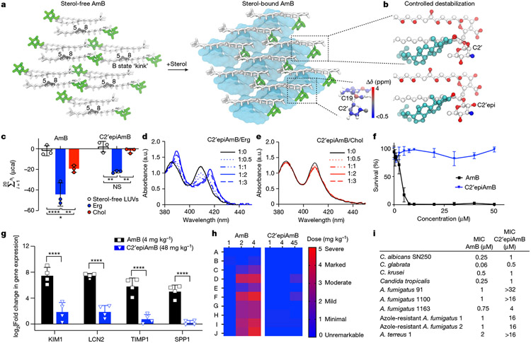
Fig. 2 ∣. Controlled destabilization of AmB–sterol clathrates results in Erg selectivity.
a, Comparison of apo-AmB and sterol-bound AmB clathrate structures highlighting changes in the mycosamine orientation, with the inset showing chemical shift perturbations relative to the apo state A. b, Expansion of the sterol-bound complex showing proximity of the sterol 3β-OH to the C2′OH and disruption of this interaction following in silico epimerization of the C2′centre. c, ITC assays comparing C2′epiAmB and AmB binding affinity to sterol-free, Erg-containing and Chol-containing large unilamellar vesicles (LUVs; n = 3). (AmB–Erg binding data reported in ref. 5.) Pairwise statistical analysis by two-way analysis of variance (ANOVA) with Tukey’s multiple comparison test; *P = 0.0245, **P = 0.0019, ****P < 0.0001, **P = 0.0022, **P = 0.0089. NS, not significant. d,e, UV–Vis spectra of C2′epiAmB following titration with increasing molar ratios (0, 0.5, 1, 2 and 3) of Erg (d) and Chol (e); a.u., arbitrary units. f, C2′epiAmB does not kill human primary renal cells in vitro up to 50 μM concentration (n = 3). g, C2′epiAmB does not elevate kidney damage biomarkers in female CD-1 mice measured 24 h post single IV dosage (n = 4 per group). Pairwise statistical analyses by two-way ANOVA with Tukey’s multiple comparison test; ****P < 0.0001. h, Heatmap representation of kidney histopathological scores (A, mixed-cell infiltrate, interstitial; B, tubular basophilia, cortex, medulla; C, Tubular cellular casts, cortex; D, tubular cellular casts, medulla; E, tubular degeneration and necrosis, cortex; F, tubular degeneration and necrosis, medulla; G, tubular dilatation, cortex; H, tubular protein casts, cortex; I, tubular protein casts, medulla; J, vascular congestion, medulla) in mice 24 h post single IV dosage of AmB and C2′epiAmB (n = 4 per group). Both AmB and C2′epiAmB were formulated as 1:2 deoxycholate to improve aqueous solubility. i, Comparison of AmB and C2′epiAmB MICs. Results are means ± s.d. Data presented in d,e are all representative of at least three independent experiments.

