eLife logoLink to eLife
. 2024 Feb 22;13:e91719. doi: 10.7554/eLife.91719

Rapid binding to protofilament edge sites facilitates tip tracking of EB1 at growing microtubule plus-ends

Samuel J Gonzalez 1, Julia M Heckel 1, Rebecca R Goldblum 2,3, Taylor A Reid 1, Mark McClellan 1, Melissa K Gardner 1,
Editors: Julie PI Welburn4, Amy H Andreotti5
PMCID: PMC10883673  PMID: 38385657

Abstract

EB1 is a key cellular protein that delivers regulatory molecules throughout the cell via the tip-tracking of growing microtubule plus-ends. Thus, it is important to understand the mechanism for how EB1 efficiently tracks growing microtubule plus-ends. It is widely accepted that EB1 binds with higher affinity to GTP-tubulin subunits at the growing microtubule tip, relative to GDP-tubulin along the microtubule length. However, it is unclear whether this difference in affinity alone is sufficient to explain the tip-tracking of EB1 at growing microtubule tips. Previously, we found that EB1 binds to exposed microtubule protofilament-edge sites at a ~70 fold faster rate than to closed-lattice sites, due to diffusional steric hindrance to binding. Thus, we asked whether rapid protofilament-edge binding could contribute to efficient EB1 tip tracking. A computational simulation with differential EB1 on-rates based on closed-lattice or protofilament-edge binding, and with EB1 off-rates that were dependent on the tubulin hydrolysis state, robustly recapitulated experimental EB1 tip tracking. To test this model, we used cell-free biophysical assays, as well as live-cell imaging, in combination with a Designed Ankyrin Repeat Protein (DARPin) that binds exclusively to protofilament-edge sites, and whose binding site partially overlaps with the EB1 binding site. We found that DARPin blocked EB1 protofilament-edge binding, which led to a decrease in EB1 tip tracking on dynamic microtubules. We conclude that rapid EB1 binding to microtubule protofilament-edge sites contributes to robust EB1 tip tracking at the growing microtubule plus-end.

Research organism: None

Introduction

Microtubules are important cellular filaments that are comprised of αβ tubulin heterodimers. The tubulin heterodimers are stacked end-to-end to form structures known as protofilaments, which associate laterally to form the hollow-tube structure of the microtubule (Mitchison and Kirschner, 1984). The α/β polarity of the tubulin dimer induces microtubule polarity, such that the microtubule end with β-tubulin exposed forms the fast-growing, dynamic ‘plus-end’ of the microtubule (Desai and Mitchison, 1997; Mitchison and Kirschner, 1984). In solution, the β-tubulin subunit binds to a GTP nucleotide, which then hydrolyzes to GDP after incorporation into the microtubule lattice. This delayed hydrolysis leads to a high concentration of GTP tubulin at the growing microtubule plus-end, commonly referred to as the ‘GTP-cap’ (Desai and Mitchison, 1997). The presence of the GTP-cap creates a distinct region that is present exclusively at growing microtubule ends (Maurer et al., 2011; Maurer et al., 2014; Zanic et al., 2009).

The localization of proteins along different regions of the microtubule is central to the role of microtubules in cell migration, intracellular transport, and cell division. EB1 is a key cellular protein that autonomously localizes to the growing ends of microtubules (‘tip tracks’) and recruits other important proteins that have little or no affinity to growing microtubule ends (Bieling et al., 2007; Dixit et al., 2009; Morrison et al., 1998; Mustyatsa et al., 2017). It has been shown that improper localization of EB1 at growing microtubule plus-ends can lead to disruptions in both cell division and cell migration (Dema et al., 2023; Dong et al., 2010; Honoré et al., 2008; Mustyatsa et al., 2017; Rogers et al., 2002; van Haren et al., 2018).

EB1 binds a small pocket within the microtubule lattice that is created by four tubulin dimers. It has been shown that EB1 binds with a higher affinity to GTP-tubulin subunits as compared to GDP-tubulin subunits. (Maurer et al., 2011; Maurer et al., 2012; Maurer et al., 2014; Zanic et al., 2009; Zhang et al., 2015). This difference in affinity likely increases the enrichment of EB1 within the GTP-cap at growing microtubule plus-ends.

Recent work has demonstrated that EB1 can bind to a partial binding pocket composed of 2–3 tubulin subunits, either at the tip of a protofilament, along the side of an exposed protofilament, or at lattice openings within the microtubule (Reid et al., 2019). We describe these exposed, partial binding pockets as ‘protofilament-edge’ sites. Specifically, we use the term ‘protofilament-edge’ to describe any partial EB1 binding site on the microtubule lattice, as opposed to closed (4-tubulin) binding sites. Importantly, we recently reported that the arrival rate of EB1 to 2-tubulin protofilament-edge sites was ~70 fold faster than to closed 4-tubulin pockets, due to a reduced diffusional steric hindrance to binding (Reid et al., 2019). Here, a partial EB1 binding site on the microtubule lattice led to a dramatic reduction in the diffusional steric hindrance that EB1 encounters in order to become properly oriented and then to slide into a closed, 4-tubulin binding pocket. In other words, the expanded physical access that is afforded by EB1 binding to a partial, 2-tubulin binding pocket (as compared to a closed 4-tubulin binding pocket) led to a ~70 fold increase in the EB1 on-rate. Because protofilament-edge sites are present at growing microtubule plus-ends (Atherton et al., 2018; Gudimchuk et al., 2020; Guesdon et al., 2016), we hypothesized that this large difference in EB1 arrival rates could have important repercussions for the efficiency of EB1 tip tracking at growing microtubule plus-ends. We thus predicted that the rapid binding of EB1 to protofilament-edge sites at the growing microtubule plus-end could increase the efficiency of EB1 plus-end tip tracking.

In this work, we generated a single-molecule stochastic simulation that incorporated the assembly and hydrolysis of individual tubulin subunits, as well as the binding and unbinding of EB1 molecules. Importantly, in our simulation, EB1 bound rapidly to protofilament-edge sites, and bound more slowly to closed-lattice sites. In addition, consistent with previous affinity measurements, the off-rate of EB1 from GTP-tubulin sites was low, with higher EB1 off-rates from GDP-tubulin sites. The simulation predicted that rapid binding to protofilament-edge sites increased the efficiency of EB1 tip-tracking at growing microtubule plus-ends. To test this prediction, we used cell-free biophysical assays, as well as live-cell imaging, in combination with a DARPin that binds exclusively to protofilament-edge sites, and whose binding site partially overlaps with the EB1 binding site (Pecqueur et al., 2012). We found that DARPin suppressed EB1 protofilament-edge binding on stabilized microtubules, and led to a disruption of EB1 tip tracking on dynamic microtubules plus-ends, both in cell-free experiments and in cells. Together, our work predicts that protofilament-edge binding, along with a differential EB1 binding affinity for GTP vs GDP tubulin, facilitates efficient EB1 tip tracking.

Results

A stochastic simulation that simultaneously incorporates tubulin assembly and EB1 on-off dynamics

In previous work, we found that the arrival rate of EB1 to exposed protofilament-edge sites on the sides and/or tips of microtubule protofilaments was ~70 fold faster than to closed four-tubulin pockets, due to a diffusional steric hindrance to binding (Reid et al., 2019). To ask whether rapid EB1 protofilament-edge binding could contribute to EB1 tip tracking, we created a stochastic simulation in which there was an increased on-rate of EB1 to protofilament-edge sites relative to closed-lattice sites. This simulation combined the assembly of individual tubulin subunits with EB1 binding and unbinding from the dynamic microtubule.

The microtubule assembly portion of the simulation utilized a previously published model, in which individual tubulin subunits were allowed to arrive and depart from the growing microtubule plus-end (Margolin et al., 2011; Margolin et al., 2012). Once a tubulin subunit arrived at the growing microtubule plus-end, a longitudinal bond was immediately formed with its penultimate tubulin dimer. Then, lateral bonds were stochastically formed in subsequent time steps (Margolin et al., 2011; Margolin et al., 2012). Finally, lattice-incorporated GTP-tubulin subunits were stochastically hydrolyzed to GDP-tubulin. In general, the on-rate of new tubulin subunits to the microtubule plus-end depended on the simulated tubulin concentration, and the off-rate of an individual tubulin subunit from the plus-end depended on its hydrolysis state and bonding state, where a GTP-tubulin subunit with two lateral bonds had the lowest off-rate in the simulation. All of the parameter values for the microtubule assembly simulation matched a previously published parameter set (Margolin et al., 2012; Supplementary file 1), with the exception of (1) the tubulin on-rate constant, which was lowered in order to match our (slow) experimental growth rates, and (2) one additional rule was added to ensure that the tip taper at the microtubule plus-end matched our experimental values (Figure 1—figure supplement 1A, B). Here, if the difference between the longest and the penultimate shortest protofilament exceeded 600 nm (75 dimers), the tubulin subunit off-rate and the lateral bond breakage rate were dramatically increased, quickly leading to a catastrophe event.

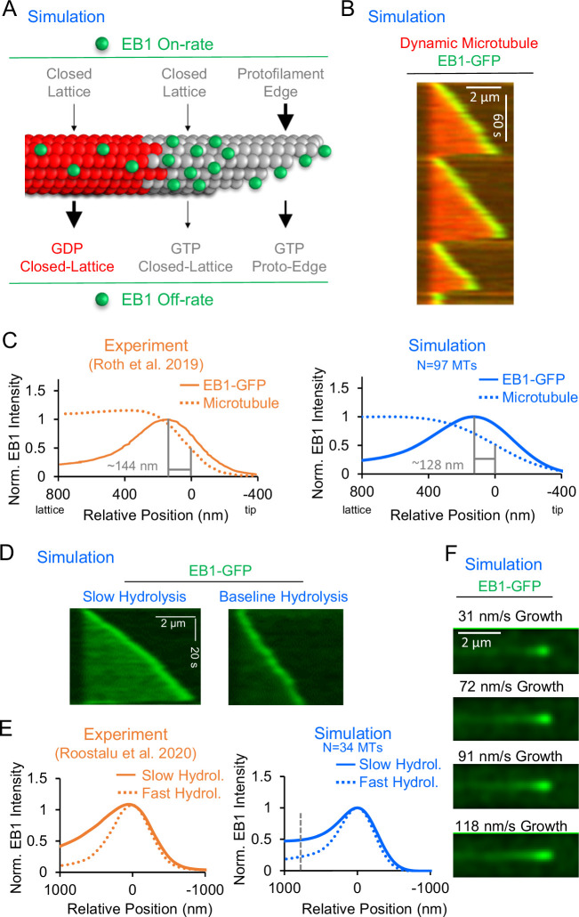
In addition to tubulin assembly, individual EB1 molecules were allowed to bind and unbind from their binding pockets at any position on the growing microtubule (Figure 1A). However, the EB1 on-rates and off-rates depended on the individual binding pocket chemistry and configuration. Specifically, the on-rates for individual EB1 molecules depended on the structure of the binding pocket, such that EB1 arrivals to protofilament-edge sites were substantially faster than to closed-lattice sites, regardless of the hydrolysis state (Figure 1A, top, see Methods) (Reid et al., 2019). In contrast, the off-rate of EB1 molecules depended on the hydrolysis state of the EB1 binding site. Here, the tubulin subunits towards the minus end of the microtubule dictated the ‘hydrolysis state’ of the EB1 binding site. If 1–2 of these tubulin subunits were hydrolyzed to GDP, the binding site was considered to be a ‘GDP’ binding site, leading to an increased EB1 off-rate (Figure 1A, bottom, see Methods). All parameter values in the EB1 model were constrained by previously published experimental values (Maurer et al., 2011; Maurer et al., 2014; Reid et al., 2019), with the exception of the EB1 off-rate from protofilament-edge sites, which has not been experimentally measured, but was constrained using bond energy arguments (see Supplementary file 2). To evaluate the uncertainty of each model parameter in impacting simulation results, the success of simulated tip tracking was plotted over a broad range of values for each parameter (see Figure 1—figure supplement 2 and Figure 1—figure supplement 3).

Figure 1. Development and validation of a stochastic simulation for EB1 tip tracking.

(A) Rules for a molecular-scale stochastic simulation that incorporates both tubulin subunit assembly and EB1 arrivals to and departures from the growing microtubule (See Methods). In the simulation, the EB1 protofilament-edge on-rate (top-right) is 50–100 fold higher than the EB1 closed-lattice on-rate (top-left and top-center). The EB1 off-rate is 6–12 fold faster for closed-lattice GDP-tubulin binding pockets (bottom-left) than for closed-lattice GTP-tubulin binding pockets (bottom-center). (B) Simulated EB1 tip tracking at growing microtubule ends. (C) Left: Line scans of EB1-GFP intensity (solid line), and microtubule intensity (dotted line) from experimentally reported data (Roth et al., 2019) (orange). Right: Line scans of EB1-GFP intensity (solid line), and microtubule intensity (dotted line) from the simulation (blue, see Methods). (D) Left: Simulated EB1 tip tracking with a slow GTP-tubulin hydrolysis rate (0.05 s–1) Right: Simulated EB1 tip tracking with the baseline GTP-tubulin hydrolysis rate (0.55 s–1). (E) Left: Experimental line scan quantification of EB3-GFP intensity along the length of the microtubule with two different hydrolysis rates (orange, Roostalu et al., 2020). Right: Simulated data line scan quantification of EB1-GFP intensity along the length of the microtubule with two different hydrolysis rates (blue). Slower hydrolysis leads to a ~ twofold increase in binding along the lattice at 768 nm distal of the peak EB1 position (dashed line). (F) Increasing the microtubule growth rate in the simulation increases the EB1 comet length, similar to reports in the literature (Bieling et al., 2007).

Figure 1—source data 1. Line scan data for EB1 intensity for Figure 1C, E (right).
elife-91719-fig1-data1.xlsx (164.6KB, xlsx)

Figure 1.

Figure 1—figure supplement 1. Additional simulation results.

Figure 1—figure supplement 1.

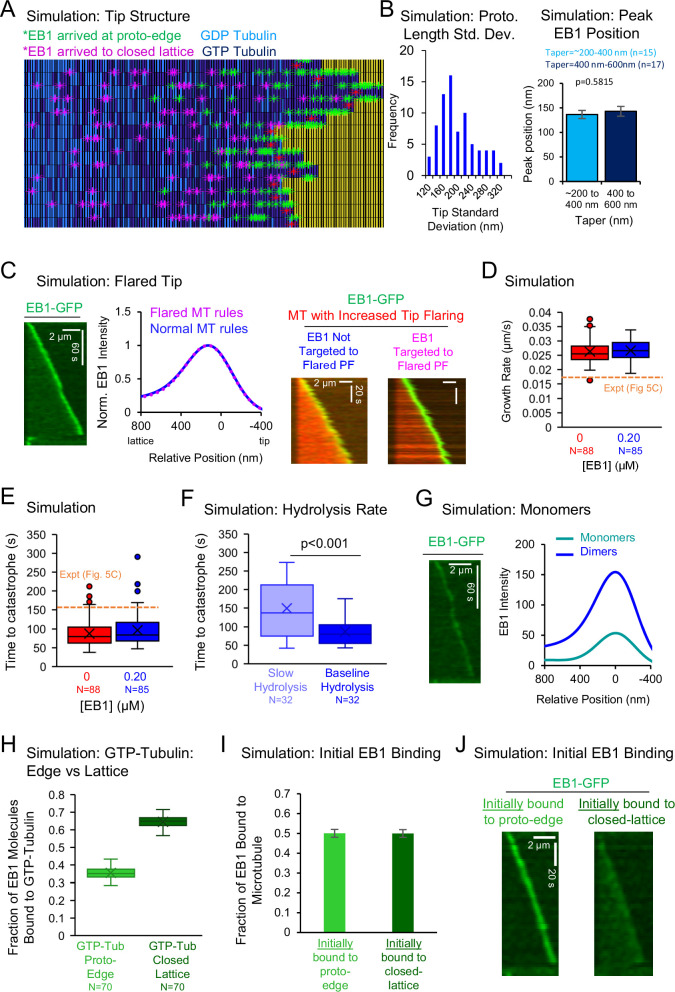
(A) Frame from animated simulated results in Video 2, with microtubule plus-end towards the right. Tubulin dimer hydrolysis state is indicated by color (GDP: light blue, GTP: dark blue). Green asterisks: EB1 molecules that originally bound to a protofilament-edge site. Purple asterisks: EB1 molecules that originally bound to a closed-lattice site. (B) Left: Tip standard deviation of simulated microtubules (Coombes et al., 2013; Demchouk et al., 2011). Simulated microtubules had a tip standard deviation of 191 ± 6 nm (Mean ± SEM). Right: Average EB1 peak position relative to microtubule tip, for averages of three simulation runs binned by microtubule taper size. (error bars, SEM; p=0.58, Mann Whitney U-test) (C) Left: A microtubule tip flaring approximation did not qualitatively alter EB1 tip tracking in the simulation. Center: The linescan of EB1 from the simulation was unaffected by whether the microtubule had tip flaring in the simulation. Right: Increased microtubule tip flaring was introduced into the simulation by reducing the lateral bond creation rate, while also increasing the tubulin on-rate to protofilaments that did not have lateral bonds with their neighbors. Blue: Simulation with increased protofilament flaring at the tip, but without EB1 targeting to flared protofilament edges. Magenta: Simulation with increased protofilament flaring at tip, and with EB1 targeting to flared protofilament edges. (D) Growth rates in the simulation were similar to experiments from Figure 5C (orange dashed line). (E) Time to catastrophe in the simulation was similar to experiments from Figure 5C (orange dashed line). (F) Decreasing the hydrolysis rate in the simulation increased the time to catastrophe (p<0.001, t-test). (G) To replicate EB1 monomer tip tracking, all off-rates were increased by fourfold (Song et al., 2020). Left: Qualitatively, higher off-rates led to a reduction in EB1 at the microtubule tip. Right: EB1 monomers (teal) in the simulation had a ~threefold reduction in the amount of EB1 at the growing microtubule end as dimers (blue). (H) Fraction of EB1 bound at GTP closed-lattice sites relative to GTP protofilament-edge sites. (I) Fraction of EB1 bound to the microtubule for EB1 molecules that initially bound to protofilament-edge sites (light green), and for EB1 molecules that initially bound to closed-lattice sites (dark green) (error bars, SEM). (J) Localization of EB1-GFP that bound originally to protofilament-edge sites (left) or to closed-lattice sites (right).
Figure 1—figure supplement 1—source data 1. Data for panels in Figure 1—figure supplement 1.
Figure 1—figure supplement 2. Parameter sensitivity testing for the EB1 tip tracking model I.

Figure 1—figure supplement 2.

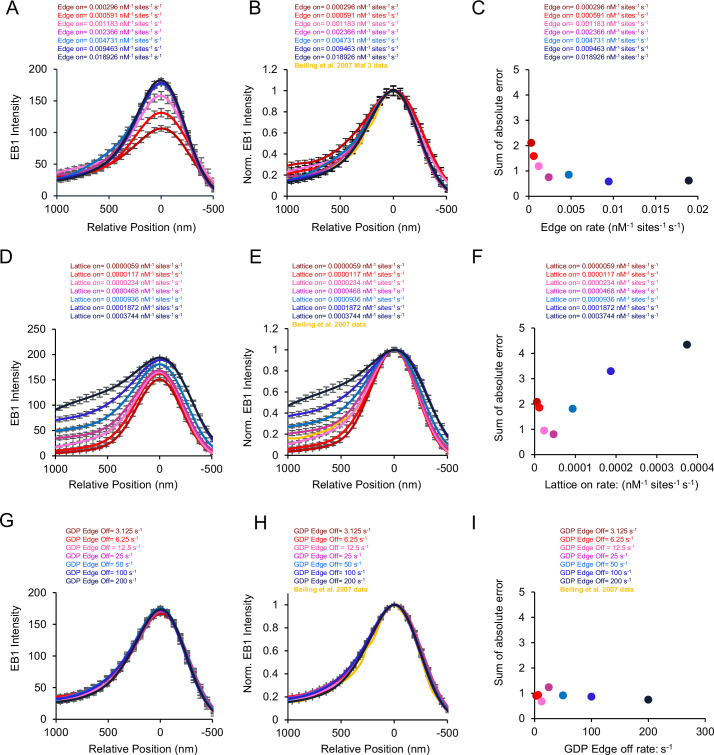
(A) Line scans of simulated EB1 tip tracking with varying protofilament-edge on-rates. (B) Normalized line scans of simulated EB1 tip tracking with varying protofilament-edge on-rates, as compared to literature data (yellow) (Bieling et al., 2007). (C) Sum of absolute error between the simulation and the literature data for each protofilament-edge on-rate. (D) Line scans of simulated EB1 tip tracking with varying closed-lattice on-rates. (E) Normalized line scans of simulated EB1 tip tracking with varying closed-lattice on-rates, as compared to literature data (yellow) (Bieling et al., 2007). (F) Sum of absolute error between the simulation and the literature data for each closed-lattice on-rate. (G) Line scans of simulated EB1 tip tracking with varying GDP protofilament-edge off-rates. (H) Normalized line scans of simulated EB1 tip tracking with varying GDP protofilament-edge off-rates, as compared to literature data (yellow) (Bieling et al., 2007). (I) Sum of absolute error between the simulation and the literature data for each GDP protofilament-edge off-rate.
Figure 1—figure supplement 2—source data 1. Data for panels in Figure 1—figure supplement 2.
Figure 1—figure supplement 3. Parameter sensitivity testing for the EB1 tip tracking model II.

Figure 1—figure supplement 3.

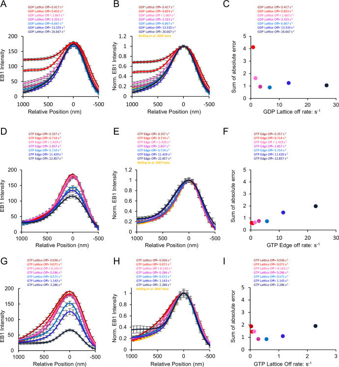
(A) Line scans of simulated EB1 tip tracking with varying GDP closed-lattice off-rates. (B) Normalized line scans of simulated EB1 tip tracking with varying GDP closed-lattice off-rates, as compared to literature data (yellow) (Bieling et al., 2007). (C) Sum of absolute error between the simulation and the literature data for each GDP closed-lattice off-rate. (D) Line scans of simulated EB1 tip tracking with varying GTP protofilament-edge off-rates. (E) Normalized line scans of simulated EB1 tip tracking with varying GTP protofilament-edge off-rates, as compared to literature data (yellow) (Bieling et al., 2007). (F) Sum of absolute error between the simulation and the literature data for each GTP protofilament-edge off-rate. (G) Line scans of simulated EB1 tip tracking with varying GTP closed-lattice off-rates. (H) Normalized line scans of simulated EB1 tip tracking with varying GTP closed-lattice off-rates, as compared to literature data (yellow) (Bieling et al., 2007). (I) Sum of absolute error between the simulation and the literature data for each GTP closed-lattice off-rate.
Figure 1—figure supplement 3—source data 1. Data for Figure 1—figure supplement 3.

Simulations with rapid binding at protofilament-edge sites can recapitulate EB1 tip tracking

We first asked whether EB1 ‘tip tracked’ growing microtubule plus-ends in the simulation, similar to experimental observations. Qualitatively, our simulated EB1 behaved similarly to experiments – strongly targeting growing microtubule plus-ends, while detaching from shortening ends (Figure 1B; Video 1). To quantitatively confirm that the simulated EB1 tip tracking was similar to experimental results, we next compared the peak EB1 position from our simulation data to results reported in the literature. The peak EB1 position refers to the distance between the highest EB1 intensity location on the microtubule, and the tip of the growing microtubule plus-end (Maurer et al., 2012; Maurer et al., 2014; Nakamura et al., 2012; Roth et al., 2019). At a microtubule growth rate of 10–30 nm/s, the peak EB1 position has been reported to be ~144 nm distal of the microtubule tip (Figure 1C, left, orange) (Roth et al., 2019). To quantify the peak EB1 position in the simulation, line scans of simulated EB1 comets were obtained and averaged over 97 simulated growth events. We found that the simulation produced a peak EB1 position of ~128 nm distal of the microtubule tip, similar to experimental observations (Figure 1C, right, blue). We note that the growth rate and time to catastrophe for the simulated microtubules were similar to experimentally reported values (Figure 1—figure supplement 1D, E), and so the simulated peak EB1 position likely reflects an appropriately sized GTP-cap. Importantly, our model with EB1 protofilament-edge binding reproduced the peak EB1 position without requiring a predetermined EB1 ‘exclusion zone,’ as has been previously hypothesized (Maurer et al., 2014). Rather, EB1 tip tracking in our current model depended solely on EB1 on/off rates and a growing microtubule plus-end.

Video 1. Simulated EB1 tip tracking.

Download video file (107.5KB, mp4)

EB1-GFP in green, Microtubule in red. 2 µm scale bar.

To ensure that the configuration of the microtubule plus-end was similar between experiments and simulation, we compared the fitted tip standard deviation in simulated microtubule images to our experimental values. Here, the ‘tip standard deviation’ reflects the range of protofilament lengths at the tip of the growing microtubule, such that a ‘tapered tip’ would have a large tip standard deviation (Coombes et al., 2013; Demchouk et al., 2011). We found that the average tip standard deviation of our simulated microtubules was 191±6 nm (mean ± SEM), similar to our experimental measurements of 180 ± 17 nm (Figure 1—figure supplement 1A, B; Video 2, mean ± SEM).

Video 2. Animated simulation output for a growth event of one microtubule.

Download video file (3.7MB, mp4)

Green asterisks: EB1 that originally bound to a protofilament-edge site. Purple asterisks: EB1 that originally bound to a closed-lattice site. Red crosses: location of most distal lateral bond for each protofilament. Protofilament #13 shares a lateral bond with protofilament #1. Dark blue rectangles: GTP-tubulin. Light blue rectangles: GDP-tubulin. A seed of 25 dimers was maintained as GTP-tubulin to represent a GMPCPP seed. The Video is updated every 1000 steps within the simulation.

It has been previously suggested that growing microtubule plus-ends could be ‘flared,’ such that they have bent protofilaments that are curved (or flared) away from the central microtubule axis (McIntosh et al., 2018). Thus, we asked how a flared microtubule tip structure would affect tip tracking in our simulation. To approximate microtubule tip flaring in the model, we assumed that, with a flared end, all EB1 binding sites in front of the most distal lateral bond would be considered protofilament-edge sites. We found that the microtubule flaring approximation in the simulation had no discernible effect on EB1 tip tracking (Figure 1—figure supplement 1C, left/center). Furthermore, we introduced increased tip flaring into the simulation by moving the most distal lateral bond farther away from the growing microtubule tip, which led to increased EB1 targeting to the flared growing microtubule plus-end (Figure 1—figure supplement 1C, right). Thus, flared microtubule tips in the simulation behaved similarly to tapered tips, both in EB1 intensity and in peak EB1 location.

It has been shown that a slower GTP-tubulin hydrolysis rate increases EB1 binding along the microtubule, likely due to an increased concentration of GTP-tubulin within the microtubule lattice (Roostalu et al., 2020). Thus, to ask whether the simulation could recapitulate this phenomenon, we ran simulations with a slower GTP-tubulin hydrolysis rate (Figure 1—figure supplement 1F). We found that a slower hydrolysis rate (0.05 s–1) led to an increased concentration of EB1 on the microtubule, as compared to the baseline simulation (0.55 s –1) (Figure 1D, Video 3). By quantifying the localization of EB1 at growing microtubule plus-ends in these simulations, we observed a ~twofold increase in EB1 binding along the lattice of simulated microtubules with a slower hydrolysis rate, relative to the baseline simulation (Figure 1E, right, blue; calculated at position 768 nm, gray dashed line), similar to previously reported experimental results (Figure 1E, left, orange; Roostalu et al., 2020). This result demonstrates that EB1 tip tracking in the simulation depends on the tubulin hydrolysis rate, similar to previous experimental results (Roostalu et al., 2020).

Video 3. Simulated EB1 tip tracking for different hydrolysis rates.

Download video file (45.2KB, mp4)

EB1-GFP in green, microtubule in red. 2 µm scale bar. Slower hydrolysis rate on left (0.05 s–1), baseline hydrolysis rate on right (0.55 s–1).

Previous work has demonstrated that EB1 monomers tip track less effectively than their dimer counterparts (Komarova et al., 2009; Skube et al., 2010). In the model, we employed experimentally determined on and off rates for EB1 (see Supplementary file 2). Therefore, because the relevant experiments were performed using EB1 in its normal state as a dimer, the baseline simulations represent the simulation results for EB1 dimers. To determine how the model results would be impacted by including monomers in the model, rather than dimers, we turned to previous work, which demonstrated that the EB1 monomer off-rate was ~fourfold larger than the off-rates for dimers (Song et al., 2020). Thus, we increased all off-rates in the model by fourfold from their baseline values, and thus ran ‘monomer’ simulations. We found that EB1 tip tracking was decreased by ~threefold in the monomer simulations (Figure 1—figure supplement 1G), consistent with previous reports (Komarova et al., 2009; Skube et al., 2010).

Finally, it has been widely reported that an increased microtubule growth rate leads to a longer EB1 ‘comet’ (Farmer et al., 2021; Maurer et al., 2014; Reid et al., 2019). Thus, we ran simulations with increasing microtubule growth rates, keeping the GTP-tubulin hydrolysis rate constant. Similar to experimental reports, we found that, as the microtubule growth rate was increased in the simulation, the comet length was increased (Figure 1F).

Protofilament-edge binding increases the efficiency and robustness of tip tracking

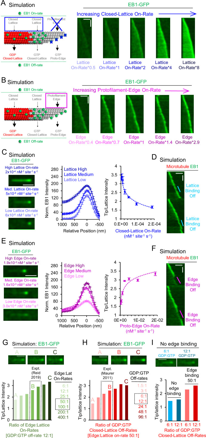
We next asked how simulated EB1 tip tracking would be affected if EB1 bound exclusively to the canonical closed-lattice sites on the microtubule. Thus, we set the EB1 protofilament-edge on-rate to zero, and then slowly increased the EB1 closed-lattice on-rate, while leaving all EB1 off-rates constant and at their baseline values (Figure 2A, left; Video 4; Supplementary file 2, see Methods). We found that, while a higher EB1 closed-lattice on-rate led to EB1 accumulation at the growing microtubule end, it also led to EB1 accumulation along the length of the microtubule (Figure 2A, right), thus reducing the specificity of EB1 localization to the growing microtubule end.

Figure 2. Simulations predict that protofilament-edge binding facilitates robust EB1 tip tracking.

Figure 2.

(A) Left: Simulations were performed in which the EB1 protofilament-edge on-rate was set to zero, and the closed-lattice on-rate was gradually increased. Right: Simulated kymographs in which the EB1 protofilament-edge on-rate was set to zero, and the on-rate at closed-lattice sites was gradually increased (scale bars: 2 µm and 10 s). (B) Left: Simulations were performed in which the closed-lattice on-rate remained constant at its baseline (non-zero) value, and the protofilament-edge on-rate was gradually increased. Right: Simulated kymographs in which the closed-lattice on-rate remained constant at its baseline (non-zero) value, and the protofilament-edge on-rate was gradually increased (scale bars: 2 µm and 10 s). (C) Left: Simulated images of EB1-GFP tip tracking over a range of closed-lattice on-rates (scale bar: 1 µm). Center: Line scans from simulated images of EB1-GFP intensity for a range of closed-lattice on-rates (error bars, SEM). Right: Tip:Lattice EB1-GFP intensity ratio vs closed-lattice on-rates in the simulation (error bars, SEM). The tip:Lattice EB1-GFP intensity ratio decreases with increasing closed-lattice on-rates. (D) Simulated kymograph in which the closed-lattice on-rate is set to zero partway through the simulation, and later returned to its baseline value (scale bars: 2 μm and 20 s). (E) Left: Simulated images of EB1-GFP tip tracking over a range of protofilament-edge on-rates (scale bar: 1 µm). Center: Line scans from simulated images of EB1-GFP intensity for a range of protofilament-edge on-rates (error bars, SEM). Right: Tip:Lattice EB1-GFP intensity ratio vs protofilament-edge on-rates in the simulation (error bars, SEM). Localization to the microtubule tip increases with increasing protofilament-edge on-rates. (F) Simulated kymograph in which the protofilament-edge on-rate is set to zero partway through the simulation and later returned to its baseline value (scale bars: 2 μm and 20 s). (G) Top: Representative images of EB1-GFP tip tracking for increasing Protofilament-edge:Closed-lattice on-rate ratios. Bottom: Tip:Lattice EB1-GFP intensity ratio for increasing Protofilament-edge:Closed-lattice on-rate ratios (GDP:GTP off-rate ratio is constant and set to 12:1). The experimentally measured Protofilament-edge:Closed-lattice on-rate ratio is 50–100 (Reid et al., 2019) (gray dashed boxes). (H) Top: Representative images of EB1-GFP tip tracking for increasing GDP:GTP closed-lattice off-rate ratios. Bottom: Tip:Lattice EB1-GFP intensity ratio for increasing GDP:GTP closed-lattice off-rate ratios (Protofilament-edge:Closed-lattice on-rate ratio is constant and set to 50:1). The experimentally measured GDP:GTP closed-lattice off-rate ratio is 6–12 (Maurer et al., 2011) (gray dashed boxes). (I) Top: Representative images of EB1-GFP tip tracking without protofilament-edge binding. Bottom: Tip:Lattice EB1-GFP intensity ratio for experimentally measured GDP:GTP closed-lattice off-rate ratios, with (red) or without (blue) EB1 binding at protofilament-edge sites.

Figure 2—source data 1. Data for Figure 2C, E, G, H, and I.
elife-91719-fig2-data1.xlsx (327.7KB, xlsx)

Video 4. Simulated EB1 tip tracking over a range of EB1 closed-lattice on rates (1.2 x 10⁻⁵ – 1.9 x 10⁻⁴ nM–1 sites –1 s–1).

Download video file (43.5KB, mp4)

EB1 protofilament-edge binding on-rate was set to 0. Then, the remaining EB1 on and off rates were kept constant except for the closed-lattice on-rate, which increases from left to right for each subset of the panel. EB1-GFP in green, microtubule in red. 2 µm scale bars.

We next explored the effect on EB1 tip tracking of increasing the EB1 protofilament-edge on-rates. Thus, we set the EB1 closed-lattice on-rate to its baseline (non-zero) value, and then slowly increased the EB1 protofilament-edge on-rate, while leaving all EB1 off-rates constant (Figure 2B, left; Video 5; Supplementary file 2, See Methods). We found that an increasingly intense EB1-GFP puncta appeared at the growing microtubule end as the protofilament-edge on-rate was increased (Figure 2B, right; Video 5).

Video 5. Simulated EB1 tip tracking over a range of protofilament-edge on-rates (5.9 x 10⁻⁴ – 4.7 x 10⁻³ nM–1 sites–1 s–1).

Download video file (99.9KB, mp4)

All EB1 on-and off-rates were kept constant except for the protofilament-edge on-rate, which increases from left to right for each subset of the panel. EB1-GFP in green, microtubule in red. 2 µm scale bars.

Specificity of EB1 targeting to growing microtubule tips is reduced with higher EB1 closed-lattice binding site on-rates

To quantitatively dissect the relative role of closed-lattice binding on EB1 localization, we ran simulations over a range of EB1 closed-lattice on-rates, while keeping all other EB1 on-rates and off-rates constant and set to their baseline values, including rapid EB1 protofilament-edge binding (Supplementary file 2). We found that a low EB1 closed-lattice on-rate led to a clear EB1 puncta at the tip of the microtubule (Figure 2C, left-bottom). Here, EB1 accumulation is dominated by protofilament-edge binding. However, increasing the EB1 closed-lattice on-rate by 32-fold led to a ~1.6 fold increase in EB1 intensity at the microtubule tip, but, importantly, also led to a ~25 fold increase in EB1 intensity along the length of the microtubule (Figure 2C, center), even in the presence of EB1 protofilament-edge binding. By plotting the ratio of Tip:Lattice EB1 intensity (see Methods), we found that, with increasing EB1 closed-lattice on-rates, the EB1 intensity at the microtubule tip was decreased relative to the lattice (Figure 2C, right). Thus, the efficiency of simulated EB1 tip tracking was reduced with faster EB1 binding to closed-lattice sites, due to increased EB1 accumulation along the length of the microtubule.

We then performed a simulation in which the closed-lattice on-rate was set to zero partway through the simulation, to observe in real-time the effect of closed-lattice binding on EB1 tip tracking. We found that EB1 tip tracking was similar whether closed-lattice binding was on or off during the dynamic microtubule simulation (Figure 2D, cyan; Video 6).

Video 6. Simulated EB1 tip tracking, with EB1 closed-lattice binding dynamically set to 0 during the run.

Download video file (94.9KB, mp4)

Simulation is run with all baseline parameter values (Supplementary file 2). At times 57 s and 209 s, EB1 closed-lattice on-rate is set to zero. Then, at times 87 s and 239 s, the EB1 closed-lattice on-rate is reset to its baseline value. EB1-GFP in green, microtubule in red. 2 µm scale bar.

Simulations with increasing EB1 protofilament-edge on-rates lead to EB1 accumulation exclusively at the growing microtubule plus-end

Next, to quantitatively assess the role of protofilament-edge binding on EB1 localization, we ran simulations over a range of protofilament-edge on-rates, while keeping all other EB1 on-rates and off-rates constant and set at their baseline values, including the closed-lattice on-rate (see Methods). We found that, by decreasing the protofilament-edge on-rate, the intensity of EB1 at the growing microtubule tip was dimmed (Figure 2E, left-bottom). Upon increasing the protofilament-edge on-rate, the intensity of EB1 at the growing tip was increased, without an increase in EB1 intensity along the length of the microtubule (Figure 2E, left-top). Here, a 32-fold increase in the protofilament-edge on-rate led to a ~2.2 fold increase in EB1 intensity at the tip of the microtubule, and, importantly, no change in the EB1 intensity along the length of the microtubule (Figure 2E, center). By plotting the ratio of Tip:Lattice EB1 intensity (see Methods), we found that, with increasing EB1 protofilament-edge on-rates, the EB1 intensity at the microtubule tip was increased relative to the lattice (Figure 2E, right). Thus, the efficiency of simulated EB1 tip tracking was enhanced by higher EB1 on-rates to incomplete, protofilament-edge binding sites.

Finally, we performed a simulation in which the protofilament-edge on rate was set to zero partway through a simulation. We found that EB1 tip tracking was rapidly diminished when protofilament-edge binding was shut off during a dynamic microtubule simulation, and returned quickly when the EB1 protofilament-edge on-rate was reset to its baseline value (Figure 2F, magenta; Video 7).

Video 7. Simulated EB1 tip tracking, with EB1 protofilament-edge binding dynamically set to 0 during the run.

Download video file (112.3KB, mp4)

Simulation is run with all baseline parameter values (Supplementary file 2). At times 56 s and 203 s, EB1 protofilament-edge on-rate is set to zero. Then, at times 85 s and 233 s, the EB1 protofilament-edge on-rate is reset to its baseline value. EB1-GFP in green, microtubule in red. 2 µm scale bar.

Dimensionless variables demonstrate key parameters that control simulated EB1 tip tracking

To quantitatively interrogate the model parameter sensitivity, we defined two key dimensionless variables that control tip tracking in the model. First, as described above, the ratio of the on-rate of EB1 at protofilament-edge sites relative to closed-lattice sites, which is independent of the hydrolysis state of the associated tubulin molecules, directly alters the EB1 tip tracking efficiency in the model (Figure 2G). Importantly, distinct tip tracking was observed using the experimentally measured on-rate ratio for protofilament-edge sites relative to closed-lattice sites (50-100:1, Reid et al., 2019; Figure 2G, image B, gray dashed boxes).

Second, as has been previously described, the ratio of the off-rate of EB1 from closed-lattice GDP-tubulin sites, relative to closed-lattice GTP-tubulin sites, also influenced EB1 tip tracking in the model (Figure 2H; note that the model is comparatively insensitive to protofilament-edge off-rates, regardless of hydrolysis state Figure 1—figure supplement 2G–I, Figure 1—figure supplement 3D–F). Similar to the on-rate ratio, clear tip tracking was observed using the experimentally measured off-rate ratio for GDP-tubulin relative to GTP-tubulin (calculated as 6–12, based on KD values reported in Maurer et al., 2011; Figure 2H, image B, gray dashed boxes).

Finally, we evaluated the relative importance of the two dimensionless variables: one that dictates relative EB1 on-rates, and the other that dictates relative EB1 off-rates, in influencing simulated EB1 tip tracking (Figure 2I). We found that, in the absence of protofilament-edge binding, the experimentally observed range of closed-lattice GDP:GTP off-rate ratios did not reproduce EB1 tip tracking (Figure 2I, top: representative simulated images; bottom: blue bars). However, by including a 50:1 protofilament-edge to closed-lattice on-rate ratio in the simulation, robust tip tracking was reproduced, with an increase in EB1 tip localization for a higher ratio of GDP:GTP off-rates (Figure 2I, red). Thus, based on the experimentally measured EB1 on and off rates, both a hydrolysis-state dependent EB1 off-rate, as well as a rapid protofilament-edge EB1 on-rate, were critical to reproduce EB1 tip tracking in the model.

Split EB1 comets have increased EB1 binding relative to single EB1 comets

It has been previously reported that EB1-GFP can split into multiple comets that track the growing microtubule end (Doodhi et al., 2016). Thus, a ‘split comet’ refers to the phenomenon in which there are two or more distinct EB1 puncta that track a growing microtubule end (Doodhi et al., 2016; Farmer et al., 2021). A split comet likely occurs when one or more protofilaments lag behind the growing microtubule tip, thus producing an extended, highly tapered tip (Figure 3A, left (gray)). In the canonical model in which tip tracking relies exclusively on a higher EB1 affinity for GTP-tubulin relative to GDP-tubulin, it is expected that, for a single microtubule growth event with a constant growth rate (and thus a constant total GTP-cap size), the total summed intensity of EB1-GFP at split-comet tips would be similar to the intensity of EB1-GFP at single-comet tips. However, in a model with preferential EB1 binding to protofilament-edge sites, we predicted that the additional protofilament-edge binding sites on the sides of exposed protofilaments, afforded by a large difference in protofilament lengths at the tip of growing microtubules with ‘split comets,’ would lead to a net increase in the summed intensity of EB1-GFP (Figure 3A, left) (Farmer et al., 2021). Thus, if EB1 binds to protofilament-edge sites, we predicted that there would be an increase in the summed EB1-GFP intensity at growing microtubule tips with split comets, due to the increased number of protofilament-edge sites that are available to recruit EB1.

Figure 3. Summed Mal3 comet intensity is increased in split comets relative to single comets.
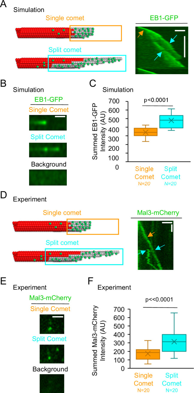
Figure 3.

(A) Left: Schematic of a single comet (top), and a split comet (bottom). Right: Simulated kymograph with a single comet (orange arrow), and split comet (cyan arrows), within one common microtubule growth event (scale bars: 2 μm and 60 s). (B) The summed comet intensity can be measured for a single simulated comet (top) and, later on, the same simulated microtubule growth event, for a split simulated comet (middle). The background (bottom) was subtracted from both the single comet and split comet summed intensity measurements. (C) Simulated split comets have a higher summed EB1 intensity at the microtubule tip than their single comet counterparts on the same growth event (p<0.0001, paired t-test,). (D) Left: Schematic of a single comet (top), and a split comet (bottom). Right: Typical experimental kymograph with a single comet (orange arrow), and split comet (cyan arrows), within one common microtubule growth event (scale bars: 2 μm and 60 s). (E) The summed comet intensity can be measured for a single comet (top) and, later on, the same microtubule growth event, for a split comet (middle). The background (bottom) was subtracted from both the single comet and split comet summed intensity measurements. (F) Split comets had a higher summed Mal3-mCherry intensity at the microtubule tip than their single comet counterparts from the same growth event (p<0.0001, paired t-test).

Figure 3—source data 1. Data for Figure 3C, F.

We first tested this prediction using our simulation. Thus, we asked whether there was an increase in the summed EB1-GFP intensity at growing microtubule tips with split comets. To generate split comets in the simulation, we altered the microtubule assembly simulation parameters to allow for an increase in taper at the growing microtubule tips (from ≤~600 nm in our standard simulation, to ≤~3 μm in the split comet simulation (see Methods)). By increasing the taper at the microtubule tip, the simulation was able to recapitulate split comets (Figure 3A, right (orange arrow: pre-split; cyan arrows: post-split)). We then asked whether there was an increase in the summed EB1-GFP intensity on individual growing microtubule tips after an EB1 comet split, relative to prior to the split. Thus, we measured the total intensity of EB1-GFP both before and after the comet split on individual simulated growing microtubules (Figure 3B, top: pre-split; middle: post-split). We subtracted the green background intensity both before and after the comet split (Figure 3B, bottom). We found that the split comets had a~40% increase in the summed intensity of EB1-GFP at the growing tip, relative to single comets on the same growth events (Figure 3C, p<0.001, paired t-test,). Therefore, consistent with our prediction, the simulation data indicates that an increase in protofilament-edge sites on the sides of exposed protoflaments during split-comet growth events leads to an increase in EB1 recruitment to the microtubule plus-end.

Next, to test this prediction experimentally, we examined experimental microtubule growth events with split comets (Figure 3D, right; orange arrow: pre-split; cyan arrows: post-split). We measured the summed Mal3-mCherry (yeast EB1-homolog) intensity both before and after the comet split on individual growing microtubules (Figure 3E; top: pre-split; middle: post-split). We subtracted the green background intensity both before and after the comet split (Figure 3E, bottom). We found that split comets had an ~80% increase in the summed intensity of Mal3 at the growing microtubule tip relative to the single comets on the same microtubule growth events (Figure 3F, p<0.0001, paired t-test,). Thus, the experimental results are consistent with the simulation results, and suggest that an increase in protofilament-edge sites on the sides of exposed protofilaments during split-comet growth events leads to an increase in EB1 recruitment to the microtubule plus-end.

DARPin suppresses EB1 binding to protofilament-edge sites on stabilized microtubules

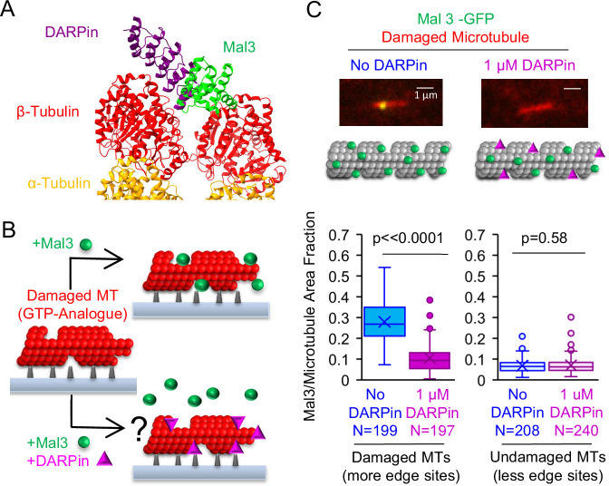
Because the simulation predicted that protofilament-edge binding is integral to EB1 tip tracking, we reasoned that EB1 tip tracking would be disrupted by a protein that could block EB1 binding to protofilament-edge sites. Thus, to test this prediction, we leveraged a DARPin D1 that binds exclusively to protofilament-edge sites, and also partially overlaps with the EB1 binding site on microtubules (Figure 4A; Video 8; Pecqueur et al., 2012). Here, DARPin could bind to protofilament-edge sites, and thus suppress EB1 binding in these locations, which would, in turn, disrupt proper EB1 tip tracking.

Figure 4. Synthetic Designed Ankyrin Repeat Protein (DARPin) peptide blocks EB binding to protofilament-edge sites.

(A) Structure of DARPin and Mal3, together with α and β tubulin at a microtubule tip was created in ChimeraX (Pettersen et al., 2021) using the crystal structures 4drx (DARPin) and 4abo (Mal3). (B) Left: To test whether DARPin blocks EB binding to protofilament-edge sites, we first generated stabilized GTP-analogue (GMPCPP) microtubules that were damaged with CaCl2 treatment, thus creating protofilament-edge sites along the length of the microtubule. Right: The damaged microtubules were incubated with Mal3-GFP in the presence (bottom) or absence (top) of 1 μM DARPin. Suppression of Mal3-GFP binding to the damaged microtubule in the presence of DARPin suggests that DARPin blocks Mal3 from binding to protofilament-edge sites (right, bottom). (C) Top: Representative images of Mal3-GFP binding to damaged microtubules, in the absence (blue) or presence (magenta) of 1 μM DARPin. Middle: Cartoons depicting the relative binding of Mal3 and DARPin in each experiment. Bottom-Left: Quantification of the fraction of microtubule area bound by Mal3-GFP for damaged microtubules in the absence or presence of DARPin (p<0.0001, t-test; sample size indicates number of images). Bottom-Right: Quantification of the fraction of microtubule area bound by Mal3-GFP for undamaged microtubules in the absence or presence of DARPin (p=0.58, t-test; sample size indicates number of images).

Figure 4—source data 1. Data for Figure 4C.

Figure 4.

Figure 4—figure supplement 1. Additional experimental data for binding of Mal3-GFP to stabilized GMPCPP microtubules.
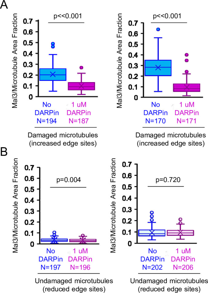
Figure 4—figure supplement 1.

(A) Quantification of the fraction of microtubule area bound by Mal3-GFP for damaged microtubules in the absence or presence of 1 μM DARPin (second and third replicates; t-tests). (B) Quantification of the fraction of microtubule area bound by Mal3-GFP for undamaged microtubules in the absence or presence of 1 μM Designed Ankyrin Repeat Protein (DARPin) (second and third replicates; t-tests).
Figure 4—figure supplement 1—source data 1. Data for Figure 4—figure supplement 1.

Video 8. Mal3 and Designed Ankyrin Repeat Protein (DARPin) overlap along an exposed tubulin plus-end.

Download video file (7.6MB, mp4)

Created in ChimeraX using the structures 4DRX (DARPin) and 4ABO (Mal3).

We first asked whether DARPin could block EB1 binding at protofilament-edge sites. Thus, we generated stabilized GTP-analogue (GMPCPP) microtubules that were damaged, such that portions of the microtubule contained openings and defects. Damaging the microtubules leads to an increased number of protofilament-edge sites along the microtubule length (Coombes et al., 2016; Gupta et al., 2013; Reid et al., 2017). Damaged microtubules can be generated by briefly exposing stabilized GMPCPP microtubules to CaCl2 (Coombes et al., 2016; Gupta et al., 2013; Reid et al., 2017). Thus, coverslip-adhered, rhodamine-labeled GMPCPP microtubules were briefly incubated in 10 mM CaCl2, followed by a wash to remove the CaCl2 (Figure 4B, left). Then, the damaged microtubules were incubated with the yeast EB1 homolog Mal3-GFP, in the absence or presence of DARPin (Figure 4B). Total Internal Reflection Fluorescence (TIRF) microscopy was used to visualize the microtubules, and Mal3 binding to the microtubules was assessed. Qualitatively, we observed a reduction in Mal3-GFP binding to the damaged microtubules in the presence of DARPin, as compared to the no-DARPin controls (Figure 4C, top). By using a custom MATLAB script to measure the Mal3-GFP binding area on the damaged microtubules (Reid et al., 2017), we found that the fraction of microtubule area bound by Mal3-GFP was ~2.7 fold lower in the presence of DARPin as compared to the no-drug controls (Figure 4C bottom-left, and Figure 4—figure supplement 1A; p<0.001, t-test). In contrast, by using undamaged GMPCPP microtubules, which did not have openings and defects to generate protofilament-edge binding sites along the microtubule length, there was no significant difference in Mal3-GFP binding in the presence and absence of DARPin (microtubules not treated with CaCl2; Figure 4C bottom-right, and Figure 4—figure supplement 1B; p=0.58, t-test). These results suggest that DARPin acts to block Mal3-GFP binding specifically on protofilament-edge sites.

In cell-free experiments, DARPin suppresses Mal3 tip tracking on dynamic microtubule plus-ends

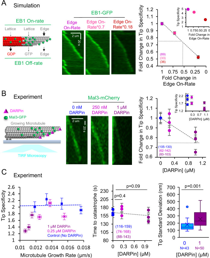
We next asked whether suppression of protofilament-edge binding would disrupt EB1 tip tracking. First, we ran simulations to quantitatively predict how EB1 tip tracking would be altered by suppressing its protofilament-edge on-rate (Figure 5A, left). Thus, we gradually reduced the protofilament-edge on-rate and generated simulated images to detect the relative localization of EB1-GFP at growing microtubule plus-ends (Figure 5A, center). To evaluate EB1-GFP localization to growing microtubule plus-ends in the simulation, we measured the EB1-GFP ‘Tip Specificity.’ Here, we defined Tip Specificity (S) as:

S=(ItipIbackground)(IlatticeIbackground) (1)

Figure 5. In cell-free experiments, Designed Ankyrin Repeat Protein (DARPin) disrupts tip tracking by blocking EB1 access to protofilament-edge sites.

Figure 5.

(A) Left: Cartoon depicting simulation rules for EB1 binding and unbinding to growing microtubules. Center: Simulated kymographs of EB1-GFP at growing microtubule tips, with decreasing protofilament-edge on-rates. Right: Simulation prediction for the fold change in the EB1 Tip Specificity as a function of the fold-change in the protofilament-edge on-rate (error bars: SEM; p<0.001, Kruskall Wallis). Gray dotted lines correspond to a twofold decrease in protofilament-edge on-rate, which leads to a ~25% decrease in predicted Tip Specificity. Inset: Absolute EB1 tip Specificity as a function of the fold-change in the protofilament-edge on-rate. (B) Left: Cartoon depicting experimental setup for dynamic microtubules with Mal3-mCherry, in the presence of DARPin, and visualized using TIRF microscopy. Center: Experimental kymographs of Mal3-mCherry tip tracking along dynamic microtubules in the presence of increasing DARPin concentrations. Right: Experimental results for the fold change in the Mal3-GFP Tip Specificity as a function of DARPin concentration (error bars: SEM; p<0.001, Kruskall Wallis). Gray dotted line represents a~25% decrease in Mal3-GFP Tip Specificity, which corresponds to a simulation prediction of a ~two-fold decrease in protofilament-edge on-rate. Inset: Absolute Mal3-GFP Tip Specificity as a function of DARPin concentration. (C) Left: The decrease in Mal3-GFP Tip Specificity in DARPin is not due to a decrease in microtubule growth rate (p<0.001, Kruskall Wallis comparing DARPin data to control data at a ~0.012 μm/s growth rate,). Center: The time to catastrophe is not altered by DARPin, suggesting that the GTP-cap size remains similar in the presence and absence of DARPin (Tukey’s post-hoc analysis after an ANOVA). Right: Tip standard deviation in the presence and absence of DARPin (p=0.001, t-test,).

Figure 5—source data 1. Data for Figure 5.

Where Itip is the EB1 intensity at the growing microtubule tip, Ilattice is the EB1 intensity on the microtubule lattice, and Ibackground is the background EB1 intensity just outside of the growing microtubule tip. By definition, a lower Tip Specificity value indicates that there is less efficient tip tracking. In addition, a Tip Specificity value equal to one (e.g. S=1) means that the EB1 intensity at the growing microtubule tip is equal to the EB1 intensity along the length of the microtubule, and therefore EB1 is not tip tracking. We found that, in the simulation, a decreased protofilament-edge on-rate led to a decrease in Tip Specificity (Figure 5A, right inset: y-axis is absolute Tip Specificity). Specifically, a ~twofold reduction in protofilament-edge on-rate led to a ~25% reduction in Tip Specificity (Figure 5A, right, large plot y-axis shows fold-change in tip specificity; gray dotted lines is ~25% reduction in tip specificity).

Thus, to test this simulation prediction, we performed a cell-free assay in which dynamic microtubules were grown from stabilized seed templates in the presence of Mal3-mCherry (Figure 5B, left). We visualized the growing microtubules using TIRF microscopy, in the presence of increasing concentrations of DARPin (Figure 5B, center). We found that Mal3 tip tracking was increasingly disrupted as the DARPin concentration was increased (p<0.001, Kruskal Wallis) (Figure 5B, right inset: y-axis is absolute Tip Specificity). Interestingly, 1 μM DARPin led to a~25% reduction in Tip Specificity, consistent with the simulation prediction of a twofold reduction in protofilament-edge on-rate (Figure 5B, right, large plot y-axis shows fold-change in tip specificity; gray dotted lines is ~25% reduction in tip specificity).

We then asked whether the suppression of tip tracking in DARPin could be due to a drop in microtubule growth rate, leading to a reduced concentration of GTP-tubulin at the growing microtubule plus-end (Farmer et al., 2021; Maurer et al., 2014; Reid et al., 2019). We found that the suppression of tip tracking was more substantial than would be predicted based on the small changes in microtubule growth rate at 1 μM DARPin (Figure 5C, left, blue dotted line: control; purple: 1 μM DARPin; p<0.001, Kruskall Wallis comparing DARPin data to Control data at ~0.012 um/s growth rate). Furthermore, we found no significant increase in the time to catastrophe with increasing DARPin concentrations, suggesting that DARPin does not affect the GTP hydrolysis rate or the associated GTP-cap size (Figure 5C, center; p=0.09–0.4, Tukey’s post-hoc test).

Finally, we asked whether DARPin could indirectly disrupt Mal3 tip tracking by altering the configuration of the growing microtubule plus-end. Here, a more blunt microtubule tip structure could reduce the number of available protofilament-edge sites, and thus indirectly disrupt tip tracking. In contrast, a more extended, tapered tip structure would naturally allow for increased numbers of protofilament-edge sites, similar to the split comet phenotype as described above (Figure 3A), which increased Mal3 targeting to the growing microtubule tip. We found that 1 μM DARPin led to a ~40% increase in tip tapering at the growing microtubule end, which reflects a moderate increase in available protofilament-edge sites (Figure 5C, right; Coombes et al., 2013; Demchouk et al., 2011). However, despite the increased availability of protofilament-edge sites, tip tracking was suppressed in DARPin (Figure 5B). Thus, DARPin does not suppress Mal3 tip tracking by indirectly reducing the number of available protofilament-edge sites. Rather, Mal3 is likely excluded from the protofilament-edge sites that are occupied by DARPin, which in turn suppresses tip tracking.

DARPin suppresses EB1 tip tracking on growing microtubules in LLC-Pk1 cells

Finally, we asked whether DARPin could block EB1 binding to protofilament-edge sites, and thus suppress EB1 tip tracking, inside of cells. Thus, we cloned the DARPin sequence into a vector with an N-terminal Turbo RFP followed by a self-cleaving P2A peptide, which allowed us to examine cells for RFP expression to detect successful plasmid transfection into the cell, while at the same time allowing DARPin to function in its native, unlabeled form.

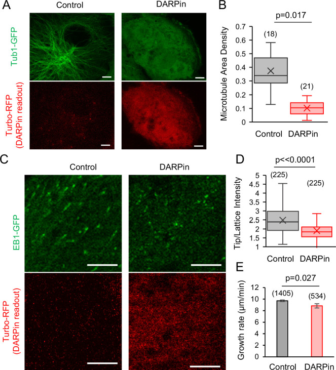
To first determine whether the DARPin protein could bind microtubule protofilament-edge sites in cells, we transfected the RFP-P2A-DARPin construct into LLC-Pk1 cells that expressed Tubulin-GFP (Rusan et al., 2001). Here, we reasoned that, if the expressed DARPin protein was binding protofilament-edge sites, a high concentration of DARPin could potentially suppress new tubulin subunit binding to growing microtubule plus-ends, and thus reduce the overall microtubule density in the transfected cells. Indeed, in comparing the microtubule density in cells that were not transfected (as identified by a lack of red fluorescence, Figure 6A, left), to transfected cells (with red fluorescence, Figure 6A, right), we observed a reduction in microtubule density in the DARPin-transfected cells (Figure 6B; p=0.017, Mann-Whitney U Test).

Figure 6. EB1 tip tracking is suppressed in cells that expressed Designed Ankyrin Repeat Protein (DARPin).

Figure 6.

(A) Left: Tubulin-GFP (top) and Turbo-RFP expression (bottom) in LLC-Pk1 cells that were not transiently transfected (scale bar: 5 μm). Right: Tubulin-GFP (top) and Turbo-RFP expression (bottom) in LLC-Pk1 cells that were transiently transfected (scale bar: 5 μm). (B) Microtubule density is lower in cells transfected with DARPin (p=0.017, Mann-Whitney U Test). (C) Left: EB1-GFP (top) and Turbo-RFP expression (bottom) in LLC-Pk1 cells that were not transiently transfected (scale bar: 5 μm). Right: EB1-GFP (top) and Turbo-RFP expression (bottom) in LLC-Pk1 cells that were transiently transfected (scale bar: 5 μm). (D) EB1-GFP Tip:Lattice intensity ratio is lower in cells transfected with DARPin as compared to control cells (p<0.0001, t-test). (E) Microtubule growth rate is similar in cells transfected with DARPin as compared to control cells (p=0.027, t-test).

Figure 6—source data 1. Data for Figure 6B, D, and E.

We then transfected the RFP-P2A-DARPin construct into LLC-Pk1 cells that overexpressed EB1-GFP, to examine the effect of the DARPin on EB1 tip tracking (Piehl et al., 2004). We observed fewer EB1 comets in the presence of DARPin, as would be expected due to a reduction in the microtubule network density (Figure 6B). However, a sufficient number of EB1 comets were present to allow for an analysis of the relative comet brightness in the presence and absence of DARPin. Cells that were not transfected had a ~31% higher average tip:lattice EB1 intensity ratio as compared to the cells that were transfected with DARPin (Figure 6C, left vs right; Video 9; Video 10; Figure 6D; p<0.0001, t-test). In examining the microtubule growth rate in cells with and without DARPin (as determined by the EB1 comet velocity), we found that the microtubule growth rate for the comets that were visible in the presence of DARPin was similar to the cells without DARPin (Figure 6E; p=0.027, t-test), suggesting that the reduced comet intensity in DARPin was not due to a slowed microtubule growth rate.

Video 9. Experimental EB1-GFP tip tracking in live LLC-PK1 cells without Designed Ankyrin Repeat Protein (DARPin) transfection.

Download video file (343.9KB, mp4)

5 µm scale bar.

Video 10. Experimental EB1-GFP tip tracking in live LLC-PK1 cells with Designed Ankyrin Repeat Protein (DARPin) transfection.

Download video file (413.1KB, mp4)

5 µm scale bar.

Discussion

In this work, we developed a molecular-scale computational simulation that incorporated both tubulin assembly dynamics, and EB1 on-off dynamics. Our simulation predicted that the binding of EB1 to protofilament-edge sites contributes to efficient tip-tracking of EB1 at growing microtubule plus-ends. To test this prediction, we used DARPin, a synthetic peptide, which binds to protofilament-edge sites on microtubules and partially overlaps the EB1 binding site. We found that DARPin blocks EB1 binding at protofilament-edge sites on stabilized microtubules, and importantly, this blocking of EB1 binding to protofilament-edge sites led to a disruption of EB1 tip tracking in dynamic microtubule cell-free assays, and in cells. We conclude that the rapid binding of EB1 to protofilament-edge sites facilitates the tip tracking of EB1 at growing microtubule ends. We note that, while we found that DARPin is not likely to indirectly suppress EB1 tip tracking by altering the plus-end tip conformation or the GTP-tubulin hydrolysis rate (Figure 5C), it remains possible that DARPin treatment could alter the conformation of the tubulin that composes the microtubule tip to indirectly suppress EB1 tip tracking. Furthermore, while the model leads to robust tip tracking by leveraging our previous experimental and simulation results that demonstrate the on-rate of EB1 molecules is rapid to protofilament-edge sites (Reid et al., 2019), we cannot exclude that another end-specific feature, that was not considered here, may be possible.

Previously, a model was developed to explain the peak position of EB1 on the growing microtubule tip, which is slightly distal from the tip of the growing microtubule (Figure 1C; Maurer et al., 2014). Because both our currently described model and the previously described model were able to reproduce the localization of EB1 on the microtubule, we sought to compare and contrast the described mechanisms in each of the two models.

In the previously described work by Maurer et al., 2014, a model was developed that relied on a constant length microtubule template with three EB1 binding ‘zones.’ Here, explicit tubulin assembly dynamics were not included in the model, but rather a constant length microtubule template was employed, in which there was an EB1 binding ‘exclusion zone’ at the tip of the microtubule. A tubulin subunit maturation rate was included in the model, which led to a second zone, slightly distal from the tip of the microtubule, in which EB1 binding was allowed. Finally, a second tubulin subunit maturation rate was employed, which led to a third zone, far from the tip and along the microtubule lattice, in which EB1 disassociation was allowed. The second tubulin subunit maturation rate likely corresponds to GTP-tubulin to GDP-tubulin hydrolysis, which is similar both in the magnitude of the hydrolysis rates employed, and in the EB1 off-rates employed, between the Maurer model and our newly described model (Supplementary file 3).

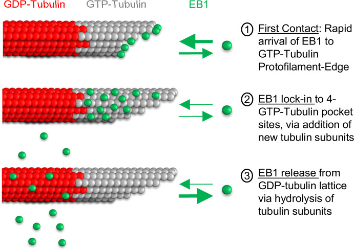
Thus, the primary difference between the two models was in the binding of EB1 to the microtubule lattice. Here, we predict that the key features of the Maurer model that involved exclusion of EB1 binding to the tip of the microtubule, along with a binding zone just distal to the microtubule tip, are incorporated into our newly described model by the ability of newly arriving tubulin subunits to ‘lock in’ protofilament-edge bound EB1 molecules into a stable 4-tubulin pocket, and by binding of EB1 to protofilament-edge sites along exposed protofilament sides that are distal from the tip of the microtubule. Specifically, in our new model, EB1 molecules arrive rapidly to easily accessible protofilament-edge sites at the growing tip of the microtubule, and to exposed protofilament sides (Figure 7, step 1). Then, upon new tubulin subunit addition, protofilament-edge bound EB1 molecules at the tip of the microtubules are ‘locked in’ to a stable, 4-GTP-tubulin pocket (Figure 7, step 2), leading to a low EB1 off-rate. Thus, in our new model, EB1 accumulates on the GTP-cap at the growing microtubule end (Maurer et al., 2014; Roth et al., 2019). Finally, upon the hydrolysis of GTP-tubulin to GDP-tubulin, the affinity of EB1 for the GDP-tubulin subunits is reduced, and EB1 dissociates from the microtubule (Figure 7, step 3). Thus, we predict that the primary difference between our new model, and the previous Maurer et al model, may be in (1) the inclusion of tubulin assembly dynamics, and (2) rapid EB1 binding to protofilament-edge sites. These features eliminate the requirement for an explicit EB1 binding ‘exclusion zone’ at the tip of the microtubule, and naturally lead to a decrease in signal at the tip of the microtubule.

Figure 7. Model for EB1 tip tracking.

Figure 7.

Step 1 (top): EB1 binds rapidly to protofilament-edge sites, with a slower on-rate to 4-tubulin closed lattice binding sites. Step 2 (middle): Incorporation of new tubulin subunits ‘locks in’ EB1 bound at protofilament-edge sites as the binding pocket transitions from a protofilament-edge site to a 4-tubulin closed-lattice site. Step 3 (bottom): As the GTP-tubulin hydrolyzes to GDP-tubulin, the EB1 dissociates from the GDP closed-lattice binding site.

The tubulin assembly portion of the model was built on earlier work, in which individual tubulin subunits were allowed to arrive and depart from the growing microtubule plus-end (Margolin et al., 2011; Margolin et al., 2012) (see Methods). Future work will involve examining the effects of a slower tubulin association rate on EB1-occupied protofilament edge sites, and whether EB1 binding to protofilament edge sites could assist in neighboring protofilament zippering on flared microtubule tips. Furthermore, microtubule targeting drugs that suppress the kinetics of tubulin assembly at the growing microtubule plus-end, such as Taxol (Castle et al., 2017), could potentially disrupt EB1 tip tracking by slowing the capture and ‘lock in’ of EB1 to 4-tubulin pocket binding sites (Figure 7, step 2), an idea that could be explored in future work.

As described above, our model predicts that rapid protofilament-edge binding increases the efficiency of EB1 tip tracking. In the simulation, the peak EB1 location is slightly distal from the tip of the growing microtubule (Figure 1C), similar to previous reports (Maurer et al., 2014). We surmise that the peak EB1 location in the simulation is heavily influenced by EB1 molecules that are stably bound to GTP-tubulin closed-lattice sites on the growing microtubule tip. Indeed, by reporting the fraction of EB1 molecules that are bound to GTP-Tubulin protofilament-edge sites as compared to GTP-Tubulin closed lattice sites, we found that there are ~twofold more EB1 molecules bound to closed-lattice GTP-tubulin sites, as compared to protofilament-edge sites, at any one time in the simulation (Figure 1—figure supplement 1H). Furthermore, the number of EB1 binding sites at the tip of each protofilament is explicitly limited by the number of protofilaments in the microtubule (13 binding sites). Thus, EB1 binding to numerous protofilament-edge sites along exposed protofilament sides that are distal from the tip of the microtubule may also contribute to the peak EB1 location. This idea is consistent with results from the ‘split comet’ simulations (Figure 3A and B). Here, by substantially increasing the taper at the tip of the simulated growing microtubule (≤~3 μm), the EB1 comet was greatly extended in length, and altered in configuration, thus shifting the location of EB1 binding (Figure 3B). However, the location of the simulated EB1 peak position was insensitive to small changes in tip taper (Figure 1—figure supplement 1B).

A key aspect of the simulation is that EB1 molecules arrive rapidly to protofilament-edge sites at the tip of the growing microtubule. We propose that, because the on-rate of new tubulin molecules is also rapid (simulated arrival rate for tubulin: ~85 s–1 at 10 μM tubulin), the simulated EB1 molecules that bind to protofilament-edge sites at the tip of the growing microtubule are quickly ‘locked in’ to a closed-lattice GTP-tubulin binding configuration (Figure 7). Thus, while most of the EB1 molecules at the microtubule tip are indeed bound to closed-lattice GTP-tubulin sites (Figure 1—figure supplement 1H), many of these EB1 molecules likely originated as arrivals to protofilament-edge sites (Figure 1—figure supplement 1A). To test this idea, we ran simulations in which we recorded the initial binding location of EB1, to determine the fraction of EB1 molecules that initially bound to protofilament-edges as compared to closed-lattice positions (Figure 1—figure supplement 1I). We found that ~50% of all EB1 binding events occurred at protofilament-edge sites (Figure 1—figure supplement 1I). However, importantly, the EB1 molecules that initially bound to protofilament-edge sites were heavily concentrated at the growing microtubule tip, while EB1 molecules that bound to closed-lattice sites were more uniformly distributed throughout the microtubule (Figure 1—figure supplement 1J and A; Video 2). This is because closed-lattice binding occurs throughout the microtubule, rather than specifically near to the growing microtubule plus-end.

While protofilament-edge binding is a key aspect of our model, it is important to emphasize that both rapid binding of EB1 to protofilament-edges (50-100:1 edge:lattice), as well as a differential GDP- to GTP-tubulin off-rate (6-12:1 GDP:GTP), were critical to produce robust tip tracking in the model. Because both of these factors contribute to tip tracking, this leads to a highly robust model, that does not require a narrow set of parameter values for either effect, in order to reproduce experimental results (Figure 2G–I). Thus, based on the experimentally measured EB1 on and off rates, a differential GDP- to GTP-tubulin off-rate, together with rapid protofilament-edge binding, was required for EB1 tip tracking in the model. Importantly, we observed robust tip tracking in the model without requiring narrow parameter sets, or by establishing an EB1 binding exclusion zone on the microtubule, as has been previously hypothesized.

Recent work has demonstrated that growing microtubule tips are less homogeneous than previously thought, such that they exhibit a wide range of protofilament lengths between the leading and lagging protofilaments, both in cells and in cell-free experiments (Atherton et al., 2018; Cleary and Hancock, 2021; Coombes et al., 2013; Gudimchuk et al., 2020; Guesdon et al., 2016; Igaev and Grubmüller, 2022). Here, a wide range of protofilament lengths at the growing microtubule end would lead to increased numbers of protofilament-edge sites on the exposed protofilament sides, which, in our model, would increase the EB1 on-rate to the tip. In addition, in our model, other tip configurations, such as partial curvature (Bechstedt et al., 2014; Farmer et al., 2021), or flaring of the growing microtubule plus-end (McIntosh et al., 2018), would also contribute to EB1 tip tracking (Figure 1—figure supplement 1C). Here, partial curvature or flaring requires opening of the closed microtubule tube – indicating that protofilaments or groups of protofilaments are separated from each other. Importantly, separation between protofilaments means that the number of protofilament-edge sites would be enriched, as new protofilament sides would be exposed. Thus, the role of protofilament-edge sites in facilitating EB1 tip tracking could apply to a wide range of growing microtubule tip configurations.

Recently published work provides support for the importance of EB1 protofilament-edge site binding in the efficiency of EB1 tip tracking. Specifically, by using the microtubule polymerase protein XMAP215 in cell-free experiments, the range of protofilament lengths between the leading and lagging protofilaments at the growing microtubule plus-end was increased (Farmer et al., 2021). Importantly, an increase in EB1-GFP intensity at the growing microtubule tip was observed with increased XMAP215-induced tip taper (Farmer et al., 2021). We note that increased tip taper would likely correspond to an increase in the number of protofilament-edge sites along exposed protofilament sides, similar to our split comet phenotype (Figure 3). Thus, XMAP215 could increase the efficiency of EB1 tip tracking by adding new protofilament-edge sites to the growing microtubule plus-end. This suggests that EB1 recruitment, and by extension the recruitment of the +Tip Complex, could be sensitive to the number of protofilament-edge sites at the tip of the growing microtubule. Correspondingly, a recent report found that EB1, and thus CLASP2, is redistributed from the plus-end onto the microtubule lattice in cells subjected to stretch and compression cycles (Li et al., 2023). This result is consistent with the idea that microtubule bending could cause openings and holes in the lattice, leading to the creation of new protofilament-edge sites along the lattice, which in turn causes a redistribution of EB1 from the plus-end tip to the lattice (Figure 4C, blue).

In conclusion, we find that protofilament-edge sites are an important contributing factor for proper EB1 tip tracking along growing microtubule ends, and that EB1 tip tracking is suppressed by blocking the protofilament-edge sites at growing microtubule ends. Therefore, altering the number of exposed protofilament-edge sites at the growing microtubule tip, or along the microtubule lattice, may provide a new mechanism to regulate EB1 localization in cells.

Materials and methods

Simulation methods

The simulation was performed in MATLAB, and all code has been deposited in GitHub (copy archived at Gonzalez, 2024a).

The microtubule assembly portion of the simulation is based on work from the Goodson lab (Margolin et al., 2012). Briefly, this model allows microtubule protofilaments to grow independently via the addition of individual tubulin subunits, and to form and break lateral bonds with neighboring protofilaments, once individual tubulin subunits are longitudinally bound to the microtubule. All of the parameter values for the microtubule assembly simulation matched a previously published parameter set (Margolin et al., 2012; Supplementary file 1), with the exception of (1) the tubulin on-rate constant, which was lowered in order to match our (slow) experimental growth rates, and (2) one additional rule was added to ensure that the tip taper at the microtubule plus-end matched our experimental values (Figure 1—figure supplement 1A and B). Here, if the difference between the longest and the penultimate shortest protofilament exceeded 600 nm (75 dimers) (Ogren et al., 2022), the tubulin subunit off-rate and the lateral bond breakage rate were dramatically increased, quickly leading to a catastrophe event.

In the EB1 binding and unbinding portion of the simulation, the model allows for EB1 to bind to and dissociate from the microtubule independently of tubulin addition/dissociation. Here, EB1 molecules bind to protofilament-edge sites and closed-lattice sites with differential on-rates, and then dissociate from GTP- and GDP-tubulin binding sites with differential off-rates (see Supplementary file 2).

At the start of every time step in the simulation, the total execution time was calculated for each potential event, which included EB1 association/dissociation, tubulin association/dissociation, and lateral bond formation/breakage between protofilaments, using the equation:

time=-lograndk (2)

where time is the total execution time required for each potential event, rand represents the built-in MATLAB function that generates a uniformly distributed random number between 0 and 1, and k is the single-molecule rate constant for each potential event (see Supplementary files 1 and 2).

Then, once all of the total execution times were calculated for each potential event, the simulation executed only one event, with the shortest time.

After the event was executed, the time to GTP-tubulin hydrolysis was calculated using Equation 2. If the hydrolysis time was shorter than the time of the executed event, then one of the GTP tubulin dimers was randomly hydrolyzed. Otherwise, no hydrolysis occurred in that time step.

After every time step in the simulation, every tubulin dimer’s hydrolysis state was recorded (GTP vs GDP), and the configuration of every potential EB1 binding site was also recorded (GTP vs GDP; Edge vs Lattice; EB1 bound or not bound). After each time step in the simulation, the tubulin dimer and EB1 binding site states and configurations were updated based on activities during the time step (e.g. tubulin association/dissociation, tubulin hydrolysis, EB1 binding/dissociation, lateral bond breakage/formation). Every thousand-time steps of the simulation (which averaged between 0.5 and 2 s of real-time in the simulation), the length of each microtubule protofilament, and the position of every bound EB1 were stored. This was repeated until the simulation ended, which in general was between 6 × 10⁴ and 4.5 × 10⁵ steps, depending on which simulation experiment was being performed. This stored data was then saved as excel files for further analysis (Crosby, 2024).

Creating model-convolved images from the simulation

To visualize the output from the simulation, 15% of the tubulin dimers were randomly labeled with a red fluorophore and plotted on an image. Then, all occupied EB1 positions were labeled with a green fluorophore and plotted. Next, uniform random noise was added to the image. Finally, the simulated image was convolved using the microscope point spread function, as would be observed on our TIRF microscope (Demchouk et al., 2011; Gardner et al., 2010; Reid et al., 2019). This model-convolution process was performed using the stored data from every 1000 steps in each simulation, and allowed for the generation of simulated videos in which the simulated EB1 localization along growing microtubules could be observed (GitHub, copy archived at Gonzalez, 2024b).

Analyzing line scans of simulated images for peak EB1 position and tip:lattice intensity

To quantify EB1 tip tracking in the simulation, line scans along the length of simulated microtubules were obtained using ImageJ. Then, each image was aligned along the peak EB1 position. Next, at least five separate simulation runs were averaged. To determine the end of the microtubule, the position where the intensity of the microtubule channel (red) was halfway between its maximum value and its minimum value was deemed as the microtubule end, and denoted with a relative position of zero nanometers. From here, the peak EB1 position could be determined by finding its relative position with respect to the microtubule end at position 0. In all simulated data that used line scans, N refers to the number of individual simulation growth events runs. To determine the tip:lattice intensity values, the maximum EB1 intensity at the tip was divided by the median EB1 intensity along the lattice, which was measured at 768–1024 nanometers distal of the microtubule plus-end.

Analyzing line scans of simulated and experimental images for microtubule tapering

To quantify the simulated and experimental microtubule tip standard deviation (Figure 1—figure supplement 1B and Figure 5C), images were loaded into a previously described program to measure tip tapering (Demchouk et al., 2011). In short, the user-defined points along the start and end of the microtubule lattice, and then the intensity of the microtubule across this distance was measured. Then, this intensity was fitted to a Gaussian survival curve to determine the tip standard deviation of the microtubule (which includes the microscope point spread function as well as the underlying protofilament length standard deviation). Each data point is the standard deviation for a single simulation run. This process was repeated for experimental microtubules.

Simulation parameter testing

To determine the robustness of tip tracking in our simulation, we ran a series of simulations in which all parameters but one were held constant, and then the parameter being tested was altered in twofold increments. The range for this parameter testing spanned from 1/16 to 16-fold times the baseline parameter value for each key parameter in the simulation. Ten simulation runs were performed for each parameter set. In each case, a simulated video was generated, and a line scan was obtained, as described above. To determine the uncertainty for a given parameter across the range we tested, we calculated the sum of absolute errors between the simulated and experimental Mal3 line scan profiles (Bieling et al., 2007).

Examining the effect of microtubule tip flaring and EB1 monomers on simulation tip tracking

In the simulation, the most distal lateral bond between protofilaments is tracked. To simulate flared microtubule tips, a rule was added in which all EB1 binding sites past the most distal lateral bond between protofilaments were considered protofilament-edge sites. Then, the effect of this rule on EB1 tip tracking was examined.

To examine the effect of EB1 monomer binding in our simulation, we course-grained monomer binding by increasing all EB1 off-rates by fourfold, as has been shown in the literature (Song et al., 2020). Then, EB1 tip tracking was examined with these faster off-rates.

Split comet simulations

To generate split comets in our simulations, we increased the maximum taper length that was allowed before increasing klateral bond break and koff (Tdimer ~400 dimers long (~3 μM)). In addition, we lowered the πbreak value in the simulation from 10 to 6, and we increased the tubulin concentration [GTP-tub] in the simulation to 30 μM. The change to max taper length and πbreak values both encouraged longer tip tapers, and by extension a higher likelihood of split comets. The increase in the tubulin concentration reduced the catastrophe frequency, to increase the lifetime of split comets. Then, simulations were run as previously, and only growth events were analyzed that produced split comets (not all growth events produced split comets). When there were split comets, images were cropped during the same growth event for the brightest single-comet frame, the brightest split-comet frame, and for a seed-only frame (no dynamic microtubule, for background). Next, the smallest region that encapsulated all the split and single comets in both images was determined. The intensity across this region was summed for all three images (single comet, split comet, no comet). Then, the background intensity (no comets) was subtracted from the single comet and split comet values, allowing for a comparison of the summed intensity of EB1 along the simulated growing microtubule with a single comet and with split comets.

Experimental methods

Tubulin purification and labeling

Tubulin was purified and labeled as previously described (Gell et al., 2010).

Preparation of GTP-analogue microtubules

GMPCPP microtubules were prepared as previously described Gell et al., 2010. In short, 3.9 µM rhodamine labeled tubulin was incubated with 1 mM GMPCPP with 1.1 mM MgCl2 in BRB80 for 5 min on ice. Then, the solution was incubated at 37 °C for 2 hr or overnight. The GMPCPP microtubules were then spun with an airfuge (Beckman Coulter, 20 psi, 5 min) and further stabilized in 10 μM Taxol in BRB80. These microtubules were stored at 37 °C and used in experiments within four days after preparation. These microtubules were used as seeds for dynamic microtubule experiments and were used for Mal3 binding along GMPCPP microtubules.

Purification of Mal3-GFP

The pETMM11-HIS6x-Mal3-GFP plasmid with a TEV cut site after the His6x tag was a kind gift from Dr. Thomas Surrey. The plasmid was transformed into Rosetta (DE3) pLysS E. coli and grown in 800 mL of LB + kan + cam at 37 °C to an OD of approximately 0.4. To induce protein expression, IPTG was added to 0.2 mM and the culture was mixed at 14 °C for 16 hr. Cells were centrifuged (30 min., 4 °C, 4400 × g) and resuspended in 25 mL lysis buffer (50 mM Tris pH7.5, 200 mM NaCl, 5% glycerol, 20 mM imidazole, 5 mM β-mercaptoethanol, 0.2% triton X-100), protease inhibitors (1 mM PMSF, 10 μM Pepstatin A, 10 μM E-64, 0.3 μM aprotinin), and DNAse I (1 U/mL). The cell suspension was sonicated on ice (90% power, 50% duty, 6 × 1 min). Cell lysates were centrifuged (1 hr, 4 °C, 14000 × g) and the soluble fraction was passed through 1 mL of Talon Metal Affinity Resin (Clontech #635509). The resin was washed for four times with 4 mL Wash Buffer (50 mM Tris pH7.5, 500 mM NaCl, 5% glycerol, 20 mM imidazole, 5 mM β-mercaptoethanol, 0.1 mM PMSF, 1 μM Pepstatin A, 1 μM E-64, 30 nM aprotinin) for 5 min each. Protein was eluted from the resin by mixing with 1 mL of Elution Buffer (50 mM Tris pH 7.5, 200 mM NaCl, 250 mM imidazole, 0.1 mM PMSF, 1 μM Pepstatin A, 1 μM E-64, 30 nM aprotinin) for 15 min followed by slow centrifugation through a fritted column to retrieve eluate. To cleave the HIS6x tag, 10 units of GST-tagged TEV enzyme (TurboTEV, #T0102M, Accelagen) and 14 mM β-mercaptoethanol were added and the eluate was dialyzed into Brb80 overnight at 4 °C. To remove the TEV enzyme, the dialysate was mixed with 100 ul of glutathione-sepharose (GE Healthcare #17-0756-01) for 30 min. at 4 °C and spun (1 min, 2000 × g). The Mal3-GFP protein was quantified by band intensity on a coomassie-stained SDS PAGE protein gel.

Purification of Mal3-mCherry

Purification of Mal3 from bacteria was based on the protocol described previously Gerson-Gurwitz et al., 2011; Hepperla et al., 2014. His6x-Mal3-mCherry is in the pETMM11 vector with a TEV cut site after the His6x tag, kindly provided by Dr. Thomas Surrey. This plasmid, in Rosetta (DE3) pLysS E. coli, was grown in 600 ml of TB + kan + cam at 37° to an OD of about 0.4, then IPTG was added to 0.2 mM to induce protein expression and growth continued at 14° for 20 hr. Cells were centrifuged and resuspended in 25 ml lysis buffer (50 mM Tris pH7.5, 200 mM NaCl, 5% glycerol, 20 mM imidazole, 5 mM B-mercaptoethanol, 0.2% triton X-100) plus protease inhibitors (1 mM PMSF, 10 uM Pepstatin A, 10 uM E-64, 0.3 uM aprotinin). DNAse I was added to 1 U/ml in the cell suspension and sonicated on ice at 90% power, 50% duty for six cycles (1 min. on, 1 min. off) to lyse. Lysate was centrifuged at 14000 × g, 4°, for 1.5 hr and the soluble fraction was mixed with 1 ml of Talon affinity resin (Clontech #635509) at 4° for 1 hr. Resin was poured into a small column and washed in 5 min. sequences with buffers with 0.1 x protease inhibitors: two times with lysis buffer, two times with lysis buffer/700 mM NaCl, one time with lysis buffer. Protein was eluted from the resin by mixing for 15 min with 2 ml of elution buffer (lysis buffer/250 mM imidazole)+0.1 x protease inhibitors, and slowly centrifuging the column to collect all the eluate. 10 units of TEV enzyme (TurboTEV, #T0102M, Accelagen) and B-mercaptoethanol to 14 mM was added and the eluate was dialyzed into Brb80 (80 mM PIPES pH6.9, 1 mM MgCl2, 1 mM EGTA) at 4°. The dialysate was mixed with four 100 ul of glutathione-sepharose (GE Healthcare #17-0756-01) for 30 min., 4° to remove the TEV enzyme, which has a GST tag. The Mal3-mCherry protein was quantified by band intensity on a coomassie-stained SDS PAGE protein gel.

Purification of DARPin D1

A plasmid containing the D1-Darpin sequence with a 6-HIS tag, a generous gift from Dr. Andreas Plückthun and Dr. Benoît Gigant (Pecqueur et al., 2012) was grown in XL1-Blue bacteria in LB + ampicillin media to an A600 of 0.6, then IPTG was added to 1 mM and grown for 21 hr. at 18°. All subsequent steps were performed at 4°. The centrifuged cell pellet was resuspended in lysis buffer (50 mM Tris pH8 /10 mM imidazole/1 mM MgCl2 /0.3 mg/ml lysozyme with cOmplete EDTA-free protease inhibitor (Sigma #4693159001)), incubated on ice for 30 min., lysed by sonication on ice and cell debris was removed by centrifugation at 18000 × g for 30 min. The soluble lysate was passed over 1 ml Talon metal affinity resin column (https://www.takarabio.com/ #635502) three times, the resin was then washed with 10 volumes of wash buffer (50 mM Tris pH8 /10 mM imidazole/1 mM MgCl2) and eluted with 50 mM Tris/300 mM imidazole/1 mM MgCl2 pH8. The eluted protein was dialyzed against Brb80 (80 mM PIPES/1 mM MgCl2 /1 mM EGTA pH6.9) and centrifuged to remove any precipitate. Purifed D1-Darpin was quantified by measuring band intensity on a Coomassie G-250-stained acrylamide protein gel.

Creation of TIRF microscopy flow chambers for cell-free assays

Imaging flow chambers were constructed as in Section VII of Gell et al., 2010, with the following modifications: two narrow strips of parafilm replaced double-sided scotch tape as chamber dividers: following placement of the smaller coverslip onto the parafilm strips, the chamber was heated to melt the parafilm and create a seal between the coverslips; typically, only three strips of parafilm were used, resulting in two chambers per holder. Chambers were prepared with an anti-rhodamine antibody (Invitrogen A6397, RRID:AB_2536196) followed by blocking with Pluronic F127, as described in Section VIII of Gell et al., 2010. Microtubules were adhered to the chamber coverslip, and the chamber was flushed gently with warm BRB80. The flow chamber was heated to 28 °C using an objective heater on the microscope stage, and then 3–4 channel volumes of imaging buffer were flushed through the chamber. Microtubules were imaged on a Nikon TiE microscope using 488 nm and 561 nm lasers sent through a Ti-TIRF-PAU for Total Internal Reflectance Fluorescence (TIRF) illumination. An Andor iXon3 EM-CCD camera fitted with or without a 2.5x projection lens depending on the experiment was used to capture images with high signal-to-noise and small pixel size (64 nm or 160 nm, respectively). Images were collected using TIRF with a Nikon CFI Apochromat 100x 1.49 NA oil objective.

Cell-free microtubule assays

For the damaged GTP-analogue microtubule assays, GMPCPP microtubules were introduced into a flow chamber as described above and allowed to incubate for 3 min before flushing out any non-adhered microtubules with BRB80. Next, 10 mM CaCl2 in warmed BRB80 was introduced into the chamber and incubated for 1–5 min, until obvious degradation occurred to the microtubules. The chamber was then washed with multiple chamber volumes of warmed BRB80. Next, the chamber was washed with one chamber volume of prewarmed imaging buffer (20 μg/mL glucose oxidase, 10 μg/mL catalase, 20 mM D-Glucose, 10 mM DTT, 80 μg/mL casein, 110 mM KCl, and 1% tween-20). Finally, a Mal3 reaction mixture with or without 1 uM DARPin (imaging buffer plus 123 nM Mal3-GFP) was introduced to the chamber and allowed to incubate for 15 min. Images of hundreds of non-overlapping fields of view were collected and used for downstream analysis.

For undamaged GTP-analogue microtubule assays, GMPCPP microtubules were introduced into a flow chamber as described above and allowed to incubate for 30 s to 3 min before flushing out any non-adhered microtubules with BRB80. Next, one chamber volume of prewarmed imaging buffer (20 μg/mL glucose oxidase, 10 μg/mL catalase, 20 mM D-Glucose, 10 mM DTT, 80 μg/mL casein, 110 mM KCl, and 1% tween-20) was added. Finally, a reaction mixture with or without 1 uM DARPin (imaging buffer plus 123 nM Mal3-GFP) was introduced to the chamber and allowed to incubate for 15 min. Finally, images of hundreds of non-overlapping fields of view were obtained and used for downstream analysis.

For the dynamic microtubule assays, GMPCPP microtubule seeds were introduced into a flow chamber as described above and allowed to incubate for ~3 min before flushing out any non-adhered microtubules with BRB80. Next, one chamber volume of prewarmed imaging buffer (20 μg/mL glucose oxidase, 10 μg/mL catalase, 20 mM D-Glucose, 10 mM DTT, 80 μg/mL casein, 110 mM KCl, and 1% tween-20) was added. Finally, a reaction mixture consisting of Imaging buffer plus 212 nM Mal3-mCherry, 11.5 μM of 12% green-labeled tubulin, and 1 mM GTP, with or without DARPin, was added. Time-lapse images were collected of dynamic microtubules growing from the GMPCPP stabilized seeds and were then used for quantification.

Analyzing microtubule area bound by Mal3

To compare the binding of Mal3-GFP on undamaged and damaged microtubules in the presence and absence of DARPin, the total length of green (Mal3-GFP) occupancy was divided by the total length of the red microtubules on each image. This was accomplished by using a previously described semi-automated MATLAB analysis code (Reid et al., 2017). Briefly, first, automatic processing of the red microtubule channel was used to determine the microtubule-positive regions, which then allowed for the conversion of the red channel into a binary image with white microtubules and a black background. The green Mal3-GFP channel was then also pre-processed to smooth high-frequency noise and to correct for TIRF illumination heterogeneity. The green channel threshold was then manually adjusted to ensure visualization of all Mal3-GFP binding areas on each microtubule. Measurements of the total Mal3-GFP coverage area were then automatically collected from the identified microtubule regions. Finally, the total coverage area of Mal3-GFP was divided by the total microtubule area in each field of view. This experiment was replicated three times, as shown in the main text and in supplemental material.

Analysis of Mal3 tip tracking

To determine the Tip Specificity (Equation 1), a custom MATLAB script was written that allowed the user to pick the brightest point of the comet, then a point on the microtubule lattice, behind the comet, and then a point alongside the comet for background. Then, a 4 × 4 pixel box was summed at the brightest point of the comet (Itip), another 4 × 4 pixel box was summed at the point behind the comet (Ilattice), and a final 4 × 4 pixel box was summed alongside the tip (Ibackground). Finally, Equation 1 was used to calculate the Tip Specificity (S) (GitHub, copy archived at Gonzalez, 2024c). Only the brightest comet per growth event was measured and every growth event in a field of view was analyzed. This was completed over three replicate experiments, and pooled together for each condition. To analyze EB1-GFP comets in LLC-Pk1 cells, the brightest comets in each frame were cropped. These comets were then not cropped in later frames to ensure each comet was only analyzed once during its lifetime.

For the growth rate and time to catastrophe measurements, kymographs were made from representative growth events. Then, the growth rate was calculated from the kymograph by determining the length of the microtubule at the start and end of each growth event and dividing by the time required to reach the end of that growth event (GitHub, copy archived at Gonzalez, 2024d). The time to catastrophe was determined by examining growth events that started from the microtubule seed, and by measuring the elapsed time until a microtubule catastrophe event occurred.

To examine split comets, dynamic microtubules were examined to determine whether split comets were present. If there were split comets, then an image was cropped during the same growth event for the brightest single-comet frame, the brightest split-comet frame, and for a seed-only frame (no dynamic microtubule, for background). Next, the smallest region that encapsulated all of the split and single comets in both images was determined. The intensity across this region was summed for all three images (single comet, split comet, no comet). Then, the background intensity (no comets) was subtracted from the single comet and split comet values, allowing for a comparison of the summed intensity of Mal3-mCherry along the growing microtubule with a single comet and with split comets.

Crystal structure diagram of DARPin and Mal3

To generate a structural schematic, the crystal structure of DARPin bound to tubulin (4drx) (Pecqueur et al., 2012) was aligned with the crystal structure of Mal3 bound to tubulin (4abo) (Maurer et al., 2014) where the β-tubulin with DARPin and Mal3 bound were used for the alignment in Chimera (Pettersen et al., 2021).

Cloning of DARPin into a Turbo-RFP-P2A vector

An RFP-P2A-DARPin plasmid was generated by isolating the DARPin sequence from the DARPin bacterial expression vector (DARPin D1 in pDST67, Pecqueur et al., 2012) via a restriction digestion with the BamHI and HindIII sites and cloning it into FLAG-HA-mRFP-pcDNA3.1 vector (plasmid #52510, Addgene) using the same BamHI and HindIII sites (RFP-DARPin vector). Proper integration of the DARPin sequence was verified with sequencing. Next, we isolated a TurboRFP and P2A from a separate vector (plasmid # 78933, Addgene) via a restriction digestion with the NHEI and BamHI sites and cloned it into the RFP-DARPin vector with the BamHI and NHEI sites leading to a mammalian expression vector with TurboRFP and P2A N-terminal to DARPin (TurboRFP—P2a—DARPin vector). Proper integration of the Turbo-RFP and P2A sequence was verified with sequencing.

Cell lines

The LLC-Pk1 cell line expressing EB1-GFP was a gift from Dr. Patricia Wadsworth (Piehl et al., 2004), and the cell line expressing GFP-Tubulin was a gift from Dr. Lynne Cassimeris (Rusan et al., 2001). The identities of the cell lines (non-human) were authenticated by microscopy observation and analysis.

Culture and imaging of LLC-PK1 cells

The LLC-Pk1 cell lines were grown in Optimem media (Thermo Fisher #31985070), 10% fetal bovine sera + penicillin/streptomycin at 37 °C and 5% CO2. Cells were grown in 14 mm glass bottom dishes for visualization by microscopy. Cells were imaged with a laser scanning confocal microscope (Nikon Ti2, 488 nm laser line) fitted with a 100 x oil objective (Nikon N2 Apochromat TIRF 100 x Oil, 1.49 NA), which allowed for a 0.16 μm pixel size.

Transfecting LLC-PK1 cells

LLC-PK1 cells were transfected with Lipofectamine 3000 following the manufacturer’s protocol, except that the transfection was performed for 16 hr before imaging rather than 2–4 days before imaging. Immediately before imaging, cells were transferred into CO2-independent imaging media.

LLC-Pk1 microtubule growth rate analysis

To analyze EB1-GFP comet velocity, which was used as a proxy for the microtubule growth rate, we employed analysis software from the Danuser lab (Applegate et al., 2011). In short, we collected multiple 100 by 100-pixel movies of LLC-Pk1 cells treated with DMSO or transfected with DARPin from three separate biological replicates and loaded them into the Danuser code software, using constant parameters for thresholding and water shedding. We then allowed the program to identify, link, and track comets over time, which provided us with EB1 comet velocities across multiple cells. We next cut off any outlier values greater than 1 μm/s, which were likely artifacts from the analysis software. The growth rates were statistically analyzed using a student t-test.

LLC-PK1 microtubule density analysis

To determine the microtubule density in LLC-Pk1 cells that were or were not transfected with DARPin, LLC-Pk1 cells overexpressing Tubulin-GFP were grouped by RFP expression, with RFP expression indicating a successful transfection of DARPin. Z-stacks were acquired across the volume of these cells using confocal microscopy. Then, maximum Z-projections were created, followed by the analysis of the area of the microtubules divided by the area of the cell, which was performed with a custom MATLAB script (GitHub, copy archived at Gonzalez, 2024e; Goldblum et al., 2021). Finally, this normalized value was compared using a Mann-Whitney U test.

Materials availability

All materials generated during this study are available by contacting the Gardner lab at klei0091@umn.edu.

Acknowledgements

The Gardner laboratory is supported by a National Institutes of Health grant NIGMS R35-GM126974. SJG was supported in part by the National Institute of Health Training Program T32GM140936. We thank members of the Gardner, Courtemanche, and Titus laboratories for helpful discussions. We thank Dr. Andreas Plückthun and Dr. Benoît Gigant for the generous gift of the DARPin construct, and Dr. Thomas Surrey for the kind gift of Mal3 constructs.

Funding Statement

The funders had no role in study design, data collection and interpretation, or the decision to submit the work for publication.

Contributor Information

Melissa K Gardner, Email: klei0091@umn.edu.

Julie PI Welburn, University of Edinburgh, United Kingdom.

Amy H Andreotti, Iowa State University, United States.

Funding Information

This paper was supported by the following grants:

  • National Institutes of Health R35-GM126974 to Melissa K Gardner.

  • National Institutes of Health T32GM140936 to Samuel J Gonzalez.

Additional information

Competing interests

No competing interests declared.

Author contributions

Software, Formal analysis, Validation, Investigation, Visualization, Methodology, Writing - original draft, Writing – review and editing.

Formal analysis, Investigation, Writing – review and editing.

Software, Writing – review and editing.

Software, Writing – review and editing.

Resources, Methodology, Writing – review and editing.

Conceptualization, Data curation, Formal analysis, Supervision, Funding acquisition, Investigation, Methodology, Project administration, Writing – review and editing.

Additional files

Supplementary file 1. Summary table with simulation parameters for the microtubule assembly portion of the simulation.

Parameters determine the on and off rates of tubulin subunits from the microtubule tip, as well as parameters that control the hydrolysis rate of GTP-tubulin subunits within the lattice.

elife-91719-supp1.docx (17KB, docx)
Supplementary file 2. Summary table with simulation parameters for single-molecule EB1 dynamics.

Parameters determine the on and off rates of EB1 molecules from the microtubule tip and lattice.

elife-91719-supp2.docx (16.6KB, docx)
Supplementary file 3. Summary table with model parameter comparisons between the EB1 on and off rates from the microtubule tip and lattice for the current study model, as compared to a model developed by Maurer et al., 2014.

In addition, tubulin ‘maturation rates’ are compared, which define the EB1 binding zones in the Maurer et al model.

elife-91719-supp3.docx (12.9KB, docx)
MDAR checklist

Data availability

All data generated or analyzed during this study are included in this manuscript and supporting files. Source data files have been provided for Figures 16.

References

  1. Applegate KT, Besson S, Matov A, Bagonis MH, Jaqaman K, Danuser G. plusTipTracker: quantitative image analysis software for the measurement of microtubule dynamics. Journal of Structural Biology. 2011;176:168–184. doi: 10.1016/j.jsb.2011.07.009. [DOI] [PMC free article] [PubMed] [Google Scholar]
  2. Atherton J, Stouffer M, Francis F, Moores CA. Microtubule architecture in vitro and in cells revealed by cryo-electron tomography. Acta Crystallographica. Section D, Structural Biology. 2018;74:572–584. doi: 10.1107/S2059798318001948. [DOI] [PMC free article] [PubMed] [Google Scholar]
  3. Bechstedt S, Lu K, Brouhard GJ. Doublecortin recognizes the longitudinal curvature of the microtubule end and lattice. Current Biology. 2014;24:2366–2375. doi: 10.1016/j.cub.2014.08.039. [DOI] [PubMed] [Google Scholar]
  4. Bieling P, Laan L, Schek H, Munteanu EL, Sandblad L, Dogterom M, Brunner D, Surrey T. Reconstitution of a microtubule plus-end tracking system in vitro. Nature. 2007;450:1100–1105. doi: 10.1038/nature06386. [DOI] [PubMed] [Google Scholar]
  5. Castle BT, McCubbin S, Prahl LS, Bernens JN, Sept D, Odde DJ. Mechanisms of kinetic stabilization by the drugs paclitaxel and vinblastine. Molecular Biology of the Cell. 2017;28:1238–1257. doi: 10.1091/mbc.E16-08-0567. [DOI] [PMC free article] [PubMed] [Google Scholar]
  6. Cleary JM, Hancock WO. Molecular mechanisms underlying microtubule growth dynamics. Current Biology. 2021;31:R560–R573. doi: 10.1016/j.cub.2021.02.035. [DOI] [PMC free article] [PubMed] [Google Scholar]
  7. Coombes CE, Yamamoto A, Kenzie MR, Odde DJ, Gardner MK. Evolving tip structures can explain age-dependent microtubule catastrophe. Current Biology. 2013;23:1342–1348. doi: 10.1016/j.cub.2013.05.059. [DOI] [PMC free article] [PubMed] [Google Scholar]
  8. Coombes C, Yamamoto A, McClellan M, Reid TA, Plooster M, Luxton GWG, Alper J, Howard J, Gardner MK. Mechanism of microtubule lumen entry for the α-tubulin acetyltransferase enzyme αTAT1. PNAS. 2016;113:46. doi: 10.1073/pnas.1605397113. [DOI] [PMC free article] [PubMed] [Google Scholar]
  9. Crosby K. Column converter for excel (https://www.mathworks.com/matlabcentral/fileexchange/28343-column-converter-for-excel), MATLAB Central File Exchange. 2024. [January 29, 2024]. https://www.mathworks.com/matlabcentral/fileexchange/28343-column-converter-for-excel
  10. Dema A, Charafeddine R, Rahgozar S, van Haren J, Wittmann T. Growth cone advance requires EB1 as revealed by genomic replacement with a light-sensitive variant. eLife. 2023;12:e84143. doi: 10.7554/eLife.84143. [DOI] [PMC free article] [PubMed] [Google Scholar]
  11. Demchouk AO, Gardner MK, Odde DJ. Microtubule tip tracking and tip structures at the nanometer scale using digital fluorescence microscopy. Cellular and Molecular Bioengineering. 2011;4:192–204. doi: 10.1007/s12195-010-0155-6. [DOI] [PMC free article] [PubMed] [Google Scholar]
  12. Desai A, Mitchison TJ. Microtubule polymerization dynamics. Annual Review of Cell and Developmental Biology. 1997;13:83–117. doi: 10.1146/annurev.cellbio.13.1.83. [DOI] [PubMed] [Google Scholar]
  13. Dixit R, Barnett B, Lazarus JE, Tokito M, Goldman YE, Holzbaur ELF. Microtubule plus-end tracking by CLIP-170 requires EB1. PNAS. 2009;106:492–497. doi: 10.1073/pnas.0807614106. [DOI] [PMC free article] [PubMed] [Google Scholar]
  14. Dong X, Liu F, Sun L, Liu M, Li D, Su D, Zhu Z, Dong JT, Fu L, Zhou J. Oncogenic function of microtubule end‐binding protein 1 in breast cancer. The Journal of Pathology. 2010;220:361–369. doi: 10.1002/path.2662. [DOI] [PubMed] [Google Scholar]
  15. Doodhi H, Prota AE, Rodríguez-García R, Xiao H, Custar DW, Bargsten K, Katrukha EA, Hilbert M, Hua S, Jiang K, Grigoriev I, Yang CPH, Cox D, Horwitz SB, Kapitein LC, Akhmanova A, Steinmetz MO. Termination of protofilament elongation by eribulin induces lattice defects that promote microtubule catastrophes. Current Biology. 2016;26:1713–1721. doi: 10.1016/j.cub.2016.04.053. [DOI] [PubMed] [Google Scholar]
  16. Farmer V, Arpağ G, Hall SL, Zanic M. XMAP215 promotes microtubule catastrophe by disrupting the growing microtubule end. The Journal of Cell Biology. 2021;220:e202012144. doi: 10.1083/jcb.202012144. [DOI] [PMC free article] [PubMed] [Google Scholar]
  17. Gardner MK, Sprague BL, Pearson CG, Cosgrove BD, Bicek AD, Bloom K, Salmon ED, Odde DJ. Model convolution: a computational approach to digital image interpretation. Cellular and Molecular Bioengineering. 2010;3:163–170. doi: 10.1007/s12195-010-0101-7. [DOI] [PMC free article] [PubMed] [Google Scholar]
  18. Gell C, Bormuth V, Brouhard GJ, Cohen DN, Diez S, Friel CT, Helenius J, Nitzsche B, Petzold H, Ribbe J, Schäffer E, Stear JH, Trushko A, Varga V, Widlund PO, Zanic M, Howard J. Microtubule dynamics reconstituted in vitro and imaged by single-molecule fluorescence microscopy. Methods in Cell Biology. 2010;95:221–245. doi: 10.1016/S0091-679X(10)95013-9. [DOI] [PubMed] [Google Scholar]
  19. Gerson-Gurwitz A, Thiede C, Movshovich N, Fridman V, Podolskaya M, Danieli T, Lakämper S, Klopfenstein DR, Schmidt CF, Gheber L. Directionality of individual kinesin-5 Cin8 motors is modulated by loop 8, ionic strength and microtubule geometry. The EMBO Journal. 2011;30:4942–4954. doi: 10.1038/emboj.2011.403. [DOI] [PMC free article] [PubMed] [Google Scholar]
  20. Goldblum RR, McClellan M, White K, Gonzalez SJ, Thompson BR, Vang HX, Cohen H, Higgins L, Markowski TW, Yang TY, Metzger JM, Gardner MK. Oxidative stress pathogenically remodels the cardiac myocyte cytoskeleton via structural alterations to the microtubule lattice. Developmental Cell. 2021;56:2252–2266. doi: 10.1016/j.devcel.2021.07.004. [DOI] [PMC free article] [PubMed] [Google Scholar]
  21. Gonzalez S. SimulatedEB1TipTracking. swh:1:rev:f368e0c9caf4dfd1517037695215dfc77d714927Software Heritage. 2024a https://archive.softwareheritage.org/swh:1:dir:7254e9007f70e3b7646f00f6acff8c0a1642f411;origin=https://github.com/SamuelGonzalez2799/SimulatedEB1TipTracking;visit=swh:1:snp:36718406ed739570041b1cf798cc4a15ea80f941;anchor=swh:1:rev:f368e0c9caf4dfd1517037695215dfc77d714927
  22. Gonzalez S. GenerateSimulatedMTImages. swh:1:rev:56ab52c6206e50ddf7e40eedf43fce73c2427e3bSoftware Heritage. 2024b https://archive.softwareheritage.org/swh:1:dir:1d92654b4a977ae22d718feb3c8655407c0b9c2c;origin=https://github.com/SamuelGonzalez2799/GenerateSimulatedMTImages;visit=swh:1:snp:9212806fd993fd53524eb95716c7fdf2e5a81848;anchor=swh:1:rev:56ab52c6206e50ddf7e40eedf43fce73c2427e3b
  23. Gonzalez S. EB1TipSpecificity. swh:1:rev:b2aa32f9055a65436c35067d74cc3d04b189c4dfSoftware Heritage. 2024c https://archive.softwareheritage.org/swh:1:dir:c1dbc20cdc1335802a47c123b415d47fc198912d;origin=https://github.com/SamuelGonzalez2799/EB1TipSpecificity;visit=swh:1:snp:ef96a8aba2aafd1078aa10e08e7d1674d81175e6;anchor=swh:1:rev:b2aa32f9055a65436c35067d74cc3d04b189c4df
  24. Gonzalez S. KymographDynamicsAnalysis. swh:1:rev:f3f125ed9badb4cba0ce9e90723fa126df097b4eSoftware Heritage. 2024d https://archive.softwareheritage.org/swh:1:dir:e1c7cddc8e0b6f9cc50d8b68c76dda9373183bde;origin=https://github.com/SamuelGonzalez2799/KymographDynamicsAnalysis;visit=swh:1:snp:7bb86b886caaa6b031238bfd835f66677ea57ad4;anchor=swh:1:rev:f3f125ed9badb4cba0ce9e90723fa126df097b4e
  25. Gonzalez S. MicrotubuleDensityAnalysis. swh:1:rev:1f9ca76cdd58df01132a96e5fd79130448627609Software Heritage. 2024e https://archive.softwareheritage.org/swh:1:dir:96ed56abaa1103c98404901aac76cb9992f0021b;origin=https://github.com/SamuelGonzalez2799/MicrotubuleDensityAnalysis;visit=swh:1:snp:14521221b33e60b83d5b4f6f63ec7297620c2221;anchor=swh:1:rev:1f9ca76cdd58df01132a96e5fd79130448627609
  26. Gudimchuk NB, Ulyanov EV, O’Toole E, Page CL, Vinogradov DS, Morgan G, Li G, Moore JK, Szczesna E, Roll-Mecak A, Ataullakhanov FI, Richard McIntosh J. Mechanisms of microtubule dynamics and force generation examined with computational modeling and electron cryotomography. Nature Communications. 2020;11:3765. doi: 10.1038/s41467-020-17553-2. [DOI] [PMC free article] [PubMed] [Google Scholar]
  27. Guesdon A, Bazile F, Buey RM, Mohan R, Monier S, García RR, Angevin M, Heichette C, Wieneke R, Tampé R, Duchesne L, Akhmanova A, Steinmetz MO, Chrétien D. EB1 interacts with outwardly curved and straight regions of the microtubule lattice. Nature Cell Biology. 2016;18:1102–1108. doi: 10.1038/ncb3412. [DOI] [PubMed] [Google Scholar]
  28. Gupta KK, Li C, Duan A, Alberico EO, Kim OV, Alber MS, Goodson HV. Mechanism for the catastrophe-promoting activity of the microtubule destabilizer Op18/stathmin. PNAS. 2013;110:20449–20454. doi: 10.1073/pnas.1309958110. [DOI] [PMC free article] [PubMed] [Google Scholar]
  29. Hepperla AJ, Willey PT, Coombes CE, Schuster BM, Gerami-Nejad M, McClellan M, Mukherjee S, Fox J, Winey M, Odde DJ, O’Toole E, Gardner MK. Minus-end-directed Kinesin-14 motors align antiparallel microtubules to control metaphase spindle length. Developmental Cell. 2014;31:61–72. doi: 10.1016/j.devcel.2014.07.023. [DOI] [PMC free article] [PubMed] [Google Scholar]
  30. Honoré S, Pagano A, Gauthier G, Bourgarel-Rey V, Verdier-Pinard P, Civiletti K, Kruczynski A, Braguer D. Antiangiogenic vinflunine affects EB1 localization and microtubule targeting to adhesion sites. Molecular Cancer Therapeutics. 2008;7:2080–2089. doi: 10.1158/1535-7163.MCT-08-0156. [DOI] [PubMed] [Google Scholar]
  31. Igaev M, Grubmüller H. Bending-torsional elasticity and energetics of the plus-end microtubule tip. PNAS. 2022;119:e2115516119. doi: 10.1073/pnas.2115516119. [DOI] [PMC free article] [PubMed] [Google Scholar]
  32. Komarova Y, De Groot CO, Grigoriev I, Gouveia SM, Munteanu EL, Schober JM, Honnappa S, Buey RM, Hoogenraad CC, Dogterom M, Borisy GG, Steinmetz MO, Akhmanova A. Mammalian end binding proteins control persistent microtubule growth. The Journal of Cell Biology. 2009;184:691–706. doi: 10.1083/jcb.200807179. [DOI] [PMC free article] [PubMed] [Google Scholar]
  33. Li Y, Kučera O, Cuvelier D, Rutkowski DM, Deygas M, Rai D, Pavlovič T, Vicente FN, Piel M, Giannone G, Vavylonis D, Akhmanova A, Blanchoin L, Théry M. Compressive forces stabilize microtubules in living cells. Nature Materials. 2023;22:913–924. doi: 10.1038/s41563-023-01578-1. [DOI] [PMC free article] [PubMed] [Google Scholar]
  34. Margolin G, Goodson HV, Alber MS. Mean-field study of the role of lateral cracks in microtubule dynamics. Physical Review E. 2011;83:041905. doi: 10.1103/PhysRevE.83.041905. [DOI] [PubMed] [Google Scholar]
  35. Margolin G, Gregoretti IV, Cickovski TM, Li C, Shi W, Alber MS, Goodson HV. The mechanisms of microtubule catastrophe and rescue: implications from analysis of a dimer-scale computational model. Molecular Biology of the Cell. 2012;23:642–656. doi: 10.1091/mbc.E11-08-0688. [DOI] [PMC free article] [PubMed] [Google Scholar]
  36. Maurer SP, Bieling P, Cope J, Hoenger A, Surrey T. GTPγS microtubules mimic the growing microtubule end structure recognized by end-binding proteins (EBs) PNAS. 2011;108:3988–3993. doi: 10.1073/pnas.1014758108. [DOI] [PMC free article] [PubMed] [Google Scholar]
  37. Maurer SP, Fourniol FJ, Bohner G, Moores CA, Surrey T. EBs recognize a nucleotide-dependent structural cap at growing microtubule ends. Cell. 2012;149:371–382. doi: 10.1016/j.cell.2012.02.049. [DOI] [PMC free article] [PubMed] [Google Scholar]
  38. Maurer SP, Cade NI, Bohner G, Gustafsson N, Boutant E, Surrey T. EB1 accelerates two conformational transitions important for microtubule maturation and dynamics. Current Biology. 2014;24:372–384. doi: 10.1016/j.cub.2013.12.042. [DOI] [PMC free article] [PubMed] [Google Scholar]
  39. McIntosh JR, O’Toole E, Morgan G, Austin J, Ulyanov E, Ataullakhanov F, Gudimchuk N. Microtubules grow by the addition of bent guanosine triphosphate tubulin to the tips of curved protofilaments. The Journal of Cell Biology. 2018;217:2691–2708. doi: 10.1083/jcb.201802138. [DOI] [PMC free article] [PubMed] [Google Scholar]
  40. Mitchison T, Kirschner M. Dynamic instability of microtubule growth. Nature. 1984;312:237–242. doi: 10.1038/312237a0. [DOI] [PubMed] [Google Scholar]
  41. Morrison EE, Wardleworth BN, Askham JM, Markham AF, Meredith DM. EB1, a protein which interacts with the APC tumour suppressor, is associated with the microtubule cytoskeleton throughout the cell cycle. Oncogene. 1998;17:3471–3477. doi: 10.1038/sj.onc.1202247. [DOI] [PubMed] [Google Scholar]
  42. Mustyatsa VV, Boyakhchyan AV, Ataullakhanov FI, Gudimchuk NB. EB-family proteins: functions and microtubule interaction mechanisms. Biochemistry. Biokhimiia. 2017;82:791–802. doi: 10.1134/S0006297917070045. [DOI] [PubMed] [Google Scholar]
  43. Nakamura S, Grigoriev I, Nogi T, Hamaji T, Cassimeris L, Mimori-Kiyosue Y, Lee KS. Dissecting the nanoscale distributions and functions of microtubule-end-binding proteins EB1 and ch-TOG in interphase HeLa cells. PLOS ONE. 2012;7:e51442. doi: 10.1371/journal.pone.0051442. [DOI] [PMC free article] [PubMed] [Google Scholar]
  44. Ogren A, Parmar S, Mukherjee S, Gonzalez SJ, Plooster M, McClellan M, Mannava AG, Davidson E, Davis TN, Gardner MK. Kinesin-14 motors participate in a force balance at microtubule plus-ends to regulate dynamic instability. PNAS. 2022;119:e2108046119. doi: 10.1073/pnas.2108046119. [DOI] [PMC free article] [PubMed] [Google Scholar]
  45. Pecqueur L, Duellberg C, Dreier B, Jiang Q, Wang C, Plückthun A, Surrey T, Gigant B, Knossow M. A designed ankyrin repeat protein selected to bind to tubulin caps the microtubule plus end. PNAS. 2012;109:12011–12016. doi: 10.1073/pnas.1204129109. [DOI] [PMC free article] [PubMed] [Google Scholar]
  46. Pettersen EF, Goddard TD, Huang CC, Meng EC, Couch GS, Croll TI, Morris JH, Ferrin TE. UCSF ChimeraX: Structure visualization for researchers, educators, and developers. Protein Science. 2021;30:70–82. doi: 10.1002/pro.3943. [DOI] [PMC free article] [PubMed] [Google Scholar]
  47. Piehl M, Tulu US, Wadsworth P, Cassimeris L. Centrosome maturation: Measurement of microtubule nucleation throughout the cell cycle by using GFP-tagged EB1. PNAS. 2004;101:1584–1588. doi: 10.1073/pnas.0308205100. [DOI] [PMC free article] [PubMed] [Google Scholar]
  48. Reid TA, Coombes C, Gardner MK. Manipulation and quantification of microtubule lattice integrity. Biology Open. 2017;6:1245–1256. doi: 10.1242/bio.025320. [DOI] [PMC free article] [PubMed] [Google Scholar]
  49. Reid TA, Coombes C, Mukherjee S, Goldblum RR, White K, Parmar S, McClellan M, Zanic M, Courtemanche N, Gardner MK. Structural state recognition facilitates tip tracking of EB1 at growing microtubule ends. eLife. 2019;8:e48117. doi: 10.7554/eLife.48117. [DOI] [PMC free article] [PubMed] [Google Scholar]
  50. Rogers SL, Rogers GC, Sharp DJ, Vale RD. Drosophila EB1 is important for proper assembly, dynamics, and positioning of the mitotic spindle. The Journal of Cell Biology. 2002;158:873–884. doi: 10.1083/jcb.200202032. [DOI] [PMC free article] [PubMed] [Google Scholar]
  51. Roostalu J, Thomas C, Cade NI, Kunzelmann S, Taylor IA, Surrey T. The speed of GTP hydrolysis determines GTP cap size and controls microtubule stability. eLife. 2020;9:e51992. doi: 10.7554/eLife.51992. [DOI] [PMC free article] [PubMed] [Google Scholar]
  52. Roth D, Fitton BP, Chmel NP, Wasiluk N, Straube A. Spatial positioning of EB family proteins at microtubule tips involves distinct nucleotide-dependent binding properties. Journal of Cell Science. 2019;132:jcs219550. doi: 10.1242/jcs.219550. [DOI] [PMC free article] [PubMed] [Google Scholar]
  53. Rusan NM, Fagerstrom CJ, Yvon AMC, Wadsworth P, Salmon T. Cell cycle-dependent changes in microtubule dynamics in living cells expressing green fluorescent protein-α tubulin. Molecular Biology of the Cell. 2001;12:971–980. doi: 10.1091/mbc.12.4.971. [DOI] [PMC free article] [PubMed] [Google Scholar]
  54. Skube SB, Chaverri JM, Goodson HV. Effect of GFP tags on the localization of EB1 and EB1 fragments in vivo. Cytoskeleton. 2010;67:1–12. doi: 10.1002/cm.20409. [DOI] [PMC free article] [PubMed] [Google Scholar]
  55. Song Y, Zhang Y, Pan Y, He J, Wang Y, Chen W, Guo J, Deng H, Xue Y, Fang X, Liang X. The microtubule end-binding affinity of EB1 is enhanced by a dimeric organization that is susceptible to phosphorylation. Journal of Cell Science. 2020;133:jcs241216. doi: 10.1242/jcs.241216. [DOI] [PubMed] [Google Scholar]
  56. van Haren J, Charafeddine RA, Ettinger A, Wang H, Hahn KM, Wittmann T. Local control of intracellular microtubule dynamics by EB1 photodissociation. Nature Cell Biology. 2018;20:252–261. doi: 10.1038/s41556-017-0028-5. [DOI] [PMC free article] [PubMed] [Google Scholar]
  57. Zanic M, Stear JH, Hyman AA, Howard J, May RC. EB1 recognizes the nucleotide state of tubulin in the microtubule lattice. PLOS ONE. 2009;4:e7585. doi: 10.1371/journal.pone.0007585. [DOI] [PMC free article] [PubMed] [Google Scholar]
  58. Zhang R, Alushin GM, Brown A, Nogales E. Mechanistic origin of microtubule dynamic instability and its modulation by EB proteins. Cell. 2015;162:849–859. doi: 10.1016/j.cell.2015.07.012. [DOI] [PMC free article] [PubMed] [Google Scholar]

Editor's evaluation

Julie PI Welburn 1

This paper represents an important study for the microtubule cytoskeleton research community. By employing computational simulation, cell-free biophysical assays, and live-cell imaging, Gonzalez et al. convincingly reveal a mechanistic insight into the EB1 tip-tracking activity at the growing microtubule plus ends, preferential binding of GTP- over GDP-microtubule protofilaments does not fully explain the plus tip tracking of EB1. The authors show a binding preference of EB1 for protofilament edges over the closed lattice, which together with the nucleotide-state dependent dissociation rate of EB1 from the closed lattice successfully recapitulates the efficiency of EB1 tip tracking.

Decision letter

Editor: Julie PI Welburn1

In the interests of transparency, eLife publishes the most substantive revision requests and the accompanying author responses.

Decision letter after peer review:

[Editors’ note: the authors submitted for reconsideration following the decision after peer review. What follows is the decision letter after the first round of review.]

Thank you for submitting the paper "Rapid binding to protofilament edge sites facilitates tip tracking of EB1 at growing microtubule plus-ends" for consideration by eLife. Your article has been reviewed by 3 peer reviewers, and the evaluation has been overseen by a Reviewing Editor and a Senior Editor. The reviewers have opted to remain anonymous.

Comments to the Authors:

We are sorry to say that, after consultation with the reviewers, we have decided that this work will not be considered further for publication by eLife.

Our decision reflects the content of individual reviews and the outcome of the consultation session. All reviewers and the editor thought that the study was potentially important and could change the way how we think about the mechanism underlying the accumulation of End Binding (EB) proteins at growing microtubule plus ends, a topic of considerable interest to the cytoskeletal community. However, significant concerns were raised both about the experimental and computational aspects of the study. Given that this is the second manuscript developing the idea of EB proteins binding to protofilaments edges, compelling experimental evidence supporting the conclusions would be needed to make this study suitable for eLife.

1. Concerns about the experimental part. The conclusions strongly rely on the use of Eribulin, which the authors propose to bind to protofilament edges and directly interfere with EB binding to protofilament edges. The evidence for this is insufficiently compelling. First, contrary to what the authors claim (but do not illustrate anywhere in the manuscript), there seems to be no steric overlap between the binding sites of Eribulin (which binds to the longitudinal interface of β-tubulin) and EB CH domain (which binds predominantly to the lateral tubulin interfaces, see attached figure). The EM data included in the manuscript confirm the fluorescence microscopy data, previously published by Doodhi et al., that Eribulin binds to microtubule ends and not to shafts. These EM data do not have sufficient resolution to pinpoint the exact binding site. Another problem is that the effect of Eribulin on EB comets can be explained in different ways. Low concentrations of Eribulin, as well as most other microtubule-depolymerizing agents, indeed have limited effects on microtubule growth rates but trigger catastrophes, presumably by affecting microtubule tip structure. Changes in microtubule tip structure can affect EB binding. Therefore, a comparison of the effects of different concentrations of different compounds, with and without steric overlap with the EB binding site, could be a useful approach. For example, Halichondrin could be a drug candidate with a binding site strongly overlapping with that of EB.

2. Concerns about the computational part. While the simulations currently represent the strongest part of the manuscript, there were also some significant criticisms, as outlined in the individual reviews. In particular, the reviewers had questions about the sensitivity of the model output to the parameter values. They also thought that it would be important to prove that the simulations reproduce microtubule dynamics, including catastrophe frequencies, successfully, and consider alternative models of microtubule tip structure (with flared, rather than tapered protofilaments – a model that is gaining popularity in the field). The potential difference in the accumulation of EB monomers vs dimers also needs to be discussed.

Since addressing these concerns would require very significant efforts and their outcome appears uncertain at the moment, we return the paper to you. However, if you think that you can address all these concerns in full, we will be happy to reconsider this manuscript. It will then be treated as a new submission, but we will do our best to send it to the same reviewers. If you decide to resubmit to eLife, please provide a point-by-point rebuttal to all comments.

Reviewer #1 (Recommendations for the authors):

Gonzalez et al. have studied the molecular mechanism of end-tracking by EB proteins. In 2019, the Gardner lab published a study of end-tracking that used Brownian dynamics simulations to argue that EB binds more rapidly to "protofilament edge sites" (Reid et al. eLife 2019). Those simulations used static lattice structures and simulated the diffusion of EB into these edge sites. The present manuscript extends this line of inquiry with a simulation of dynamic microtubules that implements an accelerated rate of binding to protofilament edge sites. They show that the simulation matches experimental data for end-tracking, particularly with regard to the gap between the microtubule tip and the EB comet position (Figure 1C).

My first comment concerns the sensitivity of the model output to the parameter values. The authors write: "Even the most sensitive parameters had at least an 8-fold range of acceptable values", with the data shown in Figure S1. But I'm confused as to how this relates to Figure 2E and 2F, where end-tracking is lost when the edge-binding parameter is turned off. The lack of sensitivity that the authors state early in the manuscript seems in conflict with a lot of the rest of the paper, where they adjust parameters and show that the model breaks. Perhaps it's because I'm having difficulty relating the actual parameter values used in the model with the ranges used in Figure S1, for example. However, elsewhere in the paper, they say "the simulation predicts that reducing the protofilament-edge on-rate by 4-fold will lead to a dramatic loss of Mal3-GFP intensity at the tips of dynamic microtubules". So: does a 4-fold change in a parameter kill the model or is there an 8-fold range at which everything is fine? The authors need to clarify which parameters of the model are important.

My second comment about the model is that there is no validation that it reproduces microtubule dynamics successfully, although the simulation is well established in the Gardner lab so I'm sure they have considered these issues. But importantly: does the simulation accurately reproduce catastrophes? Presumably, the catastrophe frequency is related to the hydrolysis rate constant, and the hydrolysis rate constant will determine the relative size of the GTP-cap. Presumably, the size of the GTP cap is significant for the model's performance, especially for the relative significance of the closed-lattice on-rate vs. edge on-rate. If I understand correctly, if there is a larger GTP zone, then a higher on-rate to the closed lattice will shift the EB signal further away from the microtubule end.

The simulation is validated by a few different types of experimental data, most notably experiments using Eribulin. The authors use a relatively low concentration of Eribulin, which does not reduce the microtubule growth rate, but which does, in their hands, cause a modest reduction in Mal3 end "tip specificity" (Figure 4C and Figure 5B). This data, while promising, is a relatively weak anchor point for their computational work at this time. Only one Eribulin concentration is used in each experiment (80 nM for the in vitro work, 50 nM for the work in cells). In comparison, Doodhi et al. went as high as 250 nM Eribulin. At these high concentrations, the microtubule growth rate starts to decrease, but presumably, this effect can also be understood within the context of their computational framework. If they observed a dose-dependence of the Eribulin response, their argument would be strengthened.

The authors claim that Eribulin blocks the EB site at protofilament edges. This point would be much clearer to the reader if the authors created a structured figure panel for their paper, e.g., one that highlights the residues that interact with Eribulin alongside the residues that interact with EB.

Lastly, the paper assumes a structure for the microtubule end that is consistent with the lab's previous work and with many people's ideas in the field, namely that the end is tapered. It's worth noting, however, that the structure of the end is not a settled manner, with the McIntosh lab and their collaborators taking a decidedly different view of the end. While McIntosh's flared growing ends would have lots of edge sites, it's the lack of a taper that prevents a problem. Without some protofilaments being longer than others, the EB signal will not be displaced back from the end of the microtubule in the same way. The paper needs to address this issue for the reader so that a less-experienced reader (e.g., an early graduate student) will not have a false sense of a settled issue. Could a McIntosh model for the microtubule end make sense in terms of EB end-tracking as these authors understand it?

The raw data on the EM is very close-cropped (Figure 3B), so it's hard to see if the gold particles are consistently edge-bound or if the examples are just a lucky few where the gold particle happened to be near the side.

The Introduction includes a "reference dump", in which a single sentence is followed by a large number of references (in this case, 12). I sympathize with the desire to cite all of our colleagues, but I consider such reference dumps to be suboptimal because the reader does not really know why each paper is being cited.

Reviewer #2 (Recommendations for the authors):

This manuscript aims to explore and understand the mechanisms by which EB1-family proteins achieve their characteristic pattern of end-recognition. The work rests heavily on kinetic simulations but also incorporates experimental data to support assumptions and/or validate predictions. I found the work to be interesting. I think it is most convincing in its demonstration that differences in binding to 'complete' GTP- vs GDP-lattice sites cannot recapitulate observed aspects of EB comets – some end-specific recognition features are required. The authors postulate a particular kind of end-specific feature ('edge sites'), but it seems others might be possible. Some moderation in language and/or more explicit acknowledgment that other end-specific features may be operating might be helpful in this regard (and would not detract from the interest of the work).

The use of kinetic simulations is a strength of the work because it allows the authors to directly test different assumptions, and explore alternative models. The computational work is generally well-done, and it was particularly helpful to see results across a range of parameter values. The conclusion that distinguishing between 'closed' GTP- and GDP- lattice sites is not sufficient to recapitulate plus-end tracking is also interesting and considered a strength. The main weakness concerns whether the Eribulin data can be interpreted in the way the authors state. Additional weaknesses include a too-brief description of the modeling in the main text and too little quantitative engagement with prior work on EB comets.

The authors state that Eribulin can interfere with the EB binding site. My understanding from the Doodhi et al. paper cited is that Eribulin binds the plus-end of ab-tubulin and when bound at the end of a protofilament effectively blocks its elongation. I think at the very least the authors should add a figure panel to show a model of the eribulin and EB binding sites, to put things into structural context and provide better support for the statements that eribulin can bind to protofilament edge sites. An alternative view might be that Eribulin is doing something to change the shape of the microtubule end or the conformation of tubulin near the microtubule end, and these latter changes are influencing EB binding. Because the eribulin data provide the main experimental support for the claims that emerge from the model, this is an important aspect of the manuscript that needs some shoring up.

The essence of the underlying polymerization model is described in one sentence in the main text ("The tubulin assembly portion …"). This is too brief. The authors should expand the description somewhat to make the models and their assumptions more obvious for someone not interested in jumping to the methods section. It would also be nice to have some cartoons illustrating what sorts of end structures their simulations are generating (how tapered are they and is there detectable protofilament splaying), and how the model parameters relate to other models such as those previously used in the Gardner lab. For example, koff(GTP)/kon = 16 nM if I calculated correctly – does that correspond to a longitudinal interaction? If so, the affinity is rather strong relative to other models in the literature.

Finally, it would be helpful for the authors to more explicitly interpret their explicit simulations in light of simpler models like those proposed in the Maurer et al. work from the Surrey group, in which a relatively simple kinetic scheme could recapitulate observed features of EB comets. Can the authors make some more or less quantitative comparison between their results and these prior simpler schemes, both in terms of the basic reactions but also the quantitative parameters used in each model (association rates, for example)? Doing so would round out the manuscript and make it more appealing.

Overall I found the manuscript to be interesting – while on one hand, it might seem obvious to state that some end-specific binding feature is important for the end-localization of EB, much of the structural explanation for EB has focused on differences between GTP and GDP lattices, which the authors show is not sufficient.

I have two additional questions.

First – would the authors consider softening or doing more explaining around 'protofilament edge sites' and what that might encompass? It's a very specific phrase and made me wonder whether other end-specific features (partial curvature, say) might also suffice to give good-looking EB-localization in simulations. Basically, the authors are postulating an awfully specific mechanism given the supporting experimental data. So, I think it would be good to discuss this more, possibly raising (or even ruling out) alternatives. Do they think their results are general in the sense that they might also apply to CAMSAP proteins at the minus end?

Second – if EB associates more slowly to 'closed' sites on the lattice, should tubulin associate more slowly to EB-occupied 'edge sites', or are those closing events mainly happening by the kind of 'isomerization' reaction mimicking protofilament:protofilament pairing? These might be useful issues to add to a more fleshed-out description of the model and what it does and does not encompass.

The authors might also consider making their summary figure (currently 5F) a new standalone. I thought its impact was diminished by being combined with cellular data.

Reviewer #3 (Recommendations for the authors):

The authors investigate the mechanism by which tip tracking proteins EB recognize and bind microtubule tips. Earlier simulations from this group suggest that EB binds much faster at the edge of the microtubule where the lattice is not yet fully formed because reduced steric hindrance allows faster and easier landing of diffusing EBs on microtubule binding sites. Authors propose that if this acceleration in binding is more significant than the acceleration of detachment from these sites (which would also always happen because the site is not complete), the overall recruitment to the edge is more efficient than the recruitment to the closed GTP lattice itself.

Thus, the authors propose that in growing microtubules binding of EB occurs predominantly at the edge. As the microtubule elongates, these EB molecules get incorporated into the lattice of the GTP cap and detach when the lattice changes from GTP to GDP.

To test this idea, the authors use clever experiments. First, they show that the drug Eribulin recognizes incomplete (edge) EB binding sites and competes with EB for binding. Moderate concentrations of Eribulin do not reduce the microtubule growth rate but do reduce the relative number of EBs on the tips. This suggests that at least partially binding to the edge does facilitate EB loading to the microtubule tips. Authors take this a step further and argue that it is in fact always the edge where EBs bind and binding directly to the GTP cap does not play any significant role. To show this, the authors use simulations. They find that at a specific set of parameters binding of EBs at the edge can reproduce observed microscopic distributions of EBs on microtubule tips and predict that their experiments are fully explained by EB binding to the edge only.

I find experiments quite solid. I also find that the model needs improvement before it can explain events at the microtubule tips as it doesn't explain some of the most fundamental EB tip tracking properties. Therefore, using the simulations to prove that it is only the edge of the microtubule where EBs bind doesn't seem too convincing. Here are more detailed comments:

1. Simulations have many parameters. It is important to understand which parameters are estimated from experimental data and which are variables. Uncertainties in parameters and which parameters are more important and which are less should be better explained. For example, the ability of EB to bind better to the edge, critical for the conclusions of the paper, is the result of two rates. The on-rate, which is increased ~ 70 times, and the off-rate, which is increased ~10 times. Where did the latter number come from and what is the associated uncertainty? If it was close to 70, there would be no overall difference between the binding to the edge or binding directly to the cap. It should also be clarified for the rate related to the closed-lattice.

2. The model presented in the text and summarized in Figure 5F proposes how monomers of EB can track microtubule tips. However, there is a number of very convincing studies showing that monomers in fact cannot track microtubule tips. EB has to be a dimer to be able to recognize and track the tip. For example, if you dissociate dimers in real-time, they can no longer track microtubule tips (https://doi.org/10.1038/s41556-017-0028-5). It is confusing that authors first find parameters that would allow monomers to tip track and validate their simulations made for monomers using the experimental data, which should represent the behaviour of dimers. It makes validation arguably difficult. Before the model can be used to make predictions about where exactly EBs bind, it should be able to explain why EB monomers do not track microtubule tips and how EB dimers do. This seems like a big difference, so it is difficult to see if or how this more realistic model would lead to the same interpretation of the experimental data.

3. The simulations that show that just the edge binding alone is sufficient to account for the profiles of microtubules observed in microscopy experiments need to be better explained. We do know that GTP caps can be long (e.g. https://doi.org/10.7554/eLife.51992) and in growing microtubules, there should be a lot more EBs sitting on the GTP lattice as compared to the number of EBs sitting on the edge simply because there are more closed-lattice sites regardless of how EB ends up there. Therefore, the shape of the experimental profile should have a much stronger contribution from the EBs sitting on the closed lattice as opposed to those sitting on the edge. If this is true, why would simulations explain the data only assuming zero closed-lattice binding and not direct binding to the GTP cap? What about the opposite experiments? It is very likely, that one could find a set of closed-lattice off-rates that would explain experimental data by assuming only direct binding to the closed lattice and no binding to the edge whatsoever. Can these explain the experimental results?

4. One prediction from only edge binding may be that microtubules growing in the presence of GTPgS should have very specific EB comets. Since incorporation at the edge is expected to be the same, the brightness at the tips should be the same as for GTP microtubules, but the comet should be significantly longer and tail off at a specific distance as the closed-lattice off rate should remain that of GTP. However, if it is only closed-lattice binding there should be no specific comet seen on GTPgS microtubules. Maybe the EB profile in these experiments can be used to extract exactly how much binding can be attributed to the lattice and how much to the edge?

5. In growing microtubules majority of EBs are expected to be at the closed-lattice of the GTP cap simply because the number of these sites should be higher than the number of the edge sites. Let's say it is 10%, 50%, or 100% of EBs that sit on the closed-lattice are incorporated by the edge binding and the rest by direct GTP closed-lattice binding. Would that have an impact on the regulation of microtubule dynamic instability of other tip interactions? Are there any other potential implications?

[Editors’ note: further revisions were suggested prior to acceptance, as described below.]

Thank you for resubmitting your work entitled "Rapid binding to protofilament edge sites facilitates tip tracking of EB1 at growing microtubule plus-ends" for further consideration by eLife. Your revised article has been evaluated by Amy Andreotti (Senior Editor) and a Reviewing Editor.

The manuscript has been improved but there are some remaining issues that need to be addressed, as outlined below:

Essential revisions:

1) Provide a detailed analysis/simulation of the split Mal3 comets

Reviewer #3 (Recommendations for the authors):

Gonzalez et al. employ an interdisciplinary approach to dissecting the molecular mechanism by which EB1 tracks the growing microtubule plus ends. In particular, the authors propose that the rapid binding to a special feature, the 'protofilament edge' and the differential binding affinity for the close lattice in GTP or GDP state facilitates efficient tip tracking activity of EB1 at growing microtubule ends. Solid experiment data support the computational simulation. As the authors have thoroughly addressed the reviewers' questions, I only have a few comments that might further improve the clarity.

1. A more detailed analysis/simulation of the split Mal3 comets

The split EB1 comets (Figure 3) are a good opportunity to test the 'protofilament edge-binding' model. The authors quantify the summed intensity of Mal3 and show an ~80% increase in the split comets, supporting additional protofilament-edge binding sites at the growing microtubules with split comets. However, as the split comets are usually quite well separated, it is counterintuitive that the continuously exposed 'protofilament edge' can cause the split comets. Is it possible to simulate the split comets? Also, it appears that the split comet in Figure 3A tracks the depolymerizing microtubules. Is it common? What is the possible explanation?

2. The mechanism by which EB1 peak is behind the very tip of microtubules.

As EB1 binds to the protofilament edge with a 5~7-fold higher affinity than to the close lattice, the location of the EB1 peak seems dependent on the protofilament density (either tapered or flared). Have the authors examined the EB1 tip tracking on microtubules with different end structures? For example, how would the EB1 comet look on microtubules with blunt but flared ends?

3. When I read the manuscript, I wondered how this current model could improve our understanding of the EB1 tip-tracking activity in the context of the model proposed by Maurer et al. 2014. From my point of view, the major conceptual advance is that the rapid binding to the 'protofilament edge' can explain the behaviors of EB1 at the growing microtubule ends without introducing an 'exclusion zone' as proposed in Maurer's model. The authors should compare Maurer's model earlier in the manuscript rather than later in the discussion.

eLife. 2024 Feb 22;13:e91719. doi: 10.7554/eLife.91719.sa2

Author response


[Editors’ note: the authors resubmitted a revised version of the paper for consideration. What follows is the authors’ response to the first round of review.]

Comments to the Authors:

We are sorry to say that, after consultation with the reviewers, we have decided that this work will not be considered further for publication by eLife.

Our decision reflects the content of individual reviews and the outcome of the consultation session. All reviewers and the editor thought that the study was potentially important and could change the way how we think about the mechanism underlying the accumulation of End Binding (EB) proteins at growing microtubule plus ends, a topic of considerable interest to the cytoskeletal community. However, significant concerns were raised both about the experimental and computational aspects of the study. Given that this is the second manuscript developing the idea of EB proteins binding to protofilaments edges, compelling experimental evidence supporting the conclusions would be needed to make this study suitable for eLife.

1. Concerns about the experimental part. The conclusions strongly rely on the use of Eribulin, which the authors propose to bind to protofilament edges and directly interfere with EB binding to protofilament edges. The evidence for this is insufficiently compelling. First, contrary to what the authors claim (but do not illustrate anywhere in the manuscript), there seems to be no steric overlap between the binding sites of Eribulin (which binds to the longitudinal interface of β-tubulin) and EB CH domain (which binds predominantly to the lateral tubulin interfaces, see attached figure). The EM data included in the manuscript confirm the fluorescence microscopy data, previously published by Doodhi et al., that Eribulin binds to microtubule ends and not to shafts. These EM data do not have sufficient resolution to pinpoint the exact binding site.

We agree that there seems to be no direct overlap between Eribulin and the EB CH domain. While diffusional hindrance to binding may explain why Eribulin acts to limit EB binding to protofilament-edge sites, even though there was perhaps not direct EB binding site overlap, we felt that this more in-depth topic would likely be better explored as future work. Therefore, we have now performed entirely new experiments, as follows:

1) We have employed a Designed Ankyrin Repeat Protein (DARPin) that exclusively targets protofilament-edge sites, and has direct overlap with the EB binding site (See Figure 4A) (Pecqueur et al., 2012). Consistent with previous reports, we found that DARPin blocked protofilament-edge sites on GTP-analogue stabilized microtubules (new Figure 4). Correspondingly, DARPin suppressed Mal3 tip tracking in cell-free dynamic microtubule experiments (Figure 5). Finally, DARPin suppressed EB1 tip tracking in cells (new Figure 6).

2) We have leveraged the appearance of “split comets” in our control experiments to further test the idea that EB1 binds rapidly to protofilament-edge sites, which increases the efficiency of tip tracking. In the canonical model in which EB1 tip tracking relies exclusively on an increased affinity for GTP tubulin relative to GDP tubulin, it is expected that, for a single microtubule growth event with a constant growth rate (and thus a constant GTP-cap size), the summed intensity of EB1 along a “split” comet would be equal to the summed intensity of EB1 in an intact comet. However, in a model with preferential EB1 binding to protofilament-edge sites, we predicted that the additional protofilamentedge binding sites in split comets, afforded by a large difference in protofilament lengths at the tip of growing microtubules, would lead to a net increase in the summed intensity of EB1. Consistent with this model, we observed a ~80% increase in summed Mal intensity on split comets relative to single comets (see Figure 3)

Another problem is that the effect of Eribulin on EB comets can be explained in different ways. Low concentrations of Eribulin, as well as most other microtubule-depolymerizing agents, indeed have limited effects on microtubule growth rates but trigger catastrophes, presumably by affecting microtubule tip structure. Changes in microtubule tip structure can affect EB binding. Therefore, a comparison of the effects of different concentrations of different compounds, with and without steric overlap with the EB binding site, could be a useful approach. For example, Halichondrin could be a drug candidate with a binding site strongly overlapping with that of EB.

Halichondrin is a natural drug, for which Eribulin is the synthetic substitute. Despite extensive efforts, we were not able find or purchase Halichondrin for our studies. However, we have now performed experiments over a range of DARPin concentrations. In addition, we have measured the effect of DARPin on microtubule growth rates, time to catastrophe, and microtubule tip structure. Finally, to test our hypothesis in the absence of other proteins, we have quantified Mal3 tip tracking intensity for “split comets”, which occur as a result of changes in tip structure, as described above (Figure 3). All details are described below, in response to individual reviewer concerns.

2. Concerns about the computational part. While the simulations currently represent the strongest part of the manuscript, there were also some significant criticisms, as outlined in the individual reviews. In particular, the reviewers had questions about the sensitivity of the model output to the parameter values. They also thought that it would be important to prove that the simulations reproduce microtubule dynamics, including catastrophe frequencies, successfully, and consider alternative models of microtubule tip structure (with flared, rather than tapered protofilaments – a model that is gaining popularity in the field). The potential difference in the accumulation of EB monomers vs dimers also needs to be discussed.

As described below, in response to individual reviewer comments, we have thoroughly addressed each of the reviewer questions.

Reviewer #1 (Recommendations for the authors):

Gonzalez et al. have studied the molecular mechanism of end-tracking by EB proteins. In 2019, the Gardner lab published a study of end-tracking that used Brownian dynamics simulations to argue that EB binds more rapidly to "protofilament edge sites" (Reid et al. eLife 2019). Those simulations used static lattice structures and simulated the diffusion of EB into these edge sites. The present manuscript extends this line of inquiry with a simulation of dynamic microtubules that implements an accelerated rate of binding to protofilament edge sites. They show that the simulation matches experimental data for end-tracking, particularly with regard to the gap between the microtubule tip and the EB comet position (Figure 1C).

My first comment concerns the sensitivity of the model output to the parameter values. The authors write: "Even the most sensitive parameters had at least an 8-fold range of acceptable values", with the data shown in Figure S1. But I'm confused as to how this relates to Figure 2E and 2F, where end-tracking is lost when the edge-binding parameter is turned off. The lack of sensitivity that the authors state early in the manuscript seems in conflict with a lot of the rest of the paper, where they adjust parameters and show that the model breaks. Perhaps it's because I'm having difficulty relating the actual parameter values used in the model with the ranges used in Figure S1, for example. However, elsewhere in the paper, they say "the simulation predicts that reducing the protofilament-edge on-rate by 4-fold will lead to a dramatic loss of Mal3-GFP intensity at the tips of dynamic microtubules". So: does a 4-fold change in a parameter kill the model or is there an 8-fold range at which everything is fine? The authors need to clarify which parameters of the model are important.

We agree that the sensitivity of the model parameter values was unclear in the previous version of the manuscript, and the wording was confusing. To simplify and clarify key model parameters, we have now established two key dimensionless variables that fundamentally control tip tracking in the model, as follows:

  • the ratio of on-rates of EB1 molecules at protofilament-edge sites relative to closed lattice sites, which is independent of the hydrolysis state of the associated tubulin molecules, and

  • the ratio of off-rates of EB1 molecules from closed-lattice GDP-tubulin sites relative to closed – lattice GTP-tubulin sites.

These two dimensionless variables control EB1 tip tracking in the model. Therefore, to succinctly address parameter sensitivity in the model, we have added three new panels to main text Figure 2 (see Figure 2G-I). These panels quantitatively demonstrate how changes in these two key dimensionless variables alter tip tracking in the model, the importance of these dimensionless variables relative to each other in the model, and, finally, a comparison of the dimensionless variable values to experimentally measured values. Following is the updated manuscript text regarding the new parameter sensitivity dimensionless variables (p. 12):

“To quantitatively interrogate the model parameter sensitivity, we defined two key dimensionless variables that control tip tracking in the model. First, as described above, the ratio of the on-rate of EB1 at protofilament-edge sites relative to closed-lattice sites, which is independent of the hydrolysis state of the associated tubulin molecules, directly alters the EB1 tip tracking efficiency in the model (Figure 2G). Importantly, clear tip tracking was observed using the experimentally measured on-rate ratio for protofilament-edge sites relative to closed-lattice sites (50-100:1, (Reid et al., 2019)) (Figure 2G, image B, grey dashed boxes).

Second, as has been previously described, the ratio of the off-rate of EB1 from closed-lattice GDP-tubulin sites, relative to closed-lattice GTP-tubulin sites, also influenced EB1 tip tracking in the model (Figure 2H; note that the model is comparatively insensitive to protofilament-edge off-rates, regardless of hydrolysis state Figure S2G-I, Figure S3D-F). Similar to the on-rate ratio, clear tip tracking was observed using the experimentally measured off-rate ratio for GDP-tubulin relative to GTP-tubulin (calculated as 6-12, based on KD values reported in (Maurer et al., 2011)) (Figure 2H, image B, grey dashed boxes).

Finally, we evaluated the relative importance of the two dimensionless variables, one that dictates relative EB1 on-rates, and the other that dictates relative EB1 off-rates, in influencing simulated EB1 tip tracking (Figure 2I). We found that, in the absence of protofilament-edge binding, the experimentally observed range of closed-lattice GDP:GTP off-rate ratios did not robustly reproduce EB1 tip tracking (Figure 2I, top: typical images; bottom: blue bars). However, by including a 50:1 protofilament-edge to closedlattice on-rate ratio in the simulation, robust tip tracking was reproduced, with an increase in EB1 tip localization for a higher ratio of GDP:GTP off-rates (Figure 2I, red). Thus, both a hydrolysis-state dependent EB1 off-rate, as well as a rapid protofilament-edge EB1 on-rate, contribute to EB1 tip tracking in the model. However, the addition of rapid protofilament-edge on-rates increased the efficiency and robustness of simulated EB1 tip tracking. “

My second comment about the model is that there is no validation that it reproduces microtubule dynamics successfully, although the simulation is well established in the Gardner lab so I'm sure they have considered these issues. But importantly: does the simulation accurately reproduce catastrophes? Presumably, the catastrophe frequency is related to the hydrolysis rate constant, and the hydrolysis rate constant will determine the relative size of the GTP-cap. Presumably, the size of the GTP cap is significant for the model's performance, especially for the relative significance of the closed-lattice on-rate vs. edge on-rate. If I understand correctly, if there is a larger GTP zone, then a higher on-rate to the closed lattice will shift the EB signal further away from the microtubule end.

We have now recorded the time to catastrophe in the simulation (see Figure S1E). While the time to catastrophe is in the range of our experimentally observed values, catastrophe events occur somewhat more rapidly in the simulation as compared to experiment. Thus, the GTP-cap is likely not larger than would be experimentally observed, and we do not expect that the EB signal is shifted further away from the microtubule end due to a larger GTP zone in the simulation. We note that the catastrophe time is similar whether or not EB1 is present in the model, which is to be expected as EB1 in the model does not currently alter the hydrolysis rate or any other tubulin assembly parameters in the model. Finally, we have now verified that, under conditions of spontaneous catastrophe, the simulated EB1 molecules dissociate from shortening microtubule tips, as has been observed experimentally (Figure 1B).

The simulation is validated by a few different types of experimental data, most notably experiments using Eribulin. The authors use a relatively low concentration of Eribulin, which does not reduce the microtubule growth rate, but which does, in their hands, cause a modest reduction in Mal3 end "tip specificity" (Figure 4C and Figure 5B). This data, while promising, is a relatively weak anchor point for their computational work at this time. Only one Eribulin concentration is used in each experiment (80 nM for the in vitro work, 50 nM for the work in cells). In comparison, Doodhi et al. went as high as 250 nM Eribulin. At these high concentrations, the microtubule growth rate starts to decrease, but presumably, this effect can also be understood within the context of their computational framework. If they observed a dose-dependence of the Eribulin response, their argument would be strengthened.

We have shifted to examining a Designed Ankyrin Repeat Protein (DARPin) that exclusively targets protofilament-edge sites, and has direct overlap with the EB binding site (Pecqueur et al., 2012). In the updated manuscript, we have now performed experiments over a range of DARPin concentrations to observe a concentration-dependent reduction in Mal3 tip tracking (Figure 5). In addition, we show that the suppression of tip tracking at 1 μM DARPin is more substantial than would be predicted based on the small reduction in microtubule growth rate (Figure 5). Finally, we have now added a more readily accessible simulation prediction figure to complement these results (Figure 5). The manuscript text is as follows (p. 15-17):

“we ran simulations to quantitatively predict how EB1 tip tracking would be altered by suppressing its protofilament-edge on-rate (Figure 5A, left). Thus, we gradually reduced the protofilament-edge on-rate and generated simulated images to detect the relative localization of EB1-GFP at growing microtubule plus-ends (Figure 5A, center). To evaluate EB1-GFP localization to growing microtubule plusends in the simulation, we measured the EB1-GFP “Tip Specificity”. Here, we defined Tip Specificity (S) as:

S=(ItipIbackground)(IlatticeIbackground)(1)

Where Itip is the EB1 intensity at the growing microtubule tip, Ilattice is the EB1 intensity on the microtubule lattice, and Ibackground is the EB1 intensity just outside of the growing microtubule tip. By definition, a lower Tip Specificity value indicates that there is less efficient tip tracking. In addition, a Tip Specificity value equal to one (e.g., S=1) means that the EB1 intensity at the growing microtubule tip is equal to the EB1 intensity along the length of the microtubule, and therefore EB1 is not tip tracking. We found that, in the simulation, Tip Specificity was correlated with the protofilament-edge on-rate. Specifically, a ~2-fold reduction in protofilament-edge on-rate led to a ~25% reduction in Tip Specificity (Figure 5A, right, grey dotted lines).

Thus, to test this simulation prediction, we performed a cell-free assay in which dynamic microtubules were grown from stabilized seed templates in the presence of Mal3-mCherry (Figure 5B, left). We visualized the growing microtubules using TIRF microscopy, in the presence of increasing concentrations of DARPin (Figure 5B, center). We found that Mal3 tip tracking was increasingly disrupted as the DARPin concentration was increased (p<<0.001, Kruskal Wallis) (Figure 5B, right). Interestingly, 1 μM DARPin led to a ~25% reduction in Tip Specificity, consistent with the simulation prediction of a 2-fold reduction in protofilament-edge on-rate (Figure 5B, right, grey dotted line).

We then asked whether the suppression of tip tracking in DARPin could be due to a drop in microtubule growth rate, leading to a reduced concentration of GTP-tubulin at the growing microtubule plus-end (Farmer et al., 2021; Maurer et al., 2014; Reid et al., 2019). We found that the suppression of tip tracking was more substantial than would be predicted based on the small changes in microtubule growth rate at 1 μM DARPin (Figure 5C, left, blue dotted line: control, purple markers: 1 μM DARPin). Furthermore, we found no significant increase in the time to catastrophe with increasing DARPin concentrations, suggesting that DARPin does not affect the GTP hydrolysis rate or the associated GTP-cap size (Figure 5C, center, Tukey’s post-hoc test).

Finally, we asked whether DARPin could indirectly disrupt Mal3 tip tracking by altering the configuration of the growing microtubule plus-end. Here, a more blunt microtubule tip structure could reduce the number of available protofilament-edge sites, and thus indirectly disrupt tip tracking. In contrast, a more extended, tapered tip structure would naturally allow for increased numbers of protofilament-edge sites, similar to the split comet phenotype as described above (Figure 3A), which increased Mal3 targeting to the growing microtubule tip. We found that 1 μM DARPin led to a ~40% increase in tip tapering at the growing microtubule end, which reflects a moderate increase in available protofilament-edge sites (C. E. Coombes et al., 2013; Demchouk et al., 2011). However, despite the increased availability of protofilament-edge sites, tip tracking was suppressed in DARPin (Figure 5B). Thus, DARPin does not suppress Mal3 tip tracking by indirectly reducing the number of available protofilament-edge sites. Rather, Mal3 is likely excluded from the protofilament-edge sites that are occupied by DARPin, which in turn suppresses tip tracking.”

The authors claim that Eribulin blocks the EB site at protofilament edges. This point would be much clearer to the reader if the authors created a structured figure panel for their paper, e.g., one that highlights the residues that interact with Eribulin alongside the residues that interact with EB.

While Eribulin binds protofilament-edge sites, it may not directly overlap the binding site of EB1. Thus, we decided to shift to DARPin D1, which binds protofilament-edge sites, and whose binding site more clearly overlaps that of EB1 (Maurer et al., 2014; Pecqueur et al., 2012; Pettersen et al., 2021). Further, we have now added a structured figure panel to the main text figures to illustrate this point (Figure 4A). Darpin directly overlaps with Mal3 at the growing microtubule ends based on crystal structures (4drx and 4abo) in ChimeraX (Maurer et al., 2012; Pecqueur et al., 2012; Pettersen et al., 2021).

Lastly, the paper assumes a structure for the microtubule end that is consistent with the lab's previous work and with many people's ideas in the field, namely that the end is tapered. It's worth noting, however, that the structure of the end is not a settled manner, with the McIntosh lab and their collaborators taking a decidedly different view of the end. While McIntosh's flared growing ends would have lots of edge sites, it's the lack of a taper that prevents a problem. Without some protofilaments being longer than others, the EB signal will not be displaced back from the end of the microtubule in the same way. The paper needs to address this issue for the reader so that a less-experienced reader (e.g., an early graduate student) will not have a false sense of a settled issue. Could a McIntosh model for the microtubule end make sense in terms of EB end-tracking as these authors understand it?

We have addressed this concern in two ways, as follows:

We have now performed additional simulations to approximate microtubule splaying in the model. Specifically, we assumed that, with a splayed end, all EB1 binding sites in front of the most distal lateral bond between two protofilaments would be considered a protofilament-edge site, in the absence of a tapered tip configuration. We found that the “microtubule splaying” approximation in the simulation had no discernible effect on tip tracking results (see Figure S1c). Further, we introduced excessive splaying into the simulation by moving the most distal lateral bond farther away from the growing microtubule tip, which led to increased EB1 targeting to the splayed growing microtubule plus-end (Figure S1c, right). This data has been added to the Results section on p. 7, as follows:

“It has been previously suggested that growing microtubule plus-ends could be “flared”, such that they have bent protofilaments that are curved (or flared) away from the central microtubule axis (McIntosh et al., 2018). Thus, we asked how a flared microtubule tip structure would affect tip tracking in our simulation. To approximate microtubule splaying in the model, we assumed that, with a splayed end, all EB1 binding sites in front of the most distal lateral bond between two protofilaments would be considered protofilament-edge sites. We found that the microtubule splaying approximation in the simulation had no discernible effect on tip tracking results (Figure S1C, left/center). Further, we introduced excessive splaying into the simulation by moving the most distal lateral bond farther away from the growing microtubule tip, which led to increased EB1 targeting to the splayed growing microtubule plus-end (Figure S1C, right).”

We have performed further analysis to better explain why the EB signal is displaced back from the end of the microtubule in the simulation. Despite the fact that EB molecules arrive quickly to protofilament-edge sites in the model, we found that the reason the peak EB1 location is slightly back from the distal tip of the microtubule is because there are substantially more EBs sitting on closed-lattice GTP sites, as compared to the number of EBs sitting on the protofilament-edge (Figure S1h). Specifically, in the previous manuscript version, we demonstrated that the peak EB1 location matched published literature (Figure 1C). However, we have now explicitly reported the fraction of EB1 molecules that are bound to GTP-Tubulin protofilament-edge sites, as compared to GTP-Tubulin closed-lattice sites, at any one time in the simulation (Figure S1H). Consistent with the idea that EB1 localization at the tip is primarily influenced by closed-lattice GTP-tubulin sites, we find that there are ~2-fold more EB1 molecules bound to closed-lattice GTP-tubulin sites, than to GTP-tubulin protofilament-edge sites, during the tiptracking simulation. We have incorporated this new data into the manuscript, as follows (p. 20-21):

“As described above, our model predicts that rapid protofilamentedge binding increases the efficiency of EB1 tip tracking. However, in the simulation, the peak EB1 location is slightly distal from the tip of the growing microtubule (Figure 1C), similar to previous reports (Maurer et al., 2014). We surmised that the peak EB1 location in the simulation is heavily influenced by EB1 molecules that are stably bound to GTP-tubulin closed-lattice sites on the growing microtubule tip. Indeed, by reporting the fraction of EB1 molecules that are bound to GTP-Tubulin protofilament-edge sites as compared to GTP-Tubulin closed lattice sites, we found that there are ~2-fold more EB1 molecules bound to closed-lattice GTP-tubulin sites, as compared to protofilament-edge sites, at any one time in the simulation (Figure S1H). “

The raw data on the EM is very close-cropped (Figure 3B), so it's hard to see if the gold particles are consistently edge-bound or if the examples are just a lucky few where the gold particle happened to be near the side.

We have now switched to using DARPin instead of Eribulin, and as a result, we have removed this figure.

The Introduction includes a "reference dump", in which a single sentence is followed by a large number of references (in this case, 12). I sympathize with the desire to cite all of our colleagues, but I consider such reference dumps to be suboptimal because the reader does not really know why each paper is being cited.

We have now carefully reviewed the references throughout the manuscript to ensure that papers are cited where appropriate.

Reviewer #2 (Recommendations for the authors):

This manuscript aims to explore and understand the mechanisms by which EB1-family proteins achieve their characteristic pattern of end-recognition. The work rests heavily on kinetic simulations but also incorporates experimental data to support assumptions and/or validate predictions. I found the work to be interesting. I think it is most convincing in its demonstration that differences in binding to 'complete' GTP- vs GDP-lattice sites cannot recapitulate observed aspects of EB comets – some end-specific recognition features are required. The authors postulate a particular kind of end-specific feature ('edge sites'), but it seems others might be possible. Some moderation in language and/or more explicit acknowledgment that other end-specific features may be operating might be helpful in this regard (and would not detract from the interest of the work).

We have moderated language throughout the manuscript, and the following specific comments have been added to the discussion to address this caveat (p. 18):

“We note that, while we found that DARPin is not likely to indirectly suppress EB1 tip tracking by altering the plus-end tip conformation or the GTP-tubulin hydrolysis rate (Figure 5C), it remains possible that DARPin treatment could alter the conformation of tubulin that composes the microtubule tip to indirectly suppress EB1 tip tracking. Further, while the model leads to robust tip tracking by leveraging our previous experimental and simulation results that demonstrate the on-rate of EB1 molecules is rapid to protofilament-edge sites (Reid et al., 2019), we cannot exclude that another end-specific feature, not considered here, may be possible.”

The use of kinetic simulations is a strength of the work because it allows the authors to directly test different assumptions, and explore alternative models. The computational work is generally well-done, and it was particularly helpful to see results across a range of parameter values. The conclusion that distinguishing between 'closed' GTP- and GDP- lattice sites is not sufficient to recapitulate plus-end tracking is also interesting and considered a strength. The main weakness concerns whether the Eribulin data can be interpreted in the way the authors state. Additional weaknesses include a too-brief description of the modeling in the main text and too little quantitative engagement with prior work on EB comets.

A detailed response to each of these concerns is included below.

The authors state that Eribulin can interfere with the EB binding site. My understanding from the Doodhi et al. paper cited is that Eribulin binds the plus-end of ab-tubulin and when bound at the end of a protofilament effectively blocks its elongation. I think at the very least the authors should add a figure panel to show a model of the eribulin and EB binding sites, to put things into structural context and provide better support for the statements that eribulin can bind to protofilament edge sites.

Previous studies have found that Eribulin binds to protofilament-edge sites, however, a structural model suggested that Eribulin may not directly overlap the binding site of EB1. Thus, we decided to shift to a Designed Ankyrin Repeat Protein (DARPin) that exclusively targets protofilamentedge sites, and has direct overlap with the EB binding site (Pecqueur et al., 2012). We have now added a structured figure panel to the main text figures to illustrate this point (Figure R9, copied from Figure 4A). DARPin directly overlaps with Mal3 at the growing microtubule ends based on crystal structures (4drx and 4abo) in ChimeraX (Maurer et al., 2012; Pecqueur et al., 2012; Pettersen et al., 2021).

An alternative view might be that Eribulin is doing something to change the shape of the microtubule end or the conformation of tubulin near the microtubule end, and these latter changes are influencing EB binding. Because the eribulin data provide the main experimental support for the claims that emerge from the model, this is an important aspect of the manuscript that needs some shoring up.

While we have switched to DARPin rather than Eribulin, the comments regarding an alternative interpretation of our experimental results are still applicable. Thus, we have addressed concerns regarding an alternative interpretation of the DARPin experimental data in four ways, as follows:

  • One way in which EB1 tip tracking could be disrupted is if the DARPin increased the GTP-tubulin hydrolysis rate, and thus reduced the size of the GTP-cap at the growing microtubule plus-end. To address this concern, we performed experiments to measure the catastrophe frequency at the growing microtubule plus-end, in the presence of increasing DARPin concentrations. Here, we reasoned that if DARPin was reducing the size of the GTP-cap by increasing the hydrolysis rate, we would observe an increased catastrophe frequency with increased DARPin concentration. However, we did not observe a change in catastrophe frequency (Figure 5). The associated manuscript text is as follows (p. 16):

“We then asked whether the suppression of tip tracking in DARPin could be due to a drop in microtubule growth rate, leading to a reduced concentration of GTP-tubulin at the growing microtubule plus-end (Farmer et al., 2021; Maurer et al., 2014; Reid et al., 2019). We found that the suppression of tip tracking was more substantial than would be predicted based on the small changes in microtubule growth rate at 1 μM DARPin (Figure 5C, left, blue dotted line: control; purple markers: 1 μM DARPin). Furthermore, we found no significant increase in the time to catastrophe with increasing DARPin concentrations, suggesting that DARPin does not affect the GTP hydrolysis rate or the associated GTP-cap size (Figure 5C, center, Tukey’s post-hoc test).”

  • Secondly, since DARPin binds to protofilament-edge sites, we reasoned that DARPin could blunt the “tip taper” at the growing microtubule tip. Here, if DARPin reduced the overall number of protofilament-edge binding sites at the growing microtubule end by blunting its natural tip taper, this could indirectly suppress EB1 binding. Thus, we performed experiments to measure the taper at the growing microtubule tip see Figure 5C, as is described in the main text on p. 16, as follows:

“Finally, we asked whether DARPin could indirectly disrupt Mal3 tip tracking by altering the configuration of the growing microtubule plus-end. Here, a more blunt microtubule tip structure could reduce the number of available protofilament-edge sites, and thus indirectly disrupt tip tracking. In contrast, a more extended, tapered tip structure would naturally allow for increased numbers of protofilament-edge sites, similar to the split comet phenotype as described above (Figure 3A), which increased Mal3 targeting to the growing microtubule tip. We found that 1 μM DARPin led to a ~40% increase in tip tapering at the growing microtubule end, which reflects a moderate increase in available protofilament-edge sites (Figure 5C, right) (C. E. Coombes et al., 2013; Demchouk et al., 2011). However, despite the increased availability of protofilament-edge sites, tip tracking was suppressed in DARPin (Figure 5B). Thus, DARPin does not suppress Mal3 tip tracking by indirectly reducing the number of available protofilament-edge sites. Rather, Mal3 is likely excluded from the protofilament-edge sites that are occupied by DARPin, which in turn suppresses tip tracking.”

  • So that our experimental results did not rely exclusively on the DARPin results, we have now also leveraged the appearance of “split comets” in our control experiments. In the canonical model in which tip tracking relies exclusively on a higher EB1 affinity for GTP-tubulin relative to GDP-tubulin, it is expected that, for a single microtubule growth event with a constant growth rate (and thus a constant GTP-cap size), the summed intensity of EB1 along a split comet would be equal to the summed intensity of EB1 in an intact comet. However, in a model with preferential EB1 binding to protofilament-edge sites, we predicted that the additional protofilamentedge binding sites afforded by a large difference in protofilament lengths at the tip of growing microtubules with “split comets” would lead to a net increase in the summed intensity of EB1. Consistent with this model, we observed an ~80% increase in summed EB1 intensity on split comets relative to single comets (see Figure 3). The new manuscript text is as follows (p.12-13):

“It has been previously reported that EB1-GFP can split into multiple comets that track the growing microtubule end (Doodhi et al., 2016). Thus, a “split comet” refers to the phenomenon in which there are two (or more) distinct EB1 puncta that track a growing microtubule end (Doodhi et al., 2016; Farmer et al., 2021) (Figure 3A, right; orange arrow: pre-split; magenta/blue arrows: post-split). A split comet likely occurs when one or more protofilaments lag behind the growing microtubule tip, thus producing leading and lagging GTP-caps (Figure 3A, left-bottom). In the canonical model in which tip tracking relies exclusively on a higher EB1 affinity for GTP-tubulin relative to GDP-tubulin, it is expected that, for a single microtubule growth event with a constant growth rate (and thus a constant GTP-cap size), the total summed intensity of EB1-GFP at split-comet tips would be expected to be similar to the intensity of EB1-GFP at single-comet tips. However, in a model with preferential EB1 binding to protofilament-edge sites, we predicted that the additional protofilament-edge binding sites afforded by a large difference in protofilament lengths at the tip of growing microtubules with “split comets” would lead to a net increase in the summed intensity of EB1 (Figure 3A, left-bottom) (Farmer et al., 2021). Thus, if EB1 binds to protofilament-edge sites, we predicted that there would be an increase in the summed EB1-GFP intensity at growing microtubule tips with split comets, due to the increased number of protofilament-edge sites that are available to recruit EB1.

To test this idea, we examined growth events that had split comets, and measured the summed Mal3 (yeast EB1-homolog) intensity before and after the comet split on single growing microtubule (Figure 3B top: orange box: pre-split; Figure 3B middle: magenta/blue box: post-split). We subtracted the background across the same area before and after the comet split (Figure 3B, bottom). We found that split comets had an ~80% increase in the summed intensity of Mal3 at the growing microtubule tip relative to the single comets on the same microtubule growth events (Figure 3C, paired t-test, p<<0.0001). Thus, these results suggest that an increase in protofilament-edge sites during splitcomet growth events lead to an increase in EB1 recruitment to the microtubule plus-end.”

  • Finally, we agree that it is not possible to rule out every possible explanation for changes in tip tracking with DARPin treatment relating to the shape of the microtubule end or the conformation of tubulin that composes the microtubule tip. Therefore, we have added the following text to the discussion to address this issue (P. 18):

“We found that DARPin blocks EB1 binding at protofilament-edge sites on stabilized microtubules, and importantly, this blocking of EB1 binding to protofilament-edge sites led to a disruption of EB1 tip tracking in dynamic microtubule cell-free assays, and in cells. We conclude that the rapid binding of EB1 to protofilament-edge sites facilitates the tip tracking of EB1 at growing microtubule ends. We note that, while we found that DARPin is not likely to indirectly suppress EB1 tip tracking by altering the plus-end tip conformation or the GTP-tubulin hydrolysis rate (Figure 5C), it remains possible that DARPin treatment could alter the conformation of tubulin that composes the microtubule tip to indirectly suppress EB1 tip tracking. Further, while the model leads to robust tip tracking by leveraging our previous experimental and simulation results that demonstrate the on-rate of EB1 molecules is rapid to protofilament-edge sites (Reid et al., 2019), we cannot exclude that another end-specific feature, that was not considered here, may be possible.”

The essence of the underlying polymerization model is described in one sentence in the main text ("The tubulin assembly portion …"). This is too brief. The authors should expand the description somewhat to make the models and their assumptions more obvious for someone not interested in jumping to the methods section.

We have now expanded on the microtubule assembly portion in the main text, as follows (p. 5-6):

“The microtubule assembly portion of the simulation utilized a previously published model, in which individual tubulin subunits were allowed to arrive and depart from the growing microtubule plus-end (Margolin et al., 2011, 2012). Once a tubulin subunit arrived to the growing microtubule plus-end, a longitudinal bond was immediately formed with its penultimate tubulin dimer. Then, lateral bonds were stochastically formed in subsequent time steps (Margolin et al., 2011, 2012). Finally, lattice-incorporated GTP-tubulin subunits were stochastically hydrolyzed to GDP-tubulin. In general, the on-rate of new tubulin subunits to the microtubule plus-end depended on the simulated tubulin concentration, and the off-rate of an individual tubulin subunit from the plus-end depended on its hydrolysis state and bonding state, where a GTP-tubulin subunit with two lateral bonds had the lowest off-rate in the simulation. All of the parameter values for the microtubule assembly simulation matched a previously published parameter set (Margolin et al., 2012) (see Table S1), with the exception of (1) the tubulin on-rate constant, which was lowered in order to match our (slow) experimental growth rates, and (2) one additional rule was added to ensure that the tip taper at the microtubule plus-end matched our experimental values (Figure S1A,B). Here, if the difference between the longest and the penultimate shortest protofilament exceeded 600 nm (75 dimers), the tubulin subunit off-rate and the lateral bond breakage rate were dramatically increased, quickly leading to a catastrophe event.”

It would also be nice to have some cartoons illustrating what sorts of end structures their simulations are generating (how tapered are they and is there detectable protofilament splaying),

We have addressed this issue in the following two ways, to address both a cartoon addition and protofilament splaying:

1) To demonstrate the range of tip structures that are generated by the simulation, we have added a new figure (see Figure S1A,B), which illustrates a typical tip structure in the simulation, as well as a histogram of tip standard deviation values at the point in the simulation in which EB1 tip specificity measurements were taken. In addition, a new Video 2, which is an animation showing the typical tip structures during a growth event of one microtubule, has been added to the manuscript submission. Finally, we have added the following text to compare simulated and experimental tip tapering measurements (p. 7):

“To ensure that the configuration of the microtubule plus-end was similar between experiment and simulation, we compared the fitted tip standard deviation in simulated microtubule images to our experimental values. Here, the “tip standard deviation” reflects the range of protofilament lengths at the tip of the growing microtubule, such that a “tapered tip” would have a large tip standard deviation. We found that the average tip standard deviation of our simulated microtubules was 191 ± 6 nm (mean ± SEM), similar to our experimental measurements of 180 ± 17 nm (Figure S1A, B; Video 2, mean ± SEM).”

2) The current simulation is based on a 2D model, published by (Margolin et al., 2012). In this model, while lateral bonds are made independently of subunit addition (e.g., there is a delay between subunit addition to the end of the lattice, and the subunit making a lateral bond), the model does not explicitly incorporate protofilament splaying, as was observed in the 3D model previously used by the Gardner laboratory. For the EB1 simulation in the current work, a simplified, course grained model was used, due to the computational intensity of the full 3D microtubule dynamics model, which would have increased the computational load in the EB1 single-molecule tip tracking simulation. Therefore, by definition, the simulated tip structures do not show obvious protofilament splaying. However, we have now leveraged the delayed lateral bonding aspect of the microtubule dynamics model (Margolin et al., 2012) to perform additional simulations that approximate microtubule splaying in the model. Specifically, we assumed that, with a splayed end, all EB1 binding sites in front of the most distal lateral bond between two protofilaments would be considered a protofilament-edge site. We found that, by including the approximation of protofilament splaying in the model, there was no discernable effect on tip tracking results (see Figure S1C). Further, we introduced excessive splaying into the simulation by moving the most distal lateral bond farther away from the growing microtubule tip, which led to increased EB1 targeting to the splayed growing microtubule plus-end (Figure S1c, right). This data has been added to the Results section on p. 7, as follows:

“It has been previously suggested that growing microtubule plus-ends could be “flared”, such that they have bent protofilaments that are curved (or flared) away from the central microtubule axis (McIntosh et al., 2018). Thus, we asked how a flared microtubule tip structure would affect tip tracking in our simulation. To approximate microtubule splaying in the model, we assumed that, with a splayed end, all EB1 binding sites in front of the most distal lateral bond between two protofilaments would be considered protofilament-edge sites. We found that the microtubule splaying approximation in the simulation had no discernible effect on tip tracking results (Figure S1C, left/center). Further, we introduced excessive splaying into the simulation by moving the most distal lateral bond farther away from the growing microtubule tip, which led to increased EB1 targeting to the splayed growing microtubule plus-end (Figure S1C, right).”

and how the model parameters relate to other models such as those previously used in the Gardner lab. For example, koff(GTP)/kon = 16 nM if I calculated correctly – does that correspond to a longitudinal interaction? If so, the affinity is rather strong relative to other models in the literature.

As noted above, the model used in this work was as published by (Margolin et al., 2012). In order to clarify the model parameters and their origin, we have now added a new Table S1, in which all of the microtubule assembly model parameters are listed, along with a reference to establish their origin. All of the parameter values used in the current work match a parameter set in the Margolin et al. 2012 paper, with the exception of (1) the tubulin on-rate constant, which was lowered in order to match our (slow) experimental growth rates, and (2) the addition of three new parameters to establish a rule that limits excessive microtubule taper lengths. Here, if the difference between the longest and the penultimate shortest protofilament exceeded 600 nm (75 dimers), the tubulin off-rate and the lateral bond breakage rate were dramatically increased, quickly leading to a catastrophe event. This rule was implemented to ensure that the model did not depend on excessively large taper lengths to recapitulate EB1 tip tracking. Further, this rule also ensured that a relatively strong longitudinal interaction energy would not lead to excessively long tip taper lengths, as very long taper lengths could have the potential to bias our EB1 simulation results.

Finally, it would be helpful for the authors to more explicitly interpret their explicit simulations in light of simpler models like those proposed in the Maurer et al. work from the Surrey group, in which a relatively simple kinetic scheme could recapitulate observed features of EB comets. Can the authors make some more or less quantitative comparison between their results and these prior simpler schemes, both in terms of the basic reactions but also the quantitative parameters used in each model (association rates, for example)? Doing so would round out the manuscript and make it more appealing.

In the Maurer et al. work from the Surrey group, a constant length microtubule template was employed, with three EB1 binding “zones”. Our new model incorporates microtubule assembly dynamics, in addition to single-molecule EB1 on/off dynamics, and in which an EB1 binding “exclusion zone” is not employed. Rather, EB1 tip tracking develops naturally as a result of the EB1 binding rules for each individually simulated EB1 molecule. Regardless, our model was able to reproduce the offset in the peak EB1 localization away from the distal end of the microtubule, as was first described in the Maurer paper (Figure 1C). In order to directly compare the two models, a new table with parameter value comparisons was added to supplemental material (see Table S3), and we have also added extensive text to the discussion, as follows (p. 34):

“Previously, a model was developed to explain the peak position of EB1 on the growing microtubule tip, which is slightly offset from the distal tip of the growing microtubule (Figure 1C) (Maurer et al., 2014). Because both our currently described model and the previously described model were able to reproduce the localization of EB1 on the microtubule, we sought to compare and contrast the described mechanisms in each of the two models.

In the previously described Maurer et al. model (Maurer et al., 2014), a model was developed that relied on a constant length microtubule template with three EB1 binding “zones”. Here, explicit tubulin assembly dynamics were not included in the model, but rather a constant length microtubule template was employed, in which there was an EB1 binding “exclusion zone” at the tip of the microtubule. A tubulin subunit maturation rate was included in the model, which led to a second zone, slightly distal from the tip of the microtubule, in which EB1 binding was allowed. Finally, a second tubulin subunit maturation rate was employed, which led to a third zone, far from the tip and along the microtubule lattice, in which EB1 disassociation was allowed. The second tubulin subunit maturation rate likely corresponds to GTP-tubulin to GDP-tubulin hydrolysis, which is similar both in the magnitude of the hydrolysis rates employed, and in the EB1 off-rates employed, between the Maurer model and our newly described model (Table S3).

Thus, the primary difference between the two models was in the binding of EB1 to the microtubule lattice. Here, we predict that the key features of the Maurer model that involved exclusion of EB1 binding to the distal tip of the microtubule, along with a binding zone just behind the distal tip, are incorporated into our newly described model by the ability of newly arriving tubulin subunits to “lock in” protofilament-edge bound EB1 molecules into a stable 4-tubulin pocket. Specifically, in our new model, EB1 molecules arrive rapidly to easily accessible protofilament-edge sites at the growing tip of the microtubule (Figure 7, step 1). However, the number of protofilament-edge sites are few relative to closed lattice sites. Thus, the EB1-GFP signal at the distal tip of the microtubule remains low. Then, upon new tubulin subunit addition, protofilament-edge bound EB1 molecules are “locked in” to a stable, 4-GTPtubulin pocket (Figure 7, step 2), leading to a low EB1 off-rate, and thus a high concentration of EB1 on GTP-tubulin closed lattice sites, slightly distal from the growing microtubule tip. Thus, in our new model,

EB1 accumulates on the GTP-cap at the growing microtubule end, with a peak EB1 position slightly distal from the growing microtubule tip, as has been previously reported (Maurer et al., 2014; Roth et al., 2019). Finally, upon the hydrolysis of GTP-tubulin to GDP-tubulin, the affinity of EB1 for the GDP-tubulin subunits is reduced, and EB1 dissociates from the microtubule (Figure 7, step 3). Therefore, the higher affinity of EB1 for GTP relative to GDP tubulin also contributes to EB1 localization just distal to the growing microtubule ends (Figure 2G-I). In summary, we predict that the primary difference between our new model, and the previous Maurer et al. model, may be in (1) the inclusion of tubulin assembly dynamics, and (2) the rapid EB1 binding to protofilament-edge sites. These features eliminate the requirement for an explicit EB1 binding “exclusion zone” at the tip of the microtubule, and naturally lead to a decrease in signal at the distal tip of the microtubule.”

Overall I found the manuscript to be interesting – while on one hand, it might seem obvious to state that some end-specific binding feature is important for the end-localization of EB, much of the structural explanation for EB has focused on differences between GTP and GDP lattices, which the authors show is not sufficient.

I have two additional questions.

First – would the authors consider softening or doing more explaining around 'protofilament edge sites' and what that might encompass? It's a very specific phrase and made me wonder whether other end-specific features (partial curvature, say) might also suffice to give good-looking EB-localization in simulations. Basically, the authors are postulating an awfully specific mechanism given the supporting experimental data. So, I think it would be good to discuss this more, possibly raising (or even ruling out) alternatives.

We have addressed this concern in three different ways, as follows:

  • We have now performed additional simulations to approximate microtubule splaying and/or protofilament curvature in the model. Specifically, we assumed that, with a splayed end, all EB1 binding sites in front of the most distal lateral bond between two protofilaments would be considered a protofilament-edge site, without a tapered tip configuration. We found that the “microtubule splaying” approximation in the simulation had no discernible effect on tip tracking results see Figure S1c. Further, we introduced excessive splaying into the simulation by moving the most distal lateral bond farther away from the growing microtubule tip, which led to increased EB1 targeting to the splayed growing microtubule plus-end (Figure S1c, right). This data has been added to the Results section on p. 7, as follows:

“It has been previously suggested that growing microtubule plus-ends could be “flared”, such that they have bent protofilaments that are curved (or flared) away from the central microtubule axis (McIntosh et al., 2018). Thus, we asked how a flared microtubule tip structure would affect tip tracking in our simulation. To approximate microtubule splaying in the model, we assumed that, with a splayed end, all EB1 binding sites in front of the most distal lateral bond between two protofilaments would be considered protofilament-edge sites. We found that the microtubule splaying approximation in the simulation had no discernible effect on tip tracking results (Figure S1C, left/center). Further, we introduced excessive splaying into the simulation by moving the most distal lateral bond farther away from the growing microtubule tip, which led to increased EB1 targeting to the splayed growing microtubule plus-end (Figure S1C, right).”

  • We agree that the term “protofilament-edge” sounds quite specific, however, we felt that this description is perhaps most illustrative of the type of binding site that is most accessible by EB1. However, to address this concern, have now added text to the introduction to more clearly explain (and generalize) the term “protofilament-edge”, and to clarify the various potential EB1 binding configurations that are encompassed by this term, as follows (p. 4):

“Recent work has demonstrated that EB1 can bind to a partial binding pocket composed of 2-3 tubulin subunits, either at the distal tip of a protofilament, along the edge of an exposed protofilament, or at lattice openings within the microtubule (Reid et al., 2019). We describe these exposed, partial binding pockets as “protofilament-edge” sites. Specifically, we use the term “protofilament-edge” to describe any partial EB1 binding site on the microtubule lattice, as opposed to closed (4-tubulin) binding sites. Importantly, we recently reported that the arrival rate of EB1 to 2-tubulin protofilament-edge sites was ~70-fold faster than to closed 4-tubulin pockets, due to a reduced diffusional steric hindrance to binding (Reid et al., 2019). Here, a partial EB1 binding site on the microtubule lattice led to a dramatic reduction in the diffusional steric hindrance that EB1 encounters in order to become properly oriented and then to slide into a closed, 4-tubulin binding pocket. In other words, the expanded physical access that is afforded by EB1 binding to a partial, 2tubulin binding pocket (as compared to a closed 4-tubulin binding pocket) led to a ~70-fold increase in the EB1 on-rate. Because protofilament-edge sites are present at growing microtubule plus-ends (Atherton et al., 2018; Gudimchuk et al., 2020; Guesdon et al., 2016), we hypothesized that this large difference in EB1 arrival rates could have important repercussions for the efficiency of EB1 tip tracking at growing microtubule plus-ends. We thus predicted that the rapid binding of EB1 to protofilament-edge sites at the growing microtubule plus-end could increase the efficiency of EB1 plus-end tip tracking.”

  • Finally, we have added text to the discussion regarding alternative configurations that could contribute to EB1 binding, as follows (p. 21):

“Recent work has demonstrated that growing microtubule tips are less homogeneous than previously thought, such that growing microtubule tips exhibit a wide range of protofilament lengths between the leading and lagging protofilaments, both in cells and in cell-free experiments (Atherton et al., 2018; Cleary and Hancock, 2021; C. E. Coombes et al., 2013; Gudimchuk et al., 2020; Guesdon et al., 2016; Igaev and Grubmüller, 2022). Here, a wide range of protofilament lengths at the growing microtubule end would lead to increased numbers of protofilament-edge sites, which, in our model, would increase the EB1 on-rate to the tip. In addition, in our model, other tip configurations, such as partial curvature (Bechstedt et al., 2014; Farmer et al., 2021), or flaring of the growing microtubule plus-end (McIntosh et al., 2018), would also contribute to EB1 tip tracking (Figure S1C). Here, partial curvature or flaring requires opening of the closed microtubule tube – indicating that protofilaments or groups of protofilaments are separated from each other. Importantly, separation between protofilaments means that the number of protofilament-edge sites would be enriched, as new protofilament sides would be exposed. Thus, the role of protofilament-edge sites in facilitating EB1 tip tracking could apply to a wide range of growing microtubule tip configurations.”

Do they think their results are general in the sense that they might also apply to CAMSAP proteins at the minus end?

CAMSAP appears to bind between α- and β-tubulin subunits rather than along the exposed α-tubulin at the minus end (Atherton et al., 2017). Therefore, while CAMSAP could potentially bind to the sides of exposed protofilaments at the minus-end, it may not rapidly bind at the extreme distal tips of minus ends as a result of a diffusional steric hindrance model.

Second – if EB associates more slowly to 'closed' sites on the lattice, should tubulin associate more slowly to EB-occupied 'edge sites', or are those closing events mainly happening by the kind of 'isomerization' reaction mimicking protofilament:protofilament pairing? These might be useful issues to add to a more fleshed-out description of the model and what it does and does not encompass.

The tubulin dynamics model does not currently encompass any potential effects of tubulin binding to EB1-occupied edge sites, as the GTP-tubulin on-rate in our simulation is constant regardless of whether or not an EB1 is already bound. Further, our simple 2D tubulin assembly model does not account for protofilament-protofilament pairing. However, to address this concern, we have added a discussion of this caveat to the Discussion section, as follows (p. 18):

“The tubulin assembly portion of the model was built on earlier work, in which individual tubulin subunits were allowed to arrive and depart from the growing microtubule plus-end (Margolin et al., 2011, 2012) (see Methods). Future work will involve examining the effects of a slower tubulin association rate to EB1occupied protofilament edge sites, and whether EB1 binding to protofilament edge sites could assist in neighboring protofilament zippering on flared microtubule tips. Further, microtubule targeting drugs that suppress the kinetics of tubulin assembly at the growing microtubule plus-end, such as Taxol (Castle et al., 2017), could potentially disrupt EB1 tip tracking by slowing the capture and “lock in” of EB1 to 4tubulin pocket binding sites (Figure 7, step 2), an idea that could be explored in future work.”

The authors might also consider making their summary figure (currently 5F) a new standalone. I thought its impact was diminished by being combined with cellular data.

We appreciate the reviewer’s comment and have incorporated their feedback. This is now Figure 7.

Reviewer #3 (Recommendations for the authors):

The authors investigate the mechanism by which tip tracking proteins EB recognize and bind microtubule tips. Earlier simulations from this group suggest that EB binds much faster at the edge of the microtubule where the lattice is not yet fully formed because reduced steric hindrance allows faster and easier landing of diffusing EBs on microtubule binding sites. Authors propose that if this acceleration in binding is more significant than the acceleration of detachment from these sites (which would also always happen because the site is not complete), the overall recruitment to the edge is more efficient than the recruitment to the closed GTP lattice itself.

Thus, the authors propose that in growing microtubules binding of EB occurs predominantly at the edge. As the microtubule elongates, these EB molecules get incorporated into the lattice of the GTP cap and detach when the lattice changes from GTP to GDP.

To test this idea, the authors use clever experiments. First, they show that the drug Eribulin recognizes incomplete (edge) EB binding sites and competes with EB for binding. Moderate concentrations of Eribulin do not reduce the microtubule growth rate but do reduce the relative number of EBs on the tips. This suggests that at least partially binding to the edge does facilitate EB loading to the microtubule tips. Authors take this a step further and argue that it is in fact always the edge where EBs bind and binding directly to the GTP cap does not play any significant role. To show this, the authors use simulations. They find that at a specific set of parameters binding of EBs at the edge can reproduce observed microscopic distributions of EBs on microtubule tips and predict that their experiments are fully explained by EB binding to the edge only.

I find experiments quite solid. I also find that the model needs improvement before it can explain events at the microtubule tips as it doesn't explain some of the most fundamental EB tip tracking properties. Therefore, using the simulations to prove that it is only the edge of the microtubule where EBs bind doesn't seem too convincing. Here are more detailed comments:

1. Simulations have many parameters. It is important to understand which parameters are estimated from experimental data and which are variables. Uncertainties in parameters and which parameters are more important and which are less should be better explained. For example, the ability of EB to bind better to the edge, critical for the conclusions of the paper, is the result of two rates. The on-rate, which is increased ~ 70 times, and the off-rate, which is increased ~10 times. Where did the latter number come from and what is the associated uncertainty? If it was close to 70, there would be no overall difference between the binding to the edge or binding directly to the cap. It should also be clarified for the rate related to the closed-lattice.

We have addressed this concern in three different ways, as follows:

1) The sensitivity of the parameter values, and, importantly, the identification of the parameters that are critical to tip tracking in the model, were unclear in the previous version of the manuscript. To simplify and clarify key model parameters, we have now established two key dimensionless variables that fundamentally control tip tracking in the model,:

  1. The ratio of on-rates of EB1 molecules at protofilament-edge sites relative to closed lattice sites, which is independent of the hydrolysis state of the associated tubulin molecules, and

  2. The ratio of off-rates of EB1 molecules from closed-lattice GDP-tubulin sites relative to closed –lattice GTP-tubulin sites.

These two dimensionless variables control EB1 tip tracking in the model. Therefore, to succinctly address parameter sensitivity in the model, we have added three new panels to main text Figure 2 (Figure 2G-I). These panels quantitatively demonstrate how changes in these two key dimensionless variables alter tip tracking in the model, the importance of these dimensionless variables relative to each other in the model, and, finally, a comparison of the dimensionless variable values to experimentally measured values. Following is the updated manuscript text regarding the new parameter sensitivity dimensionless variables (p. 12):

“To quantitatively interrogate the model parameter sensitivity, we defined two key dimensionless variables that control tip tracking in the model. First, as described above, the ratio of the on-rate of EB1 to protofilament-edge sites relative to closed-lattice sites, which is independent of the hydrolysis state of the associated tubulin molecules, directly alters the EB1 tip tracking efficiency in the model (Figure 2G). Importantly, clear tip tracking was observed using the experimentally measured on-rate ratio for protofilament-edge sites relative to closed-lattice sites (50-100:1, (Reid et al., 2019)) (Figure 2G, image B, grey dashed boxes).

Second, as has been previously described, the ratio of the off-rate of EB1 from closed-lattice GDPtubulin sites, relative to closed-lattice GTP-tubulin sites, also influenced EB1 tip tracking in the model (Figure 2H; note that the model is comparatively insensitive to protofilament-edge off-rates, regardless of hydrolysis state Figure S2G-I, Figure S3D-F). Similar to the on-rate ratio, clear tip tracking was observed using the experimentally measured off-rate ratio for GDP-tubulin relative to GTP-tubulin (calculated as 6-12, based on KD values reported in (Maurer et al., 2011)) (Figure 2H, image B, grey dashed boxes).

Finally, we evaluated the relative importance of the two dimensionless variables, one that dictates relative EB1 on-rates, and the other that dictates relative EB1 off-rates, in influencing simulated EB1 tip tracking (Figure 2I). We found that, in the absence of protofilament-edge binding, the experimentally observed range of closed-lattice GDP:GTP off-rate ratios did not robustly reproduce EB1 tip tracking (Figure 2I, top: representative simulated images; bottom: blue bars). However, by including a 50:1 protofilament-edge to closed-lattice on-rate ratio in the simulation, robust tip tracking was reproduced, with an increase in EB1 tip localization for a higher ratio of GDP:GTP offrates (Figure 2I, red). Thus, both a hydrolysis-state dependent EB1 off-rate, as well as a rapid protofilament-edge EB1 on-rate, contribute to EB1 tip tracking in the model. However, the addition of rapid protofilament-edge on-rates increased the efficiency and robustness of simulated EB1 tip tracking.”

2) Second, to estimate the uncertainty of each EB1 dynamics model parameter, the effect of changing each parameter value on the success of simulated tip tracking was plotted over a broad range of values for each parameter (see Figure S2 and S3). These tables are referenced throughout the manuscript.

3) Finally, to ensure that the value used for each parameter in the simulation was similar to previously reported experimental values, we have now included updated parameter tables for both the tubulin assembly model, and the EB1 dynamics model. The EB1 dynamics parameter table (Table S2), which also includes a bond energy justification for GTP-tubulin protofilament-edge off-rates relative to GTP-tubulin closed-lattice off-rates. All of the parameter values used in the simulation are similar to previously reported values, with the exception of the off-rate from protofilament-edge sites, which has not been experimentally measured, but was constrained relative to the closed-lattice off-rate using bond energy arguments (See Table S2). Table S2 and associated manuscript text are as follows (from p. 6):

“All parameter values in the EB1 dynamics model were constrained by previously published experimental values (Maurer et al., 2011, 2014; Reid et al., 2019), with the exception of the off-rate from protofilament-edge sites, which has not been experimentally measured, but was constrained using bond energy arguments (See Table S2). To evaluate the uncertainty of each model parameter in impacting simulation results, the success of simulated tip tracking was plotted over a broad range of values for each parameter (see Figure S2 and S3).”

2. The model presented in the text and summarized in Figure 5F proposes how monomers of EB can track microtubule tips. However, there is a number of very convincing studies showing that monomers in fact cannot track microtubule tips. EB has to be a dimer to be able to recognize and track the tip. For example, if you dissociate dimers in real-time, they can no longer track microtubule tips (https://doi.org/10.1038/s41556-017-0028-5). It is confusing that authors first find parameters that would allow monomers to tip track and validate their simulations made for monomers using the experimental data, which should represent the behaviour of dimers. It makes validation arguably difficult. Before the model can be used to make predictions about where exactly EBs bind, it should be able to explain why EB monomers do not track microtubule tips and how EB dimers do. This seems like a big difference, so it is difficult to see if or how this more realistic model would lead to the same interpretation of the experimental data.

In the model, we have employed experimentally determined on and off rates for EB1 (See Table S2). Therefore, because the relevant experiments were performed using EB1 in its normal state as a dimer, the baseline simulations represent the simulation results for EB1 dimers. To determine how the model results would be impacted by including monomers in the model, rather than dimers, we turned to previous work, which has demonstrated that the EB1 monomer off-rates from microtubules are ~4-fold faster than the off-rates for dimers (Song et al., 2020) Thus, we increased all off-rates in the model by 4-fold from their baseline values, and thus ran “monomer” simulations. We found that EB1 tip tracking was decreased by ~3-fold in the monomer simulations (Figure S1G). These results are consistent with previous work, which demonstrates that EB1 monomers tip track less effectively than their dimer counterparts (Komarova et al., 2009; Skube et al., 2010). We have now clarified that the baseline simulation parameters represent EB1 dimers, and included a discussion of the monomer simulation results, as follows, p. 8-9:

“Previous work has demonstrated that EB1 monomers tip track less effectively than their dimer counterparts (Komarova et al., 2009; Skube et al., 2010). In the model, we employed experimentally determined on and off rates for EB1 (See Table S2). Therefore, because the relevant experiments were performed using EB1 in its normal state as a dimer, the baseline simulations represent the simulation results for EB1 dimers. To determine how the model results would be impacted by including monomers in the model, rather than dimers, we turned to previous work, which demonstrated that the EB1 monomer off-rates from microtubules were ~4-fold larger than the off-rates for dimers (Song et al., 2020). Thus, we increased all off-rates in the model by 4-fold from their baseline values, and thus ran “monomer” simulations. We found that EB1 tip tracking was decreased by ~3-fold in the monomer simulations (Figure S1G), consistent with previous reports (Komarova et al., 2009; Skube et al., 2010).”

3. The simulations that show that just the edge binding alone is sufficient to account for the profiles of microtubules observed in microscopy experiments need to be better explained. We do know that GTP caps can be long (e.g. https://doi.org/10.7554/eLife.51992) and in growing microtubules, there should be a lot more EBs sitting on the GTP lattice as compared to the number of EBs sitting on the edge simply because there are more closed-lattice sites regardless of how EB ends up there. Therefore, the shape of the experimental profile should have a much stronger contribution from the EBs sitting on the closed lattice as opposed to those sitting on the edge.

We apologize for the lack of clarity in our previous manuscript version. We have addressed this comment in four ways, as follows:

  • We agree that the reason the peak EB1 location is slightly back from the distal tip of the microtubule is indeed because there are more EBs sitting on the GTP lattice as compared to the number of EBs sitting on the edge. In the previous manuscript version, we demonstrated that the peak EB1 location matched published literature (Figure 1C). However, we have now explicitly reported the fraction of EB1 molecules that are bound to protofilament-edge sites, as compared to GTP-Tubulin closed-lattice sites, at any one time in the simulation (Figure S1H). Consistent with the idea that EB1 localization at the tip is primarily influenced by binding to closed-lattice GTP-tubulin sites, we find that there are ~2-fold more EB1 molecules bound to closed-lattice GTP-tubulin sites, than to GTP-tubulin protofilament-edge sites, during the tiptracking simulation. We have incorporated this new data into the manuscript, as follows (p. 20-21):

“As described above, our model predicts that rapid protofilamentedge binding increases the efficiency of EB1 tip tracking. However, in the simulation, the peak EB1 location is slightly distal from the tip of the growing microtubule (Figure 1C), similar to previous reports (Maurer et al., 2014). We surmised that the peak EB1 location in the simulation is heavily influenced by EB1 molecules that are stably bound to GTP-tubulin closed-lattice sites on the growing microtubule tip. Indeed, by reporting the fraction of EB1 molecules that are bound to GTP-Tubulin protofilament-edge sites as compared to GTP-Tubulin closed lattice sites, we found that there are ~2-fold more EB1 molecules bound to closed-lattice GTP-tubulin sites, as compared to protofilament-edge sites, at any one time in the simulation (Figure S1H).”

  • However, a key aspect of the simulation is that EB1 molecules arrive rapidly to protofilament-edge sites at the tip of the growing microtubule (~70-fold faster on-rate to protofilament-edge sites relative to closed-lattice sites). To understand what fraction of EB1 was initially incorporated into the microtubule via protofilament-edge sites, we have now run simulations in which we recorded the initial binding location of EB1, to determine the fraction of EB1 that initially bound to protofilamentedge sites as compared to closed-lattice positions (Figure S1I). We found that ~50% of all EB1 binding events occurred at protofilament-edge sites, with ~50% binding directly to closed-lattice sites (Figure S1I).

However, importantly, we found that the EB1 molecules that initially bound to protofilament-edge sites were heavily concentrated at the growing microtubule tip, while EB1 molecules that bound to closed-lattice sites were less concentrated near the tip of the growing microtubule (Figure S1J). This is because closed-lattice binding occurs throughout the microtubule, rather than specifically near to the growing microtubule plus-end, although the increased EB1 offrate from GDP-tubulin subunits relative to GTP-tubulin subunits still leads to an increased presence of EB1 near the growing microtubule tip. A description of the new Figure S1I,J has been added to the manuscript, p. 21, as follows:

“….To test this idea, we ran simulations in which we recorded the initial binding location of EB1, to determine the fraction of EB1 molecules that initially bound to protofilament-edges as compared to closed-lattice positions (Figure S1I). We found that ~50% of all EB1 binding events occurred at protofilament-edge sites, with ~50% binding directly to closed-lattice sites (Figure S1I). However, importantly, the EB1 molecules that initially bound to protofilament-edge sites were heavily concentrated at the growing microtubule tip, while EB1 molecules that bound to closed-lattice sites were more uniformly distributed throughout the microtubule (Figure S1J, S1A, and Viseo 2). This is because closed-lattice binding occurs throughout the microtubule, rather than specifically near to the growing microtubule plus-end, although the increased EB1 off-rate from GDP-tubulin subunits relative to GTP-tubulin subunits still leads to an increased presence of EB1 near the growing microtubule tip (Figure S1J).”

  • To explain this result, we propose that because the on-rate of new tubulin molecules is also rapid (arrival rate for tubulin ~85 s-1 at 10 μM tubulin), the simulated EB1 molecules that bind to protofilament-edge sites are quickly “locked in” to a closed-lattice GTP-tubulin binding configuration (see Figure 7). Thus, while most of the EB1 molecules bound to GTP-tubulin subunits are indeed bound to closed-lattice GTP-tubulin sites (Figure S1h), these EB1 molecules likely originated as arrivals to protofilamentedge sites (Figure S1h). To demonstrate this point, we have now included a simulation output cartoon (see Figure S1A) and a new video (Video 2), both of which show the origin of each bound EB1 molecule. Here, it is clear that the majority of EB1 molecules at the tip of the growing microtubule originally arrived to protofilament-edge sites, regardless of whether they are currently bound to GTP-tubulin closed lattice sites or to GTP-tubulin protofilament-edge sites (Figure S1a, green). We have incorporated this new data into the manuscript, as follows (p. 21):

“…a key aspect of the simulation is that EB1 molecules arrive rapidly to protofilament-edge sites at the tip of the growing microtubule. We propose that, because the on-rate of new tubulin molecules is also rapid (arrival rate for tubulin ~85 s-1 at 10 μM tubulin), the simulated EB1 molecules that bind to protofilament-edge sites are quickly “locked in” to a closed-lattice GTP-tubulin binding configuration (Figure 7). Thus, while most of the EB1 molecules at the microtubule tip are indeed bound to closed-lattice GTP-tubulin sites (Figure S1H), these EB1 molecules likely originated as arrivals to protofilament-edge sites (Figure S1A).”

  • As a clarification, our model does not assume that EB1 only binds to protofilament edge sites. While the on-rate constant to protofilament-edge sites is ~70-fold higher than for closed-lattice sites, as sites does not specifically enrich GTP-tubulin binding at the growing tip of the microtubule, we find that an increased on-rate to closed-lattice sites does not efficiently enrich EB1 specifically at the growing tip of the microtubule (Figure 2A). This clarification has been added to the manuscript, p. 10-11, as follows:

“To quantitatively dissect the relative role of closed-lattice binding on EB1 localization to growing microtubules, we ran simulations over a range of EB1 closed-lattice on-rates, while keeping all other EB1 on-rates and off-rates constant and set to their baseline values, including rapid EB1 protofilament-edge binding (Table S2). We found that a low EB1 closed-lattice on-rate led to a clear EB1 puncta at the tip of the microtubule (Figure 2C, left-bottom). Here, EB1 accumulation is dominated by protofilament-edge binding. However, increasing the EB1 closed-lattice on-rate by 32fold led to a ~1.6-fold increase in EB1 intensity at the microtubule tip, but, importantly, also led to a ~25-fold increase in EB1 intensity along the length of the microtubule (Figure 2C, center), even in the presence of EB1 protofilament-edge binding. By plotting the ratio of Tip:Lattice EB1 intensity (see Methods), we found that, with increasing EB1 closed-lattice on-rates, the EB1 intensity at the microtubule tip was decreased relative to the lattice (Figure 2C, right). Thus, the efficiency of simulated EB1 tip tracking was reduced with faster EB1 binding to closed-lattice sites, due to increased EB1 accumulation along the length of the microtubule.”

If this is true, why would simulations explain the data only assuming zero closed-lattice binding and not direct binding to the GTP cap? What about the opposite experiments?

We agree that the shape of the experimental profile has a much stronger contribution from the EBs sitting on the GTP-tubulin closed-lattice sites as opposed to those sitting on the edge (see above). Further, because there are a much larger quantity of available closed-lattice binding sites than there are protofilament-edge sites, we did not feel that simulations assuming zero closed-lattice binding would represent a physically relevant scenario. Therefore, to clarify, we did not perform simulations assuming zero closed-lattice binding.

However, we did perform simulations over a range of on-rates for protofilament-edge binding, assuming an EB1 closed-lattice on-rate at its baseline value (Figure 2b). In addition, we performed simulations in which the protofilament-edge binding was assumed to be zero, and so only direct binding to closed lattice sites was allowed (Figure 2a).

In the simulation, we assume that EB1 binding can distinguish structural features (edge vs lattice), due to diffusional steric hindrance to binding (Reid et al. 2019). However, the closed-lattice EB1 on-rate to GTP or GDP tubulin subunits within the microtubule was identical, and so EB1 could bind at any closed-lattice position along the microtubule, regardless of the hydrolysis state of the tubulin subunit. Thus, increasing the direct EB1 closed-lattice on-rate tended to increase binding of EB1 over the entire length of the microtubule, rather than enriching EB1 at the microtubule tip (Figure 2a).

It is very likely, that one could find a set of closed-lattice off-rates that would explain experimental data by assuming only direct binding to the closed lattice and no binding to the edge whatsoever. Can these explain the experimental results?

We have addressed this question in the following two ways:

1) In the simulation, the off-rate of EB1 molecules bound to closed-lattice sites depended on the hydrolysis state of the tubulin subunit to which they were bound. Specifically, the previously reported difference in affinity of EB1 for GTP-tubulin as compared to GDP-tubulin (Kd) leads to an experimental ratio of GDP:GTP off-rates equal to 9 (Maurer et al., 2011). We incorporated this ratio into the model via a ~6-12-fold ratio of GDP:GTP off-rates for all simulations (Figure R2h, center).

In addition, we have now run new simulations for a 6-12 fold ratio of GDP:GTP off-rates, but with the protofilament-edge on-rate equal to zero (Figure R2i, right, blue). We found that over the experimentally observed range of GDP:GTP off-rates, robust tip tracking was not observed in the absence of protofilament-edge binding (Figure R2i, right, blue). In contrast, robust EB1 tip tracking was observed over a wide range of GDP:GTP off-rates, when combined with a 50:1 protofilamentedge:closed-lattice on-rate ratio (Figure R2h, center). Following is the updated manuscript text regarding this analysis (p. 10):

“To quantitatively interrogate the model parameter sensitivity, we defined two key dimensionless variables that control tip tracking in the model. First, as described above, the ratio of the on-rate of EB1 to protofilament-edge sites relative to closed-lattice sites, which is independent of the hydrolysis state of the associated tubulin molecules, directly alters the EB1 tip tracking efficiency in the model (Figure 2G). Importantly, clear tip tracking was observed using the experimentally measured on-rate ratio for protofilament-edge sites relative to closed-lattice sites (50-100:1, (Reid et al., 2019)) (Figure 2G, image B, grey dashed boxes).

Second, as has been previously described, the ratio of the off-rate of EB1 from closed-lattice GDPtubulin sites, relative to closed-lattice GTP-tubulin sites, also influenced EB1 tip tracking in the model (Figure 2H; note that the model is comparatively insensitive to protofilament-edge off-rates, regardless of hydrolysis state Figure S2G-I, Figure S3D-F). Similar to the on-rate ratio, clear tip tracking was observed using the experimentally measured off-rate ratio for GDP-tubulin relative to GTP-tubulin (calculated as 6-12, based on KD values reported in (Maurer et al., 2011)) (Figure 2H, image B, grey dashed boxes).

Finally, we evaluated the relative importance of the two dimensionless variables, one that dictates relative EB1 on-rates, and the other that dictates relative EB1 off-rates, in influencing simulated EB1 tip tracking (Figure 2I). We found that, in the absence of protofilament-edge binding, the experimentally observed range of closed-lattice GDP:GTP off-rate ratios did not robustly reproduce EB1 tip tracking (Figure 2I, top: representative images; bottom: blue bars). However, by including a 50:1 protofilament-edge to closed-lattice on-rate ratio in the simulation, robust tip tracking was reproduced, with an increase in EB1 tip localization for a higher ratio of GDP:GTP off-rates (Figure 2I, red). Thus, both a hydrolysis-state dependent EB1 off-rate, as well as a rapid protofilament-edge EB1 on-rate, contribute to EB1 tip tracking in the model. However, the addition of rapid protofilamentedge on-rates increased the efficiency and robustness of simulated EB1 tip tracking.”

2) While we ran a specific set of physically relevant parameter values to test whether we could find a set of closed-lattice off-rates that would explain experimental data by assuming only direct binding to the closed lattice and no binding to the protofilament-edge, we feel that a more important point regarding a protofilament-edge binding model was poorly described in the previous version of the manuscript. Here, it is important to emphasize that both rapid binding of EB1 to protofilamentedges (50:1 edge:lattice), and a high closed-lattice GDP:GTP tubulin off-rate ratio (9:1 GDP:GTP), are important for robust tip tracking in the model. Because both of these factors contribute to tip tracking, this leads to a very robust model, that does not require a narrow set of parameter values for either effect, in order to reproduce experimental results. This is clear in Figure R24 (above, copied from Figure 2G-I), in which tip tracking is evident over a large range of values for the protofilamentedge on-rates. Thus, differential GDP:GTP off-rates, together with protofilament-edge binding, robustly lead to EB1 tip tracking, without requiring narrow parameter sets or rules for a binding exclusion zone on the microtubule, as has been previously hypothesized. This point has been added to the Discussion section, p. 22, as follows:

“While protofilament-edge binding is a key aspect of our model, it is important to emphasize that both rapid binding of EB1 to protofilament-edges (50-100:1 edge:lattice), as well as a differential GDP- to GTP-tubulin off-rate (6-12:1 GDP:GTP), were important for robust tip tracking in the model. Because both of these factors contribute to tip tracking, this leads to a highly robust model, that does not require a narrow set of parameter values for either effect, in order to reproduce experimental results (Figure 2G-I). Thus, differential GDP- to GTP-tubulin off-rates, together with rapid protofilament-edge binding, robustly led to EB1 tip tracking, without requiring narrow parameter sets or an EB1 binding exclusion zone on the microtubule, as has been previously hypothesized.”

4. One prediction from only edge binding may be that microtubules growing in the presence of GTPgS should have very specific EB comets. Since incorporation at the edge is expected to be the same, the brightness at the tips should be the same as for GTP microtubules, but the comet should be significantly longer and tail off at a specific distance as the closed-lattice off rate should remain that of GTP. However, if it is only closed-lattice binding there should be no specific comet seen on GTPgS microtubules. Maybe the EB profile in these experiments can be used to extract exactly how much binding can be attributed to the lattice and how much to the edge?

GTPγS microtubules are somewhat of an enigma. Previous labs have performed EB binding experiments with GTPγS microtubules, but with widely varying results. For example, the Surrey lab found that Mal3 bound with ~3 fold higher affinity along the length of GTPγS microtubules, relative to the tip (Maurer et al., 2011). However, the Straube lab found that EB1,2, and 3 all bound to GTPγS with a 1.3-4fold lower affinity along the length of the microtubule, relative to the growing tips (Roth et al., 2019). Finally, in our previous work, we demonstrated that the lattice structure tended to be highly damaged in GTPγS microtubules, with large sections of open microtubules along the length of the microtubule (Figure 1, Reid et al., 2017). These open and damaged areas would be highly enriched in protofilamentedge sites, and so could facilitate rapid EB1 binding along the length of the lattice. Thus, due to the widely varying results using GTPγS microtubules, we were uncertain as to how to proceed with new experiments and simulations. We have therefore decided that this exploration may be more appropriately deferred for future work.

5. In growing microtubules majority of EBs are expected to be at the closed-lattice of the GTP cap simply because the number of these sites should be higher than the number of the edge sites. Let's say it is 10%, 50%, or 100% of EBs that sit on the closed-lattice are incorporated by the edge binding and the rest by direct GTP closed-lattice binding. Would that have an impact on the regulation of microtubule dynamic instability of other tip interactions? Are there any other potential implications?

In our simulation, ~50% of all microtubulebound EBs were initially incorporated through protofilament-edge binding (Figure S1i, left). However, analysis of growing microtubules in the simulation indicates that the majority of EBs that are near to the growing microtubule tip were initially incorporated by edge binding (Figure S1j, right). This result suggests that the relative number of protofilament-edge sites on the growing microtubule tip would strongly influence the targeting of EB proteins to the growing microtubule plus-end, and therefore proteins that are targeted to the plus-end via EB proteins. We now have added to following comments to the Discussion section, p. 23:

Recently published work provides support for the importance of EB1 protofilament-edge site binding in the efficiency of EB1 tip tracking. Specifically, by using the microtubule polymerase protein XMAP215 in cell-free experiments, the range of protofilament lengths between the leading and lagging protofilaments at the growing microtubule plus-end was increased (Farmer et al., 2021). Importantly, an increase in EB1-GFP intensity at the growing microtubule tip was observed with increasing XMAP215induced tip taper (Farmer et al., 2021). We note that increased tip taper would likely correspond to an increase in the number of protofilament-edge sites at the growing microtubule end, similar to our split comet phenotype (Figure 3). Thus, XMAP215 could increase the efficiency of EB1 tip tracking by adding new protofilament-edge sites to the growing microtubule plus-end. This suggests that EB1 recruitment, and by extension the recruitment of the +Tip Complex, could be sensitive to the number of protofilamentedge sites at the tip of the growing microtubule. Correspondingly, a recent report found that EB1, and thus CLASP2, is redistributed from the plus-end to the microtubule lattice in cells subjected to stretch and compression cycles (Li et al., 2023). This result is consistent with the idea that microtubule bending could cause openings and holes in the lattice, leading to the creation of new protofilament-edge sites along the lattice, which in turn causes a redistribution of EB1 from the plus-end tip to the lattice.

References

Atherton, J., Jiang, K., Stangier, M. M., Luo, Y., Hua, S., Houben, K., van Hooff, J. J. E., Joseph, A.-P., Scarabelli, G., Grant, B. J., Roberts, A. J., Topf, M., Steinmetz, M. O., Baldus, M., Moores, C. A., and Akhmanova, A. (2017). A structural model for microtubule minus-end recognition and protection by CAMSAP proteins. Nature Structural and Molecular Biology, 24(11), 931–943. https://doi.org/10.1038/nsmb.3483

Atherton, J., Stouffer, M., Francis, F., and Moores, C. A. (2018). Microtubule architecture in vitro and in cells revealed by cryo-electron tomography. Acta Crystallographica Section D Structural Biology, 74(6), 572–584. https://doi.org/10.1107/S2059798318001948

Cleary, J. M., and Hancock, W. O. (2021). Molecular mechanisms underlying microtubule growth dynamics. Current Biology, 31(10), R560–R573. https://doi.org/10.1016/j.cub.2021.02.035

Coombes, C. E., Yamamoto, A., Kenzie, M. R., Odde, D. J., and Gardner, M. K. (2013). Evolving Tip Structures Can Explain Age-Dependent Microtubule Catastrophe. Current Biology, 23(14), Article 14. https://doi.org/10.1016/j.cub.2013.05.059

Demchouk, A. O., Gardner, M. K., and Odde, D. J. (2011). Microtubule Tip Tracking and Tip Structures at the Nanometer Scale Using Digital Fluorescence Microscopy. Cellular and Molecular Bioengineering, 4(2), 192–204. https://doi.org/10.1007/s12195-010-0155-6

Doodhi, H., Prota, A. E., Rodríguez-García, R., Xiao, H., Custar, D. W., Bargsten, K., Katrukha, E. A., Hilbert, M., Hua, S., Jiang, K., Grigoriev, I., Yang, C.-P. H., Cox, D., Horwitz, S. B., Kapitein, L. C., Akhmanova, A., and Steinmetz, M. O. (2016). Termination of Protofilament Elongation by Eribulin Induces Lattice Defects that Promote Microtubule Catastrophes. Current Biology, 26(13), 1713– 1721. https://doi.org/10.1016/j.cub.2016.04.053

Farmer, V., Arpağ, G., Hall, S. L., and Zanic, M. (2021a). XMAP215 promotes microtubule catastrophe by disrupting the growing microtubule end. Journal of Cell Biology, 220(10), e202012144. https://doi.org/10.1083/jcb.202012144

Farmer, V., Arpağ, G., Hall, S. L., and Zanic, M. (2021b). XMAP215 promotes microtubule catastrophe by disrupting the growing microtubule end. Journal of Cell Biology, 220(10), e202012144. https://doi.org/10.1083/jcb.202012144

Gudimchuk, N. B., Ulyanov, E. V., O’Toole, E., Page, C. L., Vinogradov, D. S., Morgan, G., Li, G., Moore, J. K., Szczesna, E., Roll-Mecak, A., Ataullakhanov, F. I., and Richard McIntosh, J. (2020). Mechanisms of microtubule dynamics and force generation examined with computational modeling and electron cryotomography. Nature Communications, 11(1), 3765. https://doi.org/10.1038/s41467-020-17553-2

Guesdon, A., Bazile, F., Buey, R. M., Mohan, R., Monier, S., García, R. R., Angevin, M., Heichette, C., Wieneke, R., Tampé, R., Duchesne, L., Akhmanova, A., Steinmetz, M. O., and Chrétien, D. (2016). EB1 interacts with outwardly curved and straight regions of the microtubule lattice. Nature Cell Biology, 18(10), Article 10. https://doi.org/10.1038/ncb3412

Igaev, M., and Grubmüller, H. (2022). Bending-torsional elasticity and energetics of the plus-end microtubule tip. Proceedings of the National Academy of Sciences, 119(12), e2115516119. https://doi.org/10.1073/pnas.2115516119

Komarova, Y., De Groot, C. O., Grigoriev, I., Gouveia, S. M., Munteanu, E. L., Schober, J. M., Honnappa, S., Buey, R. M., Hoogenraad, C. C., Dogterom, M., Borisy, G. G., Steinmetz, M. O., and Akhmanova, A. (2009). Mammalian end binding proteins control persistent microtubule growth. Journal of Cell Biology, 184(5), 691–706. https://doi.org/10.1083/jcb.200807179

Margolin, G., Goodson, H. V., and Alber, M. S. (2011). Mean-field study of the role of lateral cracks in microtubule dynamics. Physical Review E, 83(4), Article 4. https://doi.org/10.1103/PhysRevE.83.041905

Margolin, G., Gregoretti, I. V., Cickovski, T. M., Li, C., Shi, W., Alber, M. S., and Goodson, H. V. (2012). The mechanisms of microtubule catastrophe and rescue: Implications from analysis of a dimer-scale computational model. Molecular Biology of the Cell, 23(4), Article 4. https://doi.org/10.1091/mbc.e11-08-0688

Maurer, S. P., Bieling, P., Cope, J., Hoenger, A., and Surrey, T. (2011). GTPγS microtubules mimic the growing microtubule end structure recognized by end-binding proteins (EBs). Proceedings of the National Academy of Sciences, 108(10), 3988–3993. https://doi.org/10.1073/pnas.1014758108

Maurer, S. P., Cade, N. I., Bohner, G., Gustafsson, N., Boutant, E., and Surrey, T. (2014). EB1 Accelerates Two Conformational Transitions Important for Microtubule Maturation and Dynamics. Current Biology, 24(4), 372–384. https://doi.org/10.1016/j.cub.2013.12.042

Maurer, S. P., Fourniol, F. J., Bohner, G., Moores, C. A., and Surrey, T. (2012). EBs Recognize a NucleotideDependent Structural Cap at Growing Microtubule Ends. Cell, 149(2), 371–382. https://doi.org/10.1016/j.cell.2012.02.049

Pecqueur, L., Duellberg, C., Dreier, B., Jiang, Q., Wang, C., Plückthun, A., Surrey, T., Gigant, B., and Knossow, M. (2012). A designed ankyrin repeat protein selected to bind to tubulin caps the microtubule plus end. Proceedings of the National Academy of Sciences, 109(30), 12011–12016. https://doi.org/10.1073/pnas.1204129109

Pettersen, E. F., Goddard, T. D., Huang, C. C., Meng, E. C., Couch, G. S., Croll, T. I., Morris, J. H., and Ferrin, T. E. (2021). UCSF CHIMERAX: Structure visualization for researchers, educators, and developers. Protein Science, 30(1), 70–82. https://doi.org/10.1002/pro.3943

Reid, T. A., Coombes, C., and Gardner, M. K. (2017). Manipulation and quantification of microtubule lattice integrity. Biology Open, bio.025320. https://doi.org/10.1242/bio.025320

Reid, T. A., Coombes, C., Mukherjee, S., Goldblum, R. R., White, K., Parmar, S., McClellan, M., Zanic, M., Courtemanche, N., and Gardner, M. K. (2019). Structural state recognition facilitates tip tracking of EB1 at growing microtubule ends. ELife, 8, e48117. https://doi.org/10.7554/eLife.48117

Roth, D., Fitton, B. P., Chmel, N. P., Wasiluk, N., and Straube, A. (2019). Spatial positioning of EB family proteins at microtubule tips involves distinct nucleotide-dependent binding properties. Journal of Cell Science, 132(4), jcs219550. https://doi.org/10.1242/jcs.219550

Skube, S. B., Chaverri, J. M., and Goodson, H. V. (2010). Effect of GFP tags on the localization of EB1 and EB1 fragments in vivo. Cytoskeleton, 67(1), 1–12. https://doi.org/10.1002/cm.20409

Song, Y., Zhang, Y., Pan, Y., He, J., Wang, Y., Chen, W., Guo, J., Deng, H., Xue, Y., Fang, X., and Liang, X. (2020). The dimeric organization that enhances the microtubule end-binding affinity of EB1 is susceptible to phosphorylation. Journal of Cell Science, jcs.241216. https://doi.org/10.1242/jcs.241216

VanBuren, V., Cassimeris, L., and Odde, D. J. (2005). Mechanochemical Model of Microtubule Structure and Self-Assembly Kinetics. Biophysical Journal, 89(5), 2911–2926. https://doi.org/10.1529/biophysj.105.060913

[Editors’ note: what follows is the authors’ response to the second round of review.]

The manuscript has been improved but there are some remaining issues that need to be addressed, as outlined below:

Essential revisions:

1) Provide a detailed analysis/simulation of the split Mal3 comets

As detailed below, we have now added simulation results for the split Mal3 comets, and provided a detailed analysis of simulation results relative to the experimental results.

Reviewer #3 (Recommendations for the authors):

Gonzalez et al. employ an interdisciplinary approach to dissecting the molecular mechanism by which EB1 tracks the growing microtubule plus ends. In particular, the authors propose that the rapid binding to a special feature, the 'protofilament edge' and the differential binding affinity for the close lattice in GTP or GDP state facilitates efficient tip tracking activity of EB1 at growing microtubule ends. Solid experiment data support the computational simulation. As the authors have thoroughly addressed the reviewers' questions, I only have a few comments that might further improve the clarity.

1. A more detailed analysis/simulation of the split Mal3 comets

The split EB1 comets (Figure 3) are a good opportunity to test the 'protofilament edge-binding' model. The authors quantify the summed intensity of Mal3 and show an ~80% increase in the split comets, supporting additional protofilament-edge binding sites at the growing microtubules with split comets. However, as the split comets are usually quite well separated, it is counterintuitive that the continuously exposed 'protofilament edge' can cause the split comets. Is it possible to simulate the split comets? Also, it appears that the split comet in Figure 3A tracks the depolymerizing microtubules. Is it common? What is the possible explanation?

We thank the reviewer for this suggestion, and have now added new simulation results for split comets. Interestingly, the simulations also show separated EB1 puncta in the split comets (see Figure R2 below), perhaps due to stochastic incorporation of increased concentrations of EB1 at different locations along the growing microtubule tip. In these simulations, we adjusted the tubulin assembly parameters to allow for a longer tip taper length (≤ ~3 μm as compared to ≤ ~600 nm for the remainder of the paper simulations; see methods). Similar to experimental results, the simulated split comets demonstrated a higher total EB1 intensity relative to single comets within the same growth event (Figure R2, see panel C).

With regards to the experimental split comets tracking depolymerizing ends, this is not always the case. It may be that EB1 occasionally appears to track depolymerizing ends when there are split comets because remnants of EB1 remain behind on extremely tapered tips, providing the appearance of tiptracking as microtubules begin depolymerizing.

Following is the new text in the Results section on p. 13‐14:

“We first tested this prediction using our simulation. Thus, we asked whether there was an increase in the summed EB1‐GFP intensity at growing microtubule tips with split comets. To generate split comets in the simulation, we altered the microtubule assembly simulation parameters to allow for an increase in taper at the growing microtubule tips (from ≤ ~600 nm in our standard simulation, to ≤ ~3 μm in the split comet simulation (see methods)). By increasing the taper at the microtubule tip, the simulation was able to recapitulate split comets (Figure 3A, right (orange arrow: pre‐split; cyan arrows: post‐split)). We then asked whether there was an increase in the summed EB1‐GFP intensity on individual growing microtubule tips after an EB1 comet split, relative to prior to the split. Thus, we measured the total intensity of EB1‐GFP both before and after the comet split on individual simulated growing microtubules (Figure 3B, top: pre‐split; middle: post‐split). We subtracted the green background intensity both before and after the comet split (Figure 3B, bottom). We found that the split comets had a ~40% increase in the summed intensity of EB1‐GFP at the growing tip, relative to single comets on the same growth events (Figure 3C, paired t‐test, p<0.001). Therefore, consistent with our prediction, the simulation data indicates that an increase in protofilament‐edge sites on the sides of exposed protoflaments during split‐comet growth events leads to an increase in EB1 recruitment to the microtubule plus‐end.

Next, to test this prediction experimentally, we examined experimental microtubule growth events with split comets (Figure 3D, right; orange arrow: pre‐split; cyan arrows: post‐split). We measured the summed Mal3‐mCherry (yeast EB1‐homolog) intensity both before and after the comet split on individual growing microtubules (Figure 3E; top: pre‐split; middle: post‐split). We subtracted the green background intensity both before and after the comet split (Figure 3E, bottom). We found that split comets had an ~80% increase in the summed intensity of Mal3 at the growing microtubule tip relative to the single comets on the same microtubule growth events (Figure 3F, paired t‐test, p<<0.0001). Thus, the experimental results are consistent with the simulation results, and suggest that an increase in protofilament‐edge sites on the sides of exposed protofilaments during split‐comet growth events lead to an increase in EB1 recruitment to the microtubule plus‐end.”

2. The mechanism by which EB1 peak is behind the very tip of microtubules.

As EB1 binds to the protofilament edge with a 5~7-fold higher affinity than to the close lattice, the location of the EB1 peak seems dependent on the protofilament density (either tapered or flared). Have the authors examined the EB1 tip tracking on microtubules with different end structures? For example, how would the EB1 comet look on microtubules with blunt but flared ends?

We have addressed this question in three different ways, as follows:

  • We agree that the location of the EB1 peak seems dependent on the protofilament‐edge density (either tapered or flared). As noted above, in further examining our simulation results, most of the EB1 arrivals are to the sides of the protofilaments, since the number of EB1 binding sites at the very tip of the microtubule is explicitly limited by the number of protofilaments in the microtubule (13 binding sites) (Figure 1‐ Figure supplement 1A). As the reviewer #2 notes, since protofilament‐edge sites on the sides of exposed protofilaments go deeper into the lattice, this effect may contribute to a peak EB1 location that is slightly distal from the tip of the growing microtubule. We have now updated our discussion comments on p. 21, as follows:

“…the number of EB1 binding sites at the tip of each protofilament is explicitly limited by the number of protofilaments in the microtubule (13 binding sites). Thus, EB1 binding to numerous protofilament‐edge sites along exposed protofilament sides that are distal from the tip of the microtubule may also contribute to the peak EB1 location.”

  • In regards to simulated EB1 tip tracking on microtubules with different end structures, the largest disruption to microtubule end structures was generated in the new “split comet” simulations (see above). Here, by substantially increasing the taper at the tip of the growing microtubule (≤ ~3 μm), the EB1 comet was greatly extended in length, and altered in configuration, thus shifting the location of EB1 binding. However, simulations in the remainder of the manuscript were limited to a tip taper of ≤ ~600 nm. To determine whether more subtle changes to the tip structure altered the location of the EB1 peak, we compared the peak EB1 position for simulated microtubules with tip tapers of ~200‐400 nm, to those with tip tapers of 400‐600 nm. We found that there was no significant difference in these two groups (Figure 1‐ Figure supplement 1B Right). Thus, while the protofilament‐edge density does alter the configuration and location of the EB1 comet for large changes in tip configuration (eg, for the split comet simulations), the location of the EB1 peak was robust to small changes in tip configuration. These comments have been added to the discussion, p. 21, as follows:

“Thus, EB1 binding to numerous protofilament‐edge sites along exposed protofilament sides that are distal from the tip of the microtubule may also contribute to the peak EB1 location. This idea is consistent with results from the “split comet” simulations (Figure 3A,B). Here, by substantially increasing the taper at the tip of the simulated growing microtubule (≤ ~3 μm), the EB1 comet was greatly extended in length, and altered in configuration, thus shifting the location of EB1 binding (Figure 3B). However, the location of the simulated EB1 peak position was insensitive to small changes in tip taper (Figure 1‐ Figure supplement 1B).”

Finally, the reviewer was curious as to how the EB1 comets looked on microtubules with blunt, but flared, ends. We examined the effect of flared ends on EB1 tip tracking by assuming that all protofilaments without lateral bonds were flared, and that these flared protofilaments had EB1 protofilament‐edge binding sites on their exposed protofilament sides. We found that simulated tip tracking was nearly identical for tapered tips (≤ 600 nm taper), or blunt tips with protofilament flaring (Figure 1—figure supplrment 1C, left and center). In addition, we introduced increased flaring into the simulation by reducing the lateral bond creation rate. We found that increased protofilament flaring at the tip led to very efficient tip tracking when the simulated EB1 was allowed to target flared protofilament‐edges (Figure 1—figure supplrment 1C, right, magenta), as compared to simulations in which EB1 did not bind to protofilament‐edges on flared protofilaments (Figure 1—figure supplrment 1C, right, blue). Thus, flared ends in the simulation behaved similarly to tapered tips, both in EB1 intensity and peak EB1 location. The associated manuscript text in regards to this analysis is as follows (p. 8)

“It has been previously suggested that growing microtubule plus‐ends could be “flared”, such that they have bent protofilaments that are curved (or flared) away from the central microtubule axis (McIntosh et al., 2018). Thus, we asked how a flared microtubule tip structure would affect tip tracking in our simulation. To approximate microtubule tip flaring in the model, we assumed that, with a flared end, all EB1 binding sites in front of the most distal lateral bond would be considered protofilament‐edge sites. We found that the microtubule flaring approximation in the simulation had no discernible effect on EB1 tip tracking (Figure 1‐ Figure supplement 1C, left/center). Further, we introduced increased tip flaring into the simulation by moving the most distal lateral bond farther away from the growing microtubule tip, which led to increased EB1 targeting to the flared growing microtubule plus‐end (Figure 1‐ Figure supplement 1C, right). Thus, flared microtubule tips in the simulation behaved similarly to tapered tips, both in EB1 intensity and in peak EB1 location.”

3. When I read the manuscript, I wondered how this current model could improve our understanding of the EB1 tip-tracking activity in the context of the model proposed by Maurer et al. 2014. From my point of view, the major conceptual advance is that the rapid binding to the 'protofilament edge' can explain the behaviors of EB1 at the growing microtubule ends without introducing an 'exclusion zone' as proposed in Maurer's model. The authors should compare Maurer's model earlier in the manuscript rather than later in the discussion.

In addition to the comparison of our new model to the Maurer 2014 model in the discussion near the end of the paper, we have now included additional references to the Maurer model in the Results section, as follows (p. 7):

“Importantly, our model with EB1 protofilament‐edge binding reproduced the peak EB1 position without requiring a predetermined EB1 “exclusion zone”, as has been previously hypothesized (Maurer et al., 2014). Rather, EB1 tip tracking in our current model depended solely on EB1 on/off rates and a growing microtubule plus‐end.”

Associated Data

    This section collects any data citations, data availability statements, or supplementary materials included in this article.

    Supplementary Materials

    Figure 1—source data 1. Line scan data for EB1 intensity for Figure 1C, E (right).
    elife-91719-fig1-data1.xlsx (164.6KB, xlsx)
    Figure 1—figure supplement 1—source data 1. Data for panels in Figure 1—figure supplement 1.
    Figure 1—figure supplement 2—source data 1. Data for panels in Figure 1—figure supplement 2.
    Figure 1—figure supplement 3—source data 1. Data for Figure 1—figure supplement 3.
    Figure 2—source data 1. Data for Figure 2C, E, G, H, and I.
    elife-91719-fig2-data1.xlsx (327.7KB, xlsx)
    Figure 3—source data 1. Data for Figure 3C, F.
    Figure 4—source data 1. Data for Figure 4C.
    Figure 4—figure supplement 1—source data 1. Data for Figure 4—figure supplement 1.
    Figure 5—source data 1. Data for Figure 5.
    Figure 6—source data 1. Data for Figure 6B, D, and E.
    Supplementary file 1. Summary table with simulation parameters for the microtubule assembly portion of the simulation.

    Parameters determine the on and off rates of tubulin subunits from the microtubule tip, as well as parameters that control the hydrolysis rate of GTP-tubulin subunits within the lattice.

    elife-91719-supp1.docx (17KB, docx)
    Supplementary file 2. Summary table with simulation parameters for single-molecule EB1 dynamics.

    Parameters determine the on and off rates of EB1 molecules from the microtubule tip and lattice.

    elife-91719-supp2.docx (16.6KB, docx)
    Supplementary file 3. Summary table with model parameter comparisons between the EB1 on and off rates from the microtubule tip and lattice for the current study model, as compared to a model developed by Maurer et al., 2014.

    In addition, tubulin ‘maturation rates’ are compared, which define the EB1 binding zones in the Maurer et al model.

    elife-91719-supp3.docx (12.9KB, docx)
    MDAR checklist

    Data Availability Statement

    All data generated or analyzed during this study are included in this manuscript and supporting files. Source data files have been provided for Figures 16.


    Articles from eLife are provided here courtesy of eLife Sciences Publications, Ltd

    RESOURCES