



Abstract
Developing assays that combine CRISPR/Cas and isothermal nucleic acid amplification has become a burgeoning research area due to the novelty and simplicity of CRISPR/Cas and the potential for point-of-care uses. Most current research explores various two-step assays by appending different CRISPR/Cas effectors to the end of different isothermal nucleic acid amplification methods. However, efforts in integrating both components into more ideal single-step assays are scarce, and poor-performing single-step assays have been reported. Moreover, lack of investigations into CRISPR/Cas in single-step assays results in incomplete understanding. To fill this knowledge gap, we conducted a systematic investigation by developing and comparing assays that share the identical recombinase polymerase amplification (RPA) but differ in CRISPR/Cas12a. We found that the addition of CRISPR/Cas12a indeed unlocks signal amplification but, at the same time, impedes RPA and that CRISPR/Cas12a concentration is a key parameter for attenuating RPA impediment and ensuring assay performance. Accordingly, we found that our protospacer adjacent motif (PAM)-free CRISPR/Cas12a-assisted RPA assay, which only moderately impeded RPA at its optimal CRISPR/Cas12a concentration, outperformed its counterparts in assay design, signal, sensitivity, and speed. We also discovered that a new commercial Cas12a effector could also drive our PAM-free CRISPR/Cas12a-assisted RPA assay and reduce its cost, though simultaneously lowering its signal. Our study and the new insights can be broadly applied to steer and facilitate further advances in CRISPR/Cas-based assays.
Graphical Abstract

1. INTRODUCTION
Leveraging clustered regularly interspaced short palindromic repeats (CRISPR)/CRISPR-associated systems (Cas) has in recent years become the focal point of developing novel nucleic acid detection assays.1–5 Indeed, nucleic acid-activated complexes of Cas endonuclease and CRISPR RNA (crRNA) and their subsequent cleavage of single-stranded nucleic acids provide an elegant signal amplification and detection strategy that are compatible with nucleic acid amplification testing (NAAT) methods, as evidenced by a myriad of assays combining various CRISPR/Cas methods (e.g., Cas9, Cas12a, Cas12b, Cas13a, Cas14) and various NAAT methods (e.g., recombinase polymerase amplification (RPA), loop-mediated isothermal amplification (LAMP), polymerase chain reaction (PCR)) reported in the last few years.6–18 Despite many combinations, most of the assays to date keep CRISPR/Cas methods and NAAT methods as separate steps rather than integrating them in a single step, even though single-step assays can be advantageous in requiring less manual operation, simplifying instrumentation requirement, reducing the likelihood of contamination, and accelerating the assay turnaround time. Moreover, although CRISPR/Cas methods and several isothermal NAAT methods have similar reaction temperatures and are therefore compatible for integration, only a handful of such single-step isothermal CRISPR/Cas-assisted NAAT assays have been reported to date.19–23 The paucity thus signals the need for further advance and development.
Advancing single-step isothermal CRISPR/Cas-assisted NAAT assays should ideally encompass both developing new capabilities and gaining fundamental understanding. In other words, while it is important to detect new targets (e.g., SARSCoV-221) and adopt new detection modalities (e.g., digital detection24,25 and point-of-care diagnostics26), it is equally important to conduct studies toward understanding how the components work together in these assays. In fact, to date, the function of CRISPR/Cas and their impact on single-step assays remain incompletely understood. For example, amid the development of new single-step assays, there have been reports that single-step CRISPR/Cas-assisted assays resulted in poor sensitivities and were only demonstrated at high target concentration.27,28 Although there have been explorations of reagents (e.g., glycerol) in single-step assays,22,23,29 systematic investigations of the role of CRISPR/Cas in single-step assays have been lacking. Moreover, both RPA and LAMP are fast and sensitive isothermal NAATs without the assistance of CRISPR/Cas. As such, rather than presuming the benefits of CRISPR/Cas for isothermal NAATs, it is important to systematically investigate their role and impact.
We are thus motivated to conduct such an investigation. To do so, we recognize that the role and impact of CRISPR/Cas in single-step isothermal NAAT assays can be elucidated by developing similar assays that employ the same amplification method, but differ only in CRISPR/Cas, and subsequently comparing the performance of these assays. We chose RPA and CRISPR/Cas12a as our test bed because they currently represent the most often used and most accessible (i.e., commercialized) pairing. We developed a CRISPR/Cas12a-free RPA assay, which functioned as the baseline, and two CRISPR/Cas12a-assisted RPA assays—one incorporating and one bypassing the protospacer adjacent motif (PAM)—to facilitate a more thorough investigation and to formulate a mechanistic framework of the role of CRISPR/Cas12a in these assays. For comparing the three assays, in addition to their design and operation, we focused on their signal, sensitivity, speed, and cost. In doing so, we were able to pinpoint the advantages and disadvantages of adding both types of CRISPR/Cas12a to RPA. Moreover, we found that a new commercial Cas12a enzyme presented an alternative for CRISPR/Cas12a-assisted RPA with an intriguing trade-off between the assay signal and assay cost. Thus, our study sheds new light on the role and impact of CRISPR/Cas in single-step isothermal NAAT assays.
2. EXPERIMENTAL SECTION
2.1. Assembly of the CRISPR/Cas12a-Free RPA Mix.
The assembly of the CRISPR/Cas12a-free RPA mix began by resuspending each dried pellet of the TwistAmp exo reaction mix with 29.5 μL of rehydration buffer to prepare a 1.7× rehydrated TwistAmp exo reaction mix. The optimized CRISPR/Cas12a-free RPA mix contained the following: 1× rehydrated TwistAmp exo reaction mix, 150 nM each of RPA primers, 100 nM exo probe, 14 mM MgOAc, and synthetic SARS-CoV-2 ssDNA target. All components except the synthetic SARS-CoV-2 ssDNA target were first assembled in 1.5 mL protein low-binding microcentrifuge tubes (MilliporeSigma, Burlington, MA) inside a PCR hood (AirClean Systems, Creedmoor, NC). The synthetic SARS-CoV-2 ssDNA target was then added to the mix inside a separate biosafety cabinet (The Baker Company, Sanford, ME) to prevent any carryover contamination.
2.2. Assembly of the PAM and PAM-Free CRISPR/Cas12a-Assisted RPA Mix.
The assembly of the CRISPR/Cas12a-assisted RPA mix began by resuspending each dried pellet of the TwistAmp basic reaction mix with 29.5 μL rehydration buffer to prepare a 1.7× rehydrated TwistAmp basic reaction mix. The optimized PAM-free and PAM CRISPR/Cas12a-assisted RPA mix contained the following: 1× rehydrated TwistAmp basic reaction mix, 320 nM each of RPA primers, 4 μM ssDNA fluorogenic reporters, EnGen Lba Cas12a (1280 nM for PAM one and 640 nM for PAM-free one), crRNAs (1280 nM for PAM one and 640 nM for PAM-free one), 14 mM MgOAc, and synthetic SARS-CoV-2 ssDNA target. The optimized PAM-free CRISPR/Cas12a (IDT)-assisted RPA mix contained the following: 1× rehydrated TwistAmp basic reaction mix, 320 nM each of RPA primers, 4 μM ssDNA fluorogenic reporters, 80 nM Alt-R L.b. Cas12a (Cpf1) Ultra, 80 nM crRNAs, 14 mM MgOAc, and synthetic SARS-CoV-2 ssDNA target. All components except the synthetic SARS-CoV-2 ssDNA target were first assembled in 1.5 mL protein low-binding microcentrifuge tubes (MilliporeSigma, Burlington, MA) inside a PCR hood (AirClean Systems, Creedmoor, NC). After a 10 min incubation at room temperature for Cas12a-crRNA complex formation, the synthetic SARS-CoV-2 ssDNA target was added to the mix inside a separate biosafety cabinet (The Baker Company, Sanford, ME) to prevent any carryover contamination.
2.3. Assay Incubation and Fluorescence Detection.
All assays were performed in a Bio-Rad CFX96 Touch real-time PCR detection system (Bio-Rad, Hercules, CA). The assembled CRISPR/Cas12a-free RPA mix and PAM or PAM-free CRISPR/Cas12a-assisted RPA mix were pipetted into PCR strips (Bio-Rad, Hercules, CA) at a final volume of 10 μL. All reactions were incubated at 42 °C for 60 min, and the fluorescence signals were measured every 1 min. The fluorescence signals measured by the Bio-Rad CFX96 system were displayed and showed without baseline subtraction (i.e., under “No Baseline Subtraction” mode in the CFX Manager Software).
2.4. Data Processing and Statistical Analysis.
In all experiments, real-time fluorescence intensities measured by the Bio-Rad CFX96 Touch real-time PCR detection system were exported, without background subtraction, as Microsoft Excel files, and then analyzed via Microsoft Excel (Office 365) and Origin 9.8 (OriginLab Corporation, Northampton, MA). All plots in this work were plotted in Origin 9.8 and organized in Inkscape. Complete data acquisition and analysis procedures are detailed in the Supporting Information.
3. RESULTS
3.1. Designing Comparable Assays for Investigating CRISPR/Cas12a.
As the requisite first step for our study, we employed a common RPA for the three assays so that the impact of CRISPR/Cas12a can be isolated and compared. To do so, we adopted the same pair of RPA primers that target a fragment of the SARS-CoV-2 N gene21,24,26 and used the same synthetic DNA target in all three assays (Figure 1A and Table S1). During assay incubation, the RPA primers and recombinases can form complexes and anneal to their respective complementary strands while displacing their respective opposite strands. Meanwhile, single-stranded DNA binding proteins (SSB) can bind to the displaced opposite strands to form stable loops. Subsequently, polymerases can initiate amplification and generate amplicons (Figure 1A).
Figure 1.
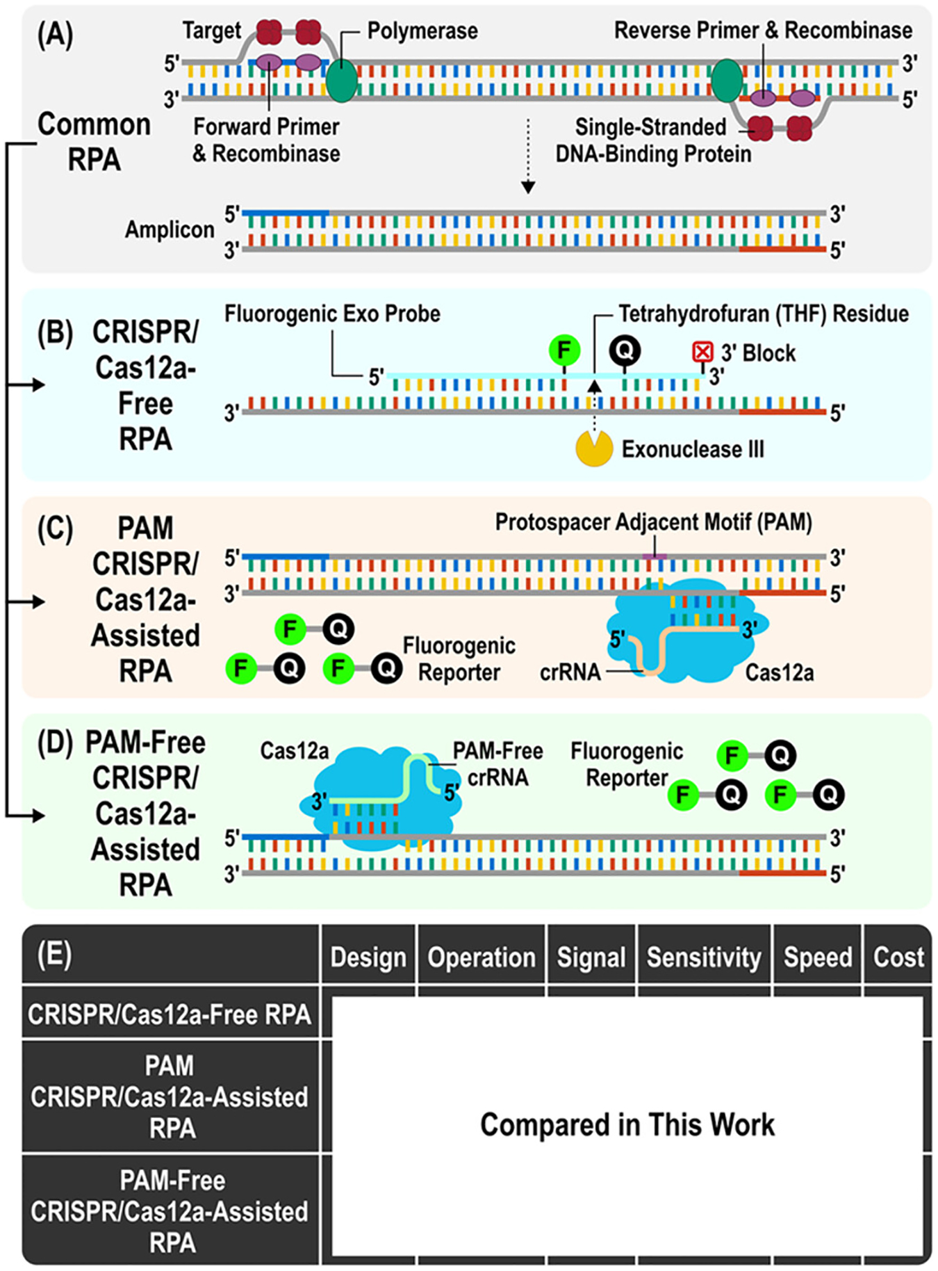
Designing and evaluating three comparable assays to elucidate the role of CRISPR/Cas12a in single-step assays. (A) The three assays share a common RPA reaction with the same pair of RPA primers, RPA reagents (e.g., recombinase, single-stranded DNA binding protein, polymerase), and DNA target. (B) The CRISPR/Cas12a-free RPA assay detects RPA amplicons through a fluorogenic exo probe—internally labeled with a fluorophore (F) and a quencher (Q) around a tetrahydrofuran (THF) residue and terminally labeled with a 3′ blocker—that hybridizes to its complementary strand of the RPA amplicon, gets cleaved at the THF residue by exonuclease III (the additional enzyme in the RPA reaction mix of this assay), and yields a fluorescence signal. (C) The PAM CRISPR/Cas12a-assisted RPA assay entails adding Cas12a, a CRISPR guide RNA (crRNA) that is immediately downstream to the protospacer adjacent motif (PAM) associated with the Cas12a, and a fluorogenic reporter that is terminally labeled with a fluorophore and a quencher. During the reaction, the presence of RPA amplicons leads to Cas12a activation and cleavage of the fluorogenic reporter, thus producing a fluorescence signal. (D) The PAM-free CRISPR/Cas12a-assisted RPA assay uses the same Cas12a and fluorogenic reporter as its PAM counterpart and fluorescently detects RPA amplicons similar to its PAM counterpart but uses a different crRNA that is without an upstream PAM. (E) Comparison of assay design, operation, signal, sensitivity, speed, and cost helps identify advantages and disadvantages of the CRISPR/Cas12a-assisted RPA assays with respect to the CRISPR/Cas12a-free assay, thereby pinpointing the role and impact of CRISPR/Cas12a in these assays.
Within the RPA amplicon region, we then designed the fluorogenic probe for the CRISPR/Cas12a-free RPA assay30,31 and crRNAs for the PAM and the PAM-free CRISPR/Cas12a RPA assays (Figure S1 and Table S1). For the CRISPR/Cas12a-free RPA assay, we followed the standard design rule and designed a 46-nucleotide fluorogenic “exo probe” that is internally labeled with a fluorophore (FAM) and a quencher (Black Hole Quencher 1, BHQ1) around a tetrahydrofuran (THF) residue. The THF residue serves as the cleavage site for exonuclease III—the additional enzyme in this RPA reaction mix of this assay (Figure 1B). During assay incubation, the exo probe hybridizes to its complementary strand of the RPA amplicon and then gets cleaved by exonuclease III, thus yielding a fluorescence signal for detection. For the PAM CRISPR/Cas12a-assisted RPA assay, we identified, within the RPA amplicon region, the 4-nucleotide PAM (i.e., TTTN) for the Cas12a used in this work and designed a crRNA to target a 20-nucleotide sequence that is immediately downstream and on the opposite strand (Figure 1C). For the PAM-free CRISPR/Cas12a-assisted RPA assay, we adopted a previously reported crRNA21,24,25 that targets a different 20-nucleotide sequence within the RPA amplicon region but lacks the upstream PAM on the opposite strand (Figure 1D). Both assays include Cas12a and a 5-nucleotide “fluorogenic reporter” that is labeled with a fluorophore (6-FAM) on the 5′ terminus and a quencher (Iowa Black Hole Quencher FQ, IABkFQ) on the 3′ terminus. During assay incubation, both crRNAs can form complexes with Cas12a, recognize their respective target sequences, become activated, and finally cleave the fluorogenic reporter to yield a fluorescence signal for detection.
We subsequently compared the three assays. Our comparison included a discussion on design and operation and also incorporated experimental assessment of signal, sensitivity, speed, and cost—all of which represent important parameters for evaluating diagnostic assays (Figure 1E). Such a comparison would help us identify advantages and disadvantages of the CRISPR/Cas12a-assisted RPA assays with respect to the CRISPR/Cas12a-free assay, thus allowing us to specify the role and impact of CRISPR/Cas12a in these assays.
3.2. Assay Optimizations for Systematic Comparisons of Assay Performances.
To facilitate an unbiased comparison among the assays, we fine-tuned the three assays for optimal fluorescence signals from the target at 200 copies/μL and the no target control (NTC). For the CRISPR/Cas12a-free RPA assay optimization, we first sought to determine the optimal exo probe concentration by affixing the RPA primer concentration at vendor-recommended 420 nM with varying exo probe concentrations. Among four different concentrations of the exo probe ranging from 50 to 150 nM, 100 nM yielded a detectable normalized end-point fluorescence signal of 0.09 at 60 min (see Experimental Section in Supporting Information for calculation; referred to hereafter as “end-point signal”) from 200 copies/μL target while maintaining a low end-point signal of 0.01 from the NTC (Figures 2A(i) and S2A). We further optimized the RPA primer concentration at 100 nM exo probe, and the result shows that 150 nM RPA primers produced the strongest end-point signal of 0.14 from 200 copies/μL target and a low end-point signal of 0.01 from the NTC (Figures 2A(ii) and S2B). Based on these results, we selected 150 nM RPA primers and 100 nM exo probe as an optimal condition for the CRISPR/Cas12a-free RPA assay in subsequent evaluation.
Figure 2.
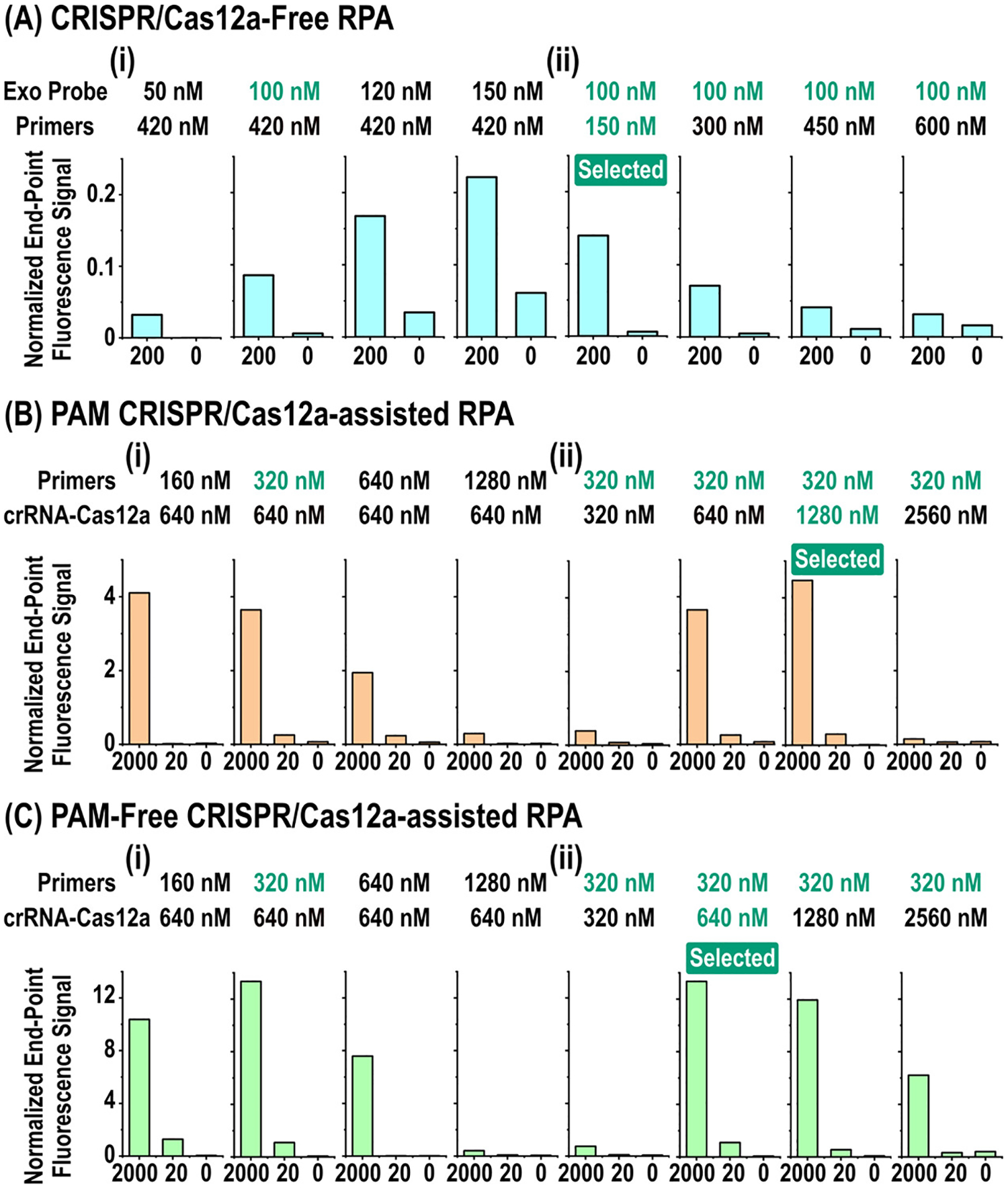
Assay optimizations for systematic comparisons between three RPA assays. (A) For the CRISPR/Cas12a-free RPA assay, combining (i) 100 nM exo probe and (ii) 150 nM RPA primers yields a strong normalized end-point fluorescence signal from 200 copies/μL (0.14) target without elevating the signal for the NTC (0.01). (B) For the PAM CRISPR/Cas12a-assisted RPA assay, combining (i) 320 nM RPA primers and (ii) 1280 nM crRNA–Cas12a results a strong end-point signal of 4.47 from 2000 copies/μL target, a detectable end-point signal of 0.30 from 20 copies/μL target, and a low end-point signal of 0.02 from the NTC. (C) For the PAM-free CRISPR/Cas12a-assisted RPA assay, combining (i) 320 nM RPA primers and (ii) 640 nM crRNA–Cas12a produces the strongest end-point signals of 13.35 and 1.10 from 2000 and 20 copies/μL target, respectively, while maintaining a low end-point signal of 0.07 from the NTC.
We next fine-tuned the PAM CRISPR/Cas12a-assisted RPA assay. For the optimization of the RPA primer concentration, we first fixed crRNA and Cas12a concentrations of 640 nM at 1:1 ratio by employing the condition from the previous report24 and assessed varying RPA primer concentrations ranging from 160 to 1280 nM. We found 320 nM RPA primers as the best condition capable of detecting both high (2000 copies/μL) and low (20 copies/μL) target concentrations, which generated a strong end-point signal of 3.65 from 2000 copies/μL target and a detectable end-point signal of 0.25 from 20 copies/μL target, as well as a low end-point signal of 0.07 from the NTC (Figure 2B(i) and S3A). To further optimize the crRNA and Cas12a concentrations, we assessed four crRNA and Cas12a concentrations ranging from 320 nM to 2560 nM with fixed RPA primer concentration at 320 nM. We found that 1280 nM crRNA–Cas12a improved the end-point signal to 4.47 from 2000 copies/μL target, while retaining a robust end-point signal of 0.30 from 20 copies/μL target and a low end-point signal of 0.02 from the NTC (Figures 2B(ii) and S3B). Importantly, the signals from this assay were stronger than those from the CRISPR/Cas12a-free RPA assay, even at a lower target concentration in the PAM CRISPR/Cas12a-assisted RPA assay (i.e., 20 copies/μL) than the CRISPR/Cas12a-free RPA assay (i.e., 200 copies/μL). The stronger signals with additional CRISPR/Cas systems are likely due to activated Cas12a cleaving the higher concentration of fluorogenic reporters, resulting in signal amplification.
When we fine-tuned the PAM-free CRISPR/Cas12a-assisted RPA assay under the same conditions as the PAM CRISPR/Cas12a-assisted RPA assay, we observed ~3- to 4-fold stronger end-point signals. Indeed, at the fine-tuned concentrations of 320 nM RPA primers and 640 nM crRNA–Cas12a, we observed the strongest end-point signals of 13.35 and 1.10 from 2000 copies/μL and 20 copies/μL targets, respectively, as well as a low end-point signal of 0.07 from the NTC (Figures 2C and S4). Moreover, as we used the same concentration of the fluorogenic reporter between the two CRISPR/Cas12a-assisted RPA assays, these results suggest that more fluorogenic reporter molecules were cleaved in the PAM-free CRISPR/Cas12a-assisted RPA assay than in the PAM CRISPR/Cas12a-assisted RPA assay, thus giving rise to the strongest signals among the three assays.
3.3. Mechanisms of CRISPR/Cas12a-Assisted RPA Assays.
The difference between the two CRISPR/Cas12a-assisted RPA assays prompted further analysis. Because they share the same RPA and fluorogenic reporter, their difference most likely originates from how their respective crRNA–Cas12a recognize the target sequence and become activated for cleaving the target (cis-cleavage) and the fluorogenic reporter (transcleavage). We therefore conducted two experiments to elucidate these interactions and reveal potential differences. First, we performed both PAM and PAM-free CRISPR/Cas12a-assisted RPA assays using 2560, 1280, 640, or 0 nM of their respective crRNA–Cas12a to amplify 2000 copies/μL DNA target and subsequently used gel electrophoresis to visualize RPA amplicons and cis-cleaved fragments from these reactions. Second, in the absence of RPA, we directly incubated 2560 nM of each crRNA–Cas12a and 4 μM fluorogenic reporter with 10, 1, or 0 nM of either double-stranded (ds) DNA activator or single-stranded (ss) DNA activator (i.e., SARS-CoV-2 dsDNA and ssDNA target, see Methods and Table S1) and compared the signals from these reactions. By revealing activation and cis-cleavage of PAM crRNA–Cas12a and PAM-free crRNA– Cas12a, these experiments could then help us formulate a mechanistic framework for both assays.
Gel electrophoresis and activator comparison indeed informed how differently the crRNA–Cas12a from the two assays interacted with RPA amplicons. Gel images revealed that, for both assays, reactions in the presence of crRNA–Cas12a generated DNA fragments shorter than RPA amplicons, which corresponded with cis-cleaved products. Moreover, as the concentration of crRNA–Cas12a decreased, both RPA amplicon band and cis-cleaved fragment band became more visible in the gel images, indicating increased amounts of both RPA amplicons and cis-cleaved fragments. Notably, we observed clearer bands reflecting RPA amplicons and cis-cleaved fragments from PAM-free CRISPR/Cas12a-assisted RPA than from PAM CRISPR/Cas12a-assisted RPA, with 640 nM PAM-free crRNA–Cas12a generating the most visible bands among all reactions (Figures 3A and S5). These results indicate that 640 nM PAM-free crRNA–Cas12a produced more amount of RPA amplicons and cis-cleaved fragments, and hence stronger Cas12a activation, than other conditions. For direct comparison of PAM crRNA–Cas12a and PAM-free crRNA–Cas12a with DNA activators, we observed that PAM crRNA–Cas12a resulted in detectable signals from both dsDNA and ssDNA activators, with stronger signals from dsDNA activators (Figures 3B and S6). These results match with the previous reports that a PAMharboring dsDNA activator can be targeted by PAM crRNA– Cas12a as well as an ssDNA activator, and crRNA–Cas12a utilizes PAM as an anchor for Cas12a binding on double-stranded target recognition, allowing Cas12a to unwind duplex, thus resulting in crRNA–target base pairing and cis-cleavage.32–34 In contrast, PAM-free crRNA–Cas12a activated by ssDNA activators yielded stronger signals than by dsDNA activators (Figures 3B and S6). These results suggest that PAM-free crRNA–Cas12a can readily hybridize to the ssDNA activator but can only access the unbound state of the dsDNA activator at the equilibrium of duplex formation, implying that PAM-free crRNA–Cas12a would preferentially hybridize to ssDNA activators for activation and subsequent trans-cleavage.
Figure 3.
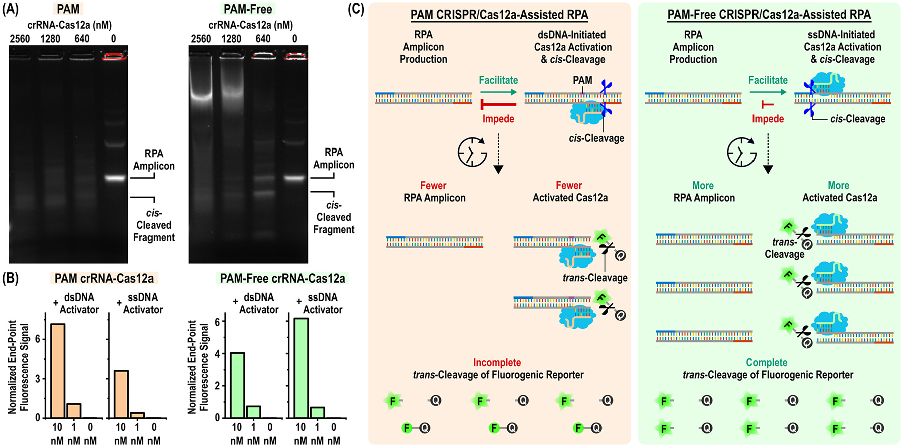
Formulating mechanistic framework for PAM and PAM-free CRISPR/Cas12a-assisted RPA assays. (A) Gel electrophoresis of reaction products from PAM and PAM-free CRISPR/Cas12a-assisted RPAs shows clearer RPA amplicon and cis-cleaved fragment bands from the PAM-free CRISPR/Cas12a-assisted RPA than from its PAM counterpart, with the brightest bands with 640 nM PAM-free crRNA–Cas12a. (B) The PAM crRNA–Cas12a is preferentially activated by dsDNA activators than by ssDNA activators, whereas the PAM-free crRNA–Cas12a is preferentially activated by ssDNA activators than by dsDNA activators. (C) The crRNA–Cas12a complex in the PAM CRISPR/Cas12a-assisted RPA assay can recognize and cis-cleave double-stranded RPA amplicons, resulting in impediment of exponential amplification. Such impediment leads to fewer activated Cas12a and incomplete trans-cleavage of the fluorogenic reporter. For the PAM-free CRISPR/Cas12a-assisted RPA, the crRNA–Cas12a complex is preferentially accessible to its single-stranded complementary region that is temporarily available during DNA polymerization, leading to less impediment of exponential amplification and thus generates a stronger signal via stronger trans-cleavage activity than its PAM counterpart.
Based on our experimental results and previous reports on CRISPR diagnostics, we suggest reaction mechanisms of PAM and PAM-free CRISPR/Cas12a-assisted RPA assays. As PAM crRNA–Cas12a preferred dsDNA as activators, the CRISPR/Cas12a reaction can initiate by leveraging PAM to directly anchor onto double-stranded RPA amplicons, resulting in the cis-cleavage of RPA amplicons (i.e., RPA template), thus impeding exponential amplification of target DNA. During the PAM CRISPR/Cas12a-assisted RPA reaction, such impediment of exponential amplification can lead to fewer RPA amplicons, fewer activated Cas12a, and lastly incomplete trans-cleavage of the fluorogenic reporter (Figure 3C). For the PAM-free CRISPR/Cas12a-assisted RPA, rather than relying on double-stranded RPA amplicons as the activator, PAM-free crRNA would hybridize to its single-stranded complementary portion within double-stranded RPA amplicons. The single-stranded portion complementary to PAM-free crRNA is likely temporarily available during DNA polymerization in RPA,21 allowing a brief window for PAM-free crRNA–Cas12a to be activated for subsequent trans-cleavage. Consequently, the impediment to exponential amplification by the CRISPR/Cas12a reaction in PAM-free CRISPR/Cas12a-assisted RPA will be weaker than its PAM counterpart, resulting in stronger Cas12a activation and therefore stronger signal generation via trans-cleavage (Figure 3C), as evidenced by stronger and plateauing signals observed in our experimental results (Figures 2C and S4).
3.4. Systematic Comparisons of Assay Sensitivity, Speed, and Cost.
Finally, we compared the sensitivity, speed, and cost of the three assays. To do so, we set up the three assays at their fine-tuned reagent concentrations from 2000 to 2 target copies/μL (in 10-fold dilutions) and the NTC in triplicate. For sensitivity comparison, we compared the limit of detection (LOD) of each assay based on Student’s t-tests between the end-point signals of each target concentration and the NTC at 60 min. For speed comparison, we calculated and compared the time-to-positive—the time at which the signal from the target becomes detectable above the NTC (see Methods for calculation)—at each target concentration for each assay. Finally, for cost comparison, we summed up and compared the reagent cost of each assay at 10 μL—the standard reaction volume in this work—and at its fine-tuned reagent concentrations.
The CRISPR/Cas12a-free RPA assay served as the baseline for our comparison. For this assay, the end-point signals from 2000, 200, 20, and 2 target copies/μL and the NTC were 0.21 ± 0.03, 0.17 ± 0.03, 0.04 ± 0.01, 0.01 ± 0.01, and 0.01 ± 0.01, respectively (mean ± SD, n = 3; Figures 4A(i) and S7A). The LOD was 20 copies/μL, as it was significantly differentiated from the NTC (p < 0.05). For the detectable target concentrations of 2000, 200, and 20 copies/μL, the times-to-positive were 6.5 ± 1.0, 9.2 ± 0.6, and 16.5 ± 6 min, respectively (mean ± SD, n = 3; Figure 4A(ii)). The reagent cost of the assay was $0.87, where the exo RPA kit represented most of the total reagent cost ($0.85), while the exo probe ($0.02) and the RPA primers (<$0.01) constituted the rest (Figure 4A(iii) and Table S2).
Figure 4.
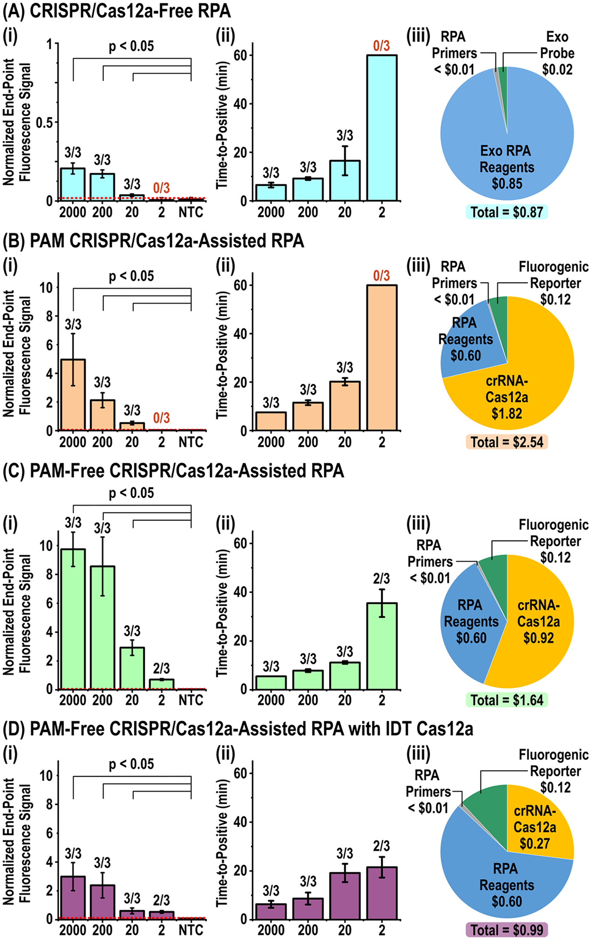
Comparison of assay sensitivity, speed, and cost. For each assay, (i) sensitivity is evaluated by measuring the end-point signals from 2000, 200, 20, and 2 target copies/μL and the no target control (NTC) in triplicate (n = 3) and performing Student’s t-tests, (ii) speed is evaluated by measuring the “times-to-positive”—the time at which the signal from the target becomes detectable above the NTC—in triplicate (n = 3), and (iii) cost is evaluated by totaling the reagent cost of a 10 μL reaction at the fine-tuned reagent concentrations. (A) The CRISPR/Cas12a-free assay (i) produces low but detectable end-point signals from only 2000, 200, and 20 copies/μL (p < 0.05) while (ii) reporting fast times-to-positive and (iii) costing $0.87. (B) The PAM CRISPR/Cas12a-assisted assay (i) generates stronger end-point signals among 2000, 200, and 20 copies/μL (p < 0.05) with (ii) longer times-to-positive, and (iii) a nearly 3-fold higher cost of $2.54 due to its high concentration of crRNA–Cas12a. (C) The PAM-free CRISPR/Cas12a-assisted assay (i) yields strongest end-point signals from 2000, 200, and 20 copies/μL (p < 0.05) and can detect 2 copies/μL (signal =0.71 ± 0.07, n = 2) while (ii) posting the fastest times-to-positive, although (iii) its cost of $1.64 due to its reduced concentration of crRNA–Cas12a remains high. (D) The PAM-free CRISPR/Cas12a-assisted assay with an alternative Cas12a from Integrated DNA Technology (IDT) (i) produces weaker but detectable end-point signals from 2000, 200, and 20 copies/μL (p < 0.05) and also manages to detect 2 copies/μL (signal = 0.54 ± 0.09, n = 2). Despite (ii) longer times-to-positive, its (iii) cost is only $0.99 due to its low concentration of crRNA–Cas12a. Data presented are in (i) and (ii) as mean ± SD. Red dashed line represents signal thresholds at mean + 3SD of the NTCs.
Compared to the CRISPR/Cas12a-free RPA assay, the PAM CRISPR/Cas12a-assisted RPA assay achieved comparable sensitivity but at a slightly slower speed and a significantly higher cost. The end-point signals from 2000, 200, 20, and 2 target copies/μL and the NTC were 4.95 ± 1.82, 2.12 ± 0.52, 0.53 ± 0.12, 0.04 ± 0.01, and 0.04 ± 0.01, respectively (mean ± SD, n = 3; Figures 4B(i) and S7B). Despite producing >10-fold stronger end-point signals from the detectable target concentrations, this assay still reported an LOD of 20 copies/μL (p < 0.05) as it failed to detect 2 copies/μL. The times-to-positive for the detectable target concentrations of 2000, 200, and 20 copies/μL slowed to 7.5 ± 0, 11.5 ± 1.0, and 20.2 ± 1.5 min, respectively (Figure 4B(ii)). Finally, the reagent cost of the assay was $2.54—nearly tripling the cost of the CRISPR/Cas12a-free RPA assay (Figure 4B(iii) and Table S3). The bulk of the cost stemmed from the high crRNA–Cas12a concentration in this assay ($1.82) rather than the RPA kit ($0.60), the fluorogenic reporter ($0.12), or the RPA primers (<$0.01). These results show that the PAM CRISPR/Cas12a-assisted RPA assay in fact failed to offer advantages over the CRISPR/Cas12a-free RPA assay.
Compared to the CRISPR/Cas12a-free RPA assay, the PAM-free CRISPR/Cas12a-assisted RPA assay was more sensitive (albeit only slightly) and faster but costed more. The end-point signals from 2000, 200, 20, and 2 target copies/μL and the NTC were 9.75 ± 1.18, 8.56 ± 2.04, 2.92 ± 0.53, 0.71 ± 0.07, and 0.04 ± 0.02, respectively (mean ± SD, n = 3 for 2000, 200, and 20 copies/μL and the NTC, n = 2 for 2 copies/μL; Figures 4C(i) and S7C), which were the strongest among the three assays. Although this assay could detect 2 copies/μL in 2 out of 3 replicates—an improvement over the other two assays—we still defined its LOD as 20 copies/μL (p < 0.05). The times-to-positive of this assay were also the fastest among the three assays—5.5 ± 0, 7.8 ± 0.6, 11.2 ± 0.6, and 35.5 ± 5.7 min for 2000, 200, 20, and 2 copies/μL, respectively (mean ± SD, n = 3 for 2000, 200, 20 copies/μL, and the NTC, n = 2 for 2 copies/μL; Figure 4C(ii)). Finally, the reagent cost was $1.64 (Figure 4C(iii) and Table S4)—between the costs of the other two assays—due to a reduced concentration (and hence cost) of crRNA–Cas12a ($0.92) while retaining the same costs of the other components. These results pinpoint not only the advantages of the PAM-free CRISPR/Cas12a-assisted RPA assay in sensitivity and speed over the CRISPR/Cas12a-free RPA assay but also its disadvantage in cost.
3.5. Alternative Cas12a in PAM-Free CRISPR/Cas12a-Assisted RPA.
The advantages of the PAM-free CRISPR/Cas12a-assisted RPA assay motivated us to explore an alternative Cas12a as means to reducing the reagent cost. The new Cas12a derived from Lachnospiraceae bacterium ND2006 from Integrated DNA Technology (IDT) emerged as a candidate. This Cas12a is derived from the same strain of bacteria as its counterpart from New England Biolabs (NEB)—the commercial Cas12a used throughout this work—and could therefore serve as a direct replacement in the PAM-free CRISPR/Cas12a-assisted RPA assay. For this assay, we discovered that the fine-tuned concentrations were 320 nM RPA primers and 80 nM crRNA–Cas12a, 8-fold lower than the fine-tuned concentration with NEB Cas12a, and the resulting end-point signals decreased to 2.98, 1.30, and 0.10 from 2000 copies/μL target, 20 copies/μL target, and the NTC, respectively (Figure S8). We subsequently discovered that the 8-fold-reduced concentration of crRNA–Cas12a was possible because this IDT Cas12a could more rapidly become activated and initiate cis-cleavage by both ssDNA and dsDNA activators without RPA (Figure S9) when compared to the NEB Cas12a. These findings supported our mechanistic framework of CRISPR/Cas12a-assisted RPA assays, as more rapid cis-cleavage of RPA amplicons would lead to fewer RPA amplicons, fewer activated Cas12a, incomplete trans-cleavage of the fluorogenic reporter, and ultimately decreased signals.
Despite decreased signals, the IDT PAM-free CRISPR/Cas12a-assisted RPA assay retained comparable sensitivity and speed as its NEB counterpart, while indeed reducing the cost. The end-point signals from 2000, 200, 20, and 2 target copies/μL and the NTC were 2.99 ± 0.97, 2.39 ± 0.88, 0.61 ± 0.20, 0.54 ± 0.09, and 0.09 ± 0.01, respectively (mean ± SD, n = 3 for 2000, 200, and 20 copies/μL and the NTC, n = 2 for 2 copies/μL; Figures 4D(i) and S10). Like its NEB counterpart, the IDT PAM-free CRISPR/Cas12a-assisted RPA assay detected 2 copies/μL in 2 out of 3 replicates, but we still defined its LOD as 20 copies/μL (p < 0.05). The times-to-positive slightly slowed to 6.3 ± 1.4, 8.7 ± 2.5, 19.2 ± 3.8, and 21.5 ± 4.2 min for 2000, 200, 20, and 2 copies/μL, respectively (mean ± SD, n = 3 for 2000, 200, and 20 copies/μL and the NTC, n = 2 for 2 copies/μL; Figure 4D(ii)). Most notably, the reagent cost was significantly lowered to $0.99 (Figure 4D(iii) and Table S5)—comparable to the CRISPR/Cas12a-free RPA assay—due to the significant cost reduction of crRNA–Cas12a ($0.27). These results illustrate that the IDT Cas12a offers an alternative option for the Cas12a enzyme for the PAM-free CRISPR/Cas12a-assisted RPA assay but introduces a trade-off between the signal and the reagent cost. Finally, to our knowledge, our study represents the first time that this IDT Cas12a can be coupled to RPA in a single-step assay.
4. DISCUSSION
Assay comparison in this work allowed us to specify advantages and disadvantages of the three assays (Table 1). Among the three assays, CRISPR/Cas12a-free RPA employs an exo probe with the most stringent design rules but has the simplest operational procedure and the lowest cost because it only requires low concentrations of the exo probe and RPA primers to be added to lyophilized reagents. Although this assay produced the weakest end-point signal in our experimental result (Figure 4A), it still achieved comparable sensitivity and speed as PAM CRISPR/Cas12a-assisted RPA. PAM CRISPR/Cas12a-assisted RPA needs a crRNA, which is easier to design than the exo probe but is still restricted by the PAM. As PAM CRISPR/Cas12a-assisted RPA utilizes a Cas12a enzyme and a fluorogenic reporter, it has a more involved operational procedure and a significantly higher cost. Despite generating a much stronger end-point signal than CRISPR/Cas12a-free RPA, it failed to improve the sensitivity or the speed (Figure 4B). PAM-free CRISPR/Cas12a-assisted RPA utilizes a crRNA that obviates the PAM and therefore has the simplest design, while involving a comparable operational procedure as PAM CRISPR/Cas12a-assisted RPA. This assay slightly outperformed the other two assays in sensitivity and speed (Figure 4C). Finally, depending on the Cas12a enzyme, the PAM-free CRISPR/Cas12a-assisted RPA assay either generated the strongest end-point signal or had comparable cost as the CRISPR/Cas12a-free RPA assay (Figure 4C,D). Based on the best overall performance of the PAM-free CRISPR/Cas12a-assisted RPA assay, combined with the possibility of lyophilizing crRNA, Cas12a, and fluorogenic reporter, we foresee the PAM-free CRISPR/Cas12a-assisted RPA assay as a new detection companion for the single-step RPA assay.
Table 1.
Summary of Assay Comparison
| assay | design | operation | signal | sensitivity | speed | cost |
|---|---|---|---|---|---|---|
| CRISPR/Cas12a-free RPA | + | +++ | + | ++ | ++ | +++ |
| PAM CRISPR/Cas12a-assisted RPA | ++ | ++ | ++ | ++ | + | + |
| PAM-free CRISPR/Cas12a-assisted RPA | +++ | ++ | +++ | ++ | +++ | ++ |
| PAM-free CRISPR/Cas12a-assisted RPA (IDT) | +++ | ++ | ++ | ++ | +++ | +++ |
Our work suggests the mechanistic difference between PAM and PAM-free CRISPR/Cas12a-assisted RPA, where weaker impediment to RPA amplification leads to stronger CRISPR/Cas12a activation and signal generation. For PAM CRISPR/Cas12a-assisted RPA, the PAM crRNA–Cas12a complex relies on PAM to directly anchor onto double-stranded RPA amplicons, resulting in the cis-cleavage of double-stranded RPA amplicons (i.e., RPA template), thus impeding the exponential amplification of the target DNA. On the other hand, for PAM-free CRISPR/Cas12a-assisted RPA, the PAM-free crRNA–Cas12a complex hybridizes to the single-stranded complementary portion within the double-stranded RPA amplicon, which is likely opened during DNA polymerization in RPA. Due to the short window for cis-cleavage of RPA amplicon, the impediment to the RPA amplification would be weaker, ultimately resulting in stronger Cas12a activation and stronger signal generation (Figure 3C). We highly expect that such mechanistic understanding on the single-step CRISPR/Cas12a-assisted assay brings insight to further advance the assay performance and new assay design.
More broadly, our work illustrates that the most significant improvement from adding crRNA–Cas12a and the fluorogenic reporter at a high concentration to RPA is amplified signals. This CRISPR/Cas12a-enabled signal amplification, however, concurrently impeded RPA and therefore only led to minor improvements in assay sensitivity and speed. Thus, for routine diagnostic assays performed in research and clinical laboratories using benchtop instruments with sensitive optics, CRISPR/Cas-enabled signal amplification would only provide a marginal benefit. Instead, the benefit would be more apparent in developing point-of-care diagnostics by reducing the need for sensitive detection optics. Leveraging CRISPR/Cas-enabled signal amplification in digital assays24,25 can significantly improve the assay performance, as amplified signals would ease the identification of positive (i.e., fluorescent) digital wells. Moreover, our work suggests that, to maximize CRISPR/Cas-enabled signal amplification while retaining a one-pot assay, it may be sensible after all to separate CRISPR/Cas from isothermal NAAT in the one-pot two-step format,6,35,36 though we note that the additional step of adding CRISPR/Cas reagents to completed isothermal NAAT would require either careful manual handling or additional reagent handling components in diagnostic devices.
5. CONCLUSIONS
In this work, we shed new light on the role and impact of CRISPR/Cas12a when coupled to RPA in single-step assays. We first designed three assays that shared a common RPA but differed in CRISPR/Cas12a, CRISPR/Cas12a-free RPA, PAM CRISPR/Cas12a-assisted RPA, and PAM-free CRISPR/Cas12a-assisted RPA, and then fine-tuned their key reagent concentrations for systematic comparison. Assay fine-tuning, along with gel electrophoresis and DNA activator comparison, helped us formulate a mechanistic framework for both PAM and PAM-free CRISPR/Cas12a-assisted RPA assays. Most notably, we pinpointed that the activation of Cas12a by RPA amplicons and subsequent cis-cleavage of RPA amplicons impeded RPA, suggesting that the rate of RPA amplicon generation relative to the rate of cis-cleavage, which was affected by the Cas12a enzyme and the crRNA, was important for assay performance. Our comparison of assay design, operation, signal, sensitivity, speed, and cost showed that, with respect to our CRISPR/Cas12a-free assay, our PAM CRISPR/Cas12a-assisted RPA assay in fact only elevated the signal (without improving either sensitivity or speed) at the expense of significantly increased cost, whereas our PAM-free CRISPR/Cas12a-assisted RPA assay further elevated the signal and slightly improved both sensitivity and speed at a moderately increased cost. Moreover, we discovered that a new IDT Cas12a with rapid cis-cleavage offered an alternative to our PAM-free CRISPR/Cas12a-assisted RPA assay but introduced a trade-off between the signal and the cost.
To the best of our knowledge, our work represents the first such investigative comparison for elucidating the role and impact of CRISPR/Cas12a in single-step isothermal nucleic acid amplification testing assays. We therefore also see ample room for follow-up studies. For example, investigations on reaction conditions (e.g., pH37) for improving the performance of the single-step CRISPR/Cas-assisted RPA assay can significantly boost the effect of the CRISPR/Cas12a system for enhanced signal, sensitivity, and speed. In addition, we focused on CRISPR/Cas12a and RPA in this work, but additional combinations of CRISPR systems and isothermal NAATs could be explored. Likewise, for demonstration, we focused on detecting a synthetic DNA sequence as the target in this work, but other DNA and RNA targets could be tested. Finally, we demonstrated that PAM-free CRISPR/Cas12a-assisted RPA, despite significantly elevating the assay signal, only offered a minor improvement in assay sensitivity. As sensitivity represents one of the most important requirements for diagnostic assay development, we envision building upon this work and further improving assay sensitivity. Taken together, our work not only presents a mechanistic framework and pinpoints advantages and disadvantages of CRISPR/Cas12a when coupled to RPA in a single-step assay but also opens new directions for future research. Our work thus offers new and useful knowledge that can steer and facilitate advances of CRISPR/Cas-based detection assays.
Supplementary Material
ACKNOWLEDGMENTS
This research was financially supported by the National Institutes of Health (R01CA260628, R01AI137272, R61AI154628, UH3CA211457). K.H. is grateful for a developmental grant from the Johns Hopkins University Center for AIDS Research, a National Institutes of Health funded program (P30AI094189). G.Z. acknowledges financial support from Visiting Scholar Program administered by the China Scholarship Council.
Footnotes
Supporting Information
The Supporting Information is available free of charge at https://pubs.acs.org/doi/10.1021/acs.analchem.2c05632.
Experimental section (reagents and materials, exo probe design, assay optimization experiments, comparison of three RPA assays on sensitivity and speed, synthetic ssDNA and dsDNA activator experiment, gel electrophoresis, and data processing and statistical analysis), Supplementary Figures (target regions of RPA primers, exo probe, and crRNAs; reaction real-time curves of CRISPR/Cas12a-free RPA assays for optimizing concentrations of the exo probe and RPA primers; reaction real-time curves of PAM CRISPR/Cas12a-assisted RPA assays for optimizing concentrations of RPA primers and crRNA–Cas12a; reaction real-time curves of PAM-free CRISPR/Cas12a-assisted RPA assays for optimizing concentrations of RPA primers and crRNA–Cas12a; original gel electrophoresis images of reaction products from (A) PAM CRISPR/Cas12a-assisted RPA assays and (B) PAM-free CRISPR/Cas12a-assisted RPA assays; reaction real-time curves of crRNA–Cas12a activation assays in the absence of RPA; reaction real-time curves of three assays for assessing sensitivity; reaction real-time curves of IDT Cas12a-based PAM-free CRISPR/Cas12a-assisted RPA assays for optimizing the concentration of crRNA–Cas12a; reaction real-time curves of IDT Cas12a-based PAM-free crRNA–Cas12a activation assays in the absence of RPA; and reaction real-time curves of the IDT Cas12a-based PAM-free CRISPR/IDT Cas12a-assisted RPA assay for assessing assay sensitivity), and Supplementary Tables (sequences of RPA primers, fluorogenic exo probe, Cas12a crRNAs, and fluorogenic reporter; reagent cost breakdown of CRISPR/Cas12a-free RPA; reagent cost breakdown of PAM CRISPR/Cas12a-assisted RPA; and reagent cost breakdown of PAM-free CRISPR/Cas12a-assisted RPA; reagent cost breakdown of IDT Cas12a-based PAM-free CRISPR/Cas12a-assisted RPA) (PDF)
Notes
The authors declare no competing financial interest.
Complete contact information is available at: https://pubs.acs.org/10.1021/acs.analchem.2c05632
Contributor Information
Fangchi Shao, Department of Biomedical Engineering, Johns Hopkins University, Baltimore, Maryland 21218, United States;.
Joon Soo Park, Department of Biomedical Engineering, Johns Hopkins University, Baltimore, Maryland 21218, United States;.
Guojie Zhao, Department of Mechanical Engineering, Johns Hopkins University, Baltimore, Maryland 21218, United States;.
Kuangwen Hsieh, Department of Mechanical Engineering, Johns Hopkins University, Baltimore, Maryland 21218,United States;.
Tza-Huei Wang, Department of Biomedical Engineering, Johns Hopkins University, Baltimore, Maryland 21218,United States; Department of Mechanical Engineering, Johns Hopkins University, Baltimore, Maryland 21218, United States;.
REFERENCES
- (1).Kaminski MM; Abudayyeh OO; Gootenberg JS; Zhang F; Collins JJ Nat. Biomed. Eng 2021, 5, 643–656. [DOI] [PubMed] [Google Scholar]
- (2).Wang M; Zhang R; Li J Biosens. Bioelectron 2020, 165, No. 112430. [DOI] [PMC free article] [PubMed] [Google Scholar]
- (3).Kostyusheva A; Brezgin S; Babin Y; Vasil’eva I; Kostyushev D; Chulanov V CRISPR-Cas systems for diagnosing infectious diseases, 2020, DOI: 10.20944/preprints202002.0007.v1. [DOI] [PMC free article] [PubMed] [Google Scholar]
- (4).Kumar P; Malik YS; Ganesh B; Rahangdale S; Saurabh S; Natesan S; Srivastava A; Sharun K; Yatoo M; Tiwari R; et al. Front. Cell. Infect. Microbiol 2020, 10, 639. [DOI] [PMC free article] [PubMed] [Google Scholar]
- (5).Hsieh K; Zhao G; Wang T-H Analyst 2020, 145, 4880–4888. [DOI] [PMC free article] [PubMed] [Google Scholar]
- (6).Sun Y; Yu L; Liu C; Ye S; Chen W; Li D; Huang WJ Transl. Med 2021, 19, 1–10. [DOI] [PMC free article] [PubMed] [Google Scholar]
- (7).Xiong D; Dai W; Gong J; Li G; Liu N; Wu W; Pan J; Chen C; Jiao Y; Deng H; et al. PLoS Biol 2020, 18, No. e3000978. [DOI] [PMC free article] [PubMed] [Google Scholar]
- (8).Liu H; Wang J; Zeng H; Liu X; Jiang W; Wang Y; Ouyang W; Tang X Food Chem 2021, 334, No. 127608. [DOI] [PubMed] [Google Scholar]
- (9).Teng F; Guo L; Cui T; Wang X-G; Xu K; Gao Q; Zhou Q; Li W Genome Biol 2019, 20, 132. [DOI] [PMC free article] [PubMed] [Google Scholar]
- (10).Ali Z; Aman R; Mahas A; Rao GS; Tehseen M; Marsic T; Salunke R; Subudhi AK; Hala SM; Hamdan SM; et al. Virus Res 2020, 288, No. 198129. [DOI] [PMC free article] [PubMed] [Google Scholar]
- (11).Agrawal S; Fanton A; Chandrasekaran SS; Charrez B; Escajeda AM; Son S; Mcintosh R; Bhuiya A; de León Derby MD; Switz NA Rapid, point-of-care molecular diagnostics with Cas13. medRxiv 2021, 2020.2012. 2014.20247874. [Google Scholar]
- (12).Gootenberg JS; Abudayyeh OO; Kellner MJ; Joung J; Collins JJ; Zhang F Science 2018, 360, 439–444. [DOI] [PMC free article] [PubMed] [Google Scholar]
- (13).Aquino-Jarquin G Nanomedicine 2019, 18, 428–431. [DOI] [PubMed] [Google Scholar]
- (14).Li S-Y; Cheng Q-X; Wang J-M; Li X-Y; Zhang Z-L; Gao S; Cao R-B; Zhao G-P; Wang J Cell Discovery 2018, 4, 20. [DOI] [PMC free article] [PubMed] [Google Scholar]
- (15).Gootenberg JS; Abudayyeh OO; Lee JW; Essletzbichler P; Dy AJ; Joung J; Verdine V; Donghia N; Daringer NM; Freije CA; et al. Science 2017, 356, 438–442. [DOI] [PMC free article] [PubMed] [Google Scholar]
- (16).Zhang Y; Chen M; Liu C; Chen J; Luo X; Xue Y; Liang Q; Zhou L; Tao Y; Li M; et al. Sens. Actuators, B 2021, 345, No. 130411. [DOI] [PMC free article] [PubMed] [Google Scholar]
- (17).Cao Y; Wu J; Pang B; Zhang H; Le XC Chem. Commun 2021, 57, 6871. [DOI] [PubMed] [Google Scholar]
- (18).Zhang WS; Pan J; Li F; Zhu M; Xu M; Zhu H; Yu Y; Su G Anal. Chem 2021, 93, 4126–4133. [DOI] [PubMed] [Google Scholar]
- (19).Arizti-Sanz J; Freije CA; Stanton AC; Petros BA; Boehm CK; Siddiqui S; Shaw BM; Adams G; Kosoko-Thoroddsen T-SF; Kemball ME; et al. Nat. Commun 2020, 11, No. 5921. [DOI] [PMC free article] [PubMed] [Google Scholar]
- (20).Guanghui T; Gong J; Kan L; Zhang X; He Y; Pan J; Zhao L; Tian J; Lin S; Lu Z A general onepot-method for nucleic acid detection with CRISPR-Cas12a, 2020, DOI: 10.21203/rs.3.rs-44613/v1. [DOI] [PMC free article] [PubMed] [Google Scholar]
- (21).Ding X; Yin K; Li Z; Lalla RV; Ballesteros E; Sfeir MM; Liu C Nat. Commun 2020, 11, No. 4711. [DOI] [PMC free article] [PubMed] [Google Scholar]
- (22).Feng W; Peng H; Xu J; Liu Y; Pabbaraju K; Tipples G; Joyce MA; Saffran HA; Tyrrell DL; Babiuk S; et al. Anal. Chem 2021, 93, 12808–12816. [DOI] [PubMed] [Google Scholar]
- (23).Malcı K; Walls LE; Rios-Solis L ACS Synth. Biol 2022, 11, 1555–1567. [DOI] [PMC free article] [PubMed] [Google Scholar]
- (24).Park JS; Hsieh K; Chen L; Kaushik A; Trick AY; Wang TH Adv. Sci 2021, 8, No. 2003564. [DOI] [PMC free article] [PubMed] [Google Scholar]
- (25).Ding X; Yin K; Li Z; Sfeir MM; Liu C Biosens. Bioelectron 2021, 184, No. 113218. [DOI] [PMC free article] [PubMed] [Google Scholar]
- (26).Chen F-E; Lee P-W; Trick AY; Park JS; Chen L; Shah K; Mostafa H; Carroll KC; Hsieh K; Wang T-H Biosens. Bioelectron 2021, 190, No. 113390. [DOI] [PMC free article] [PubMed] [Google Scholar]
- (27).Kellner MJ; Koob JG; Gootenberg JS; Abudayyeh OO; Zhang F Nat. Protoc 2019, 14, 2986–3012. [DOI] [PMC free article] [PubMed] [Google Scholar]
- (28).Aman R; Mahas A; Marsic T; Hassan N; Mahfouz MM Front. Microbiol 2020, 11, 3277. [DOI] [PMC free article] [PubMed] [Google Scholar]
- (29).Lin M; Yue H; Tian T; Xiong E; Zhu D; Jiang Y; Zhou X Anal. Chem 2022, 94, 8277. [DOI] [PubMed] [Google Scholar]
- (30).Piepenburg O; Williams CH; Stemple DL; Armes NA PLoS Biol 2006, 4, No. e204. [DOI] [PMC free article] [PubMed] [Google Scholar]
- (31).Daher RK; Stewart G; Boissinot M; Bergeron MG Clin. Chem 2016, 62, 947–958. [DOI] [PMC free article] [PubMed] [Google Scholar]
- (32).Swarts DC; Jinek M Mol. Cell 2019, 73, 589–600. [DOI] [PMC free article] [PubMed] [Google Scholar]
- (33).Swarts DC Biochem. Soc. Trans 2019, 47, 1499–1510. [DOI] [PubMed] [Google Scholar]
- (34).Paul B; Montoya G Biomed. J 2020, 43, 8–17. [DOI] [PMC free article] [PubMed] [Google Scholar]
- (35).Pang B; Xu J; Liu Y; Peng H; Feng W; Cao Y; Wu J; Xiao H; Pabbaraju K; Tipples G; et al. Anal. Chem 2020, 92, 16204–16212. [DOI] [PubMed] [Google Scholar]
- (36).Xiong Y; Cao G; Chen X; Yang J; Shi M; Wang Y; Nie F; Huo D; Hou C Appl. Microbiol. Biotechnol 2022, 106, 4607–4616. [DOI] [PMC free article] [PubMed] [Google Scholar]
- (37).Lv H; Wang J; Zhang J; Chen Y; Yin L; Jin D; Gu D; Zhao H; Xu Y; Wang J Front. Microbiol 2021, 12, 3663. [DOI] [PMC free article] [PubMed] [Google Scholar]
Associated Data
This section collects any data citations, data availability statements, or supplementary materials included in this article.