Abstract
Simple Summary
Hepatitis B virus (HBV) affects around 300 million people worldwide and is a significant risk factor for the development of hepatocellular carcinoma (HCC). Nucleos(t)ide analog therapy has aided in decreasing mortality from HBV. However, no cure for HBV currently exists. Despite adequate treatment based on the undetectable viral load or absence of surface protein, there has been much research demonstrating persistent risk for HBV-associated HCC. The aim of this paper is to review the related factors, pathophysiology, and evidence for why this risk exists. Further clarification of the relationship and risk factors for HBV-related HCC is necessary for appropriate screening and the eventual development of a cure.
Abstract
Chronic hepatitis B virus (HBV) infection is the largest global cause of hepatocellular carcinoma (HCC). Current HBV treatment options include pegylated interferon-alpha and nucleos(t)ide analogues (NAs), which have been shown to be effective in reducing HBV DNA levels to become undetectable. However, the literature has shown that some patients have persistent risk of developing HCC. The mechanism in which this occurs has not been fully elucidated. However, it has been discovered that HBV’s covalently closed circular DNA (cccDNA) integrates into the critical HCC driver genes in hepatocytes upon initial infection; additionally, these are not targets of current NA therapies. Some studies suggest that HBV undergoes compartmentalization in peripheral blood mononuclear cells that serve as a sanctuary for replication during antiviral therapy. The aim of this review is to expand on how patients with HBV may develop HCC despite years of HBV viral suppression and carry worse prognosis than treatment-naive HBV patients who develop HCC. Furthermore, HCC recurrence after initial surgical or locoregional treatment in this setting may cause carcinogenic cells to behave more aggressively during treatment. Curative novel therapies which target the life cycle of HBV, modulate host immune response, and inhibit HBV RNA translation are being investigated.
Keywords: hepatitis B virus, hepatocellular carcinoma, nucleoside analog, antiviral therapy, hepatitis cure, cccDNA
1. Introduction
Hepatitis B virus (HBV) is a global public health problem with an estimated 350 million chronic hepatitis B carriers, causing 820,000 deaths in 2019 alone [1,2,3]. HBV is endemic in many parts of the world, including Southeast Asia, China, and Africa [4]. It belongs to a family of DNA viruses called hepadnaviruses, which is composed of at least ten genotypes, A–J [2,5,6]. The virion is an enveloped nucleocapsid that delivers an incomplete circular DNA genome into the host cell, initiating viral replication [7]. HBV is a dynamic, hepatotropic virus and its infection has a wide spectrum of clinical manifestations [7]. Fifteen–forty percent of HBV-infected patients develop cirrhosis, liver failure, or hepatocellular carcinoma (HCC) [1,8]. In fact, HBV is the most common hepatocarcinogen, being accountable for 25% of HCC cases in developed countries and 60% in developing countries [9,10,11,12]. There is a limited understanding of the pathogenesis, prognosis, and treatment strategies for HBV-associated HCC, which we highlight in this review paper.
2. HBV Pathophysiology and Hepatocarcinogenesis
HBV is transmitted by percutaneous inoculation or transmission of infectious bodily fluids. In high-prevalence areas, mother-to-child transmission is the predominant mode of transmission, while unprotected sex and injection drug use are the common modes of transmission in low-prevalence areas [13,14]. The incubation period of HBV is between 30 and 180 days. During infection, complete and incomplete viral particles are released into the serum of the host, facilitating viral replication [15].
HBV is a non-cytopathic virus, and the liver damage associated with HBV is caused by the host immune response [16,17]. During acute infection, host immune cells, most prominently CD8 T cells, kill infected cells, inducing hepatic inflammation [4,18]. Around 70% of patients with acute HBV have subclinical or anicteric hepatitis, while 30% have icteric hepatitis [19]. The clearance of HBV is mediated by the adaptive immune system and HBV utilizes multiple strategies to evade this line of defense [19,20,21]. The hypo-responsiveness of HBV-specific T cells may also contribute to persistent HBV infection [21]. While recovery commonly occurs in immunocompetent individuals, a small proportion of those infected can progress to chronic HBV infection, which is defined as the presence of HBsAg for greater than six months [22].
There are four phases of chronic hepatitis B (CHB) infection: immune tolerance, immune clearance, immune control, and immune escape/reactivation [23]. In the immune tolerance phase, ALT levels are still low, viral DNA levels are high (usually at least 2,000,000 IU/mL), and there is minimal or no inflammation on liver biopsy [4,18,23,24,25,26]. This phase may last for a few years to around 30 years [27,28]. In the immune clearance phase, hepatitis B e antigen (HBeAg) is positive, there is intermittent or persistent elevation of ALT levels, elevated HBV DNA levels (at least 2000 IU/mL), and some degree of inflammation or fibrosis on liver biopsy [23,28]. In this phase, HBV-specific CD8 T cells directly attack infected hepatocytes and recruit other immune cells to the liver, which further exacerbate hepatic injury [4,17,18,26,29]. During immune clearance, patients may present with flares, which—while often being asymptomatic—may be characterized by signs of acute hepatitis. During flares, ALT levels may be elevated to greater than five times the upper limit of normal [23,30]. The end of the immune clearance phase and beginning of immune control is marked by seroconversion, which is the loss of HBeAg and development of antibodies to hepatitis B e antigen (HBeAb). Intriguingly, the duration of the immune clearance phase has a critical association with the development of complications; those who seroconvert after the age of 40 have a significantly higher risk of cirrhosis, HCC, and CHB compared to those who seroconverted before the age of 30 [23,31]. In addition, HBeAg positivity is a known risk factor for HCC [26,32]. The immune control phase is characterized by lower levels of HBV DNA (usually <2000 IU/mL) and ALT, although HBsAg remains [4,18,23]. Some patients, even after seroconversion, may continue to have moderate levels of viral replication with associated abnormal ALT, leading to eventual reactivation of the immune active phase; this phenomenon is known as immune escape [23,28,33,34]. Resolution of infection is indicated by disappearance of HBsAg [4,18].
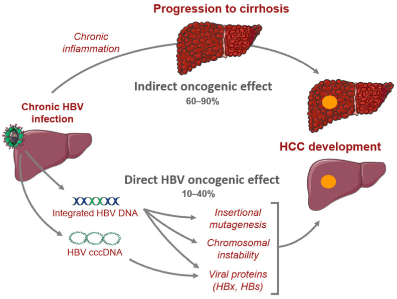
In CHB, there are crucial changes in immune cell activity and function involving both the innate and adaptive immune systems that lead to hepatic inflammation and hepatocyte killing [17]. Chronic infection may progress to liver fibrosis, cirrhosis, and HCC [17,35]. HBV-mediated carcinogenesis is a complex process that involves viral DNA integration into the host genome, ultimately leading to viral manipulation of cell-signaling and proliferation. This leads to a cascade of events which converts normal hepatocytes into malignant cells [36,37,38,39]. Oxidative stress associated with viral hepatitis changes the cellular environment in such a way that promotes carcinogenesis. Overproduction of free radicals and reactive oxidative species leads to the upregulation of inflammatory pathways that result in hepatocyte release of cytokines and chemokines that recruit neutrophils, monocytes, and lymphocytes [18,37,40,41]. As inflammation persists, immune cells, including macrophages and myeloid-derived suppressor cells, become dysfunctional, further amplifying the pro-inflammatory environment. The chronic inflammatory state leads to compensatory hepatocyte proliferation, which leads to accumulation of mutations that promote cell growth and proliferation which predisposes the host to developing HCC (Figure 1) [40,42].
Figure 1.
Mechanisms of hepatocarcinogenesis from chronic hepatitis B infection. Sourced from Péneau et al. [42].
Moreover, it was found that the incidence of HCC is fivefold higher among HBV-infected patients with cirrhosis compared to asymptomatic carriers, indicating that cirrhosis may be a pre-malignant condition [38]. Indeed, fibrosis of the liver disrupts the normal architecture which leads to modification of cell–cell interactions and ultimately, loss of regulation over cell proliferation [38].
Furthermore, it is important to note that numerous factors including host characteristics, HBV genotype, viral mutations, viral load, and HBsAg levels all influence the clinical manifestations of HBV infection [6]. Research has shown that there are differences between the various genotypes of HBV in tendency of chronicity, primary mode of transmission, timing of seroconversion, timing of HBsAg clearance, and clinical outcomes [6]. For example, multiple studies have shown that genotype C tends to cause more severe liver disease, including cirrhosis and HCC, compared to other genotypes [6,43,44]. Genotype C also has higher serum HBV levels and was shown to cause DNA mutations more frequently than Genotype B [6,45]. Further research investigating the utility of routine HBV genotyping is needed before implementation into clinical practice.
3. HCC Risk Factors and Surveillance
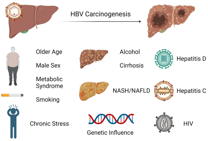
HCC is one of the major malignant diseases in the world today and ranks fifth in overall frequency. Its incidence is high in Eastern Asia and sub-Saharan Africa and is increasing in many parts of the Western world [46]. Among cancers, the annual mortality from HCC is relatively high because of its rapid progression and poor prognosis. Unfortunately, the diagnosis is typically made later in its course when therapeutic interventions are generally ineffective. Thus, the focus has been on early screening and treatment of known causes, including chronic hepatitis B which is the most frequent underlying cause of HCC. There are several major risk factors for development of HCC in chronic HBV infection. While having a first-degree relative with HCC, metabolic syndrome, type 2 diabetes, and central obesity are all host factors that have been linked to HCC development in HBV-infected patients or carriers, it appears that liver cirrhosis has been consistently identified to be the most significant risk factor for development of HCC during nucleos(t)ide therapy [47,48,49,50,51]. Notably, there is increasing evidence that non-alcoholic fatty liver disease (NAFLD) is becoming one of the largest causes of HCC, particularly in industrialized countries [52]. In these patients, HCC can develop even in the absence of cirrhosis. As there are no specific pharmacological therapies for NAFLD, adoption of healthy lifestyle changes including weight loss and regular aerobic physical activity remain the mainstay treatment. Interestingly, bariatric surgery is known to provide durable weight loss and has been identified to be associated with a decreased risk of HCC. A meta-analysis involving nearly 20 million patients showed that bariatric surgery has a protective effect on risk of HCC occurrence and incidence compared to those subjects who did not undergo the procedure [53]. Studies indicate that the presence of male gender, increasing age, higher HBV DNA level, and core promoter mutations are also independently associated with HCC risk [54,55]. It is postulated that androgens may play a role in the observed difference in incidence of HCC based on gender [56]. Among patients with chronic hepatitis B infection treated with nucleos(t)ide therapy, those with hepatitis D coinfection have been shown to have a nearly sixfold risk of HCC development compared to patients without hepatitis D coinfection [57]. While less investigated, concurrent HCV or HIV infection has also been identified to have an association with increased incidence of HCC [58,59]. The risk factors for hepatocarcinogenesis can be seen in Figure 2.
Figure 2.
Exogenous and endogenous risk factors for hepatocarcinogenesis from chronic hepatitis B infection. Created with BioRender.com (accessed on 21 January 2024).
Surveillance with ultrasound imaging and measurement of alpha-fetoprotein levels has been directed towards these populations to help with early detection of HCC. Guidelines suggest surveillance for HCC in high-risk groups including patients with cirrhosis, noncirrhotic patients with HBV and any of the following characteristics: active hepatitis, family history of HCC, Africans and African Americans, Asian males over 40 years of age, and Asian females over 50 years of age [60,61,62,63]. Other populations that undergo surveillance include patients with chronic hepatitis C virus and advanced liver fibrosis in the absence of cirrhosis, although this is not a consistent recommendation across all societies and the cost effectiveness has not been verified [61].
Currently a knowledge gap remains regarding host factors that contribute to the vagaries of HBV infection outcomes. A group of researchers attempted to explore this by investigating the diverse manifestations of CHB in three families that were observed over decades. Block et al. showed how only one case of HBV-related HCC occurred within every family cluster despite each having the same virus given perinatal transmission from mother to offspring [64]. Furthermore, one of the families had monozygotic twins in which only one sibling developed HBV-related HCC, while the other remained a chronic HBV carrier. The same finding is presenting in a case series by Noverati et al., which presented four family clusters in which patients had very variable courses, some with indolent chronic HBV infection, some requiring treatment, and others who developed HCC or cirrhosis [65]. It is postulated that inheritable immuno-genetic alleles that affect CHB differ from those that influence HBV-related HCC development, which may explain the discrepancy in manifestations in this case series [66]. The study was limited by lack of host and viral genomic analysis, as previous studies have shown certain host polymorphisms and HBV mutations are associated with HBV-related HCC. Nonetheless, this highlights that there are genetic and non-genetic host factors that play a role in development of HCC.
Exogenous, non-genetic factors such as chronic stress have been implicated in the incidence and outcomes of HCC, which may also have contributed to the differing presentations in the case outlined above [18]. One of the first published studies highlighting the significance of stress was by Russ et al., in which the group found in a large UK meta-analysis that those with higher scores on a general health questionnaire measuring psychological distress had a higher mortality from liver disease [67]. Joung et al. outlined how stress can increase inflammatory, oxidative reactions including hypoxia–reoxygenation, overactivation of Kupffer cells, influx of gut-derived lipopolysaccharide and norepinephrine, and overproduction of stress hormones in the sympathetic drive, which cause hepatocellular damage and promote mutagenesis [68]. Studies have shown how the tumor milieu of existing HCC undergoes changes that make it immunosuppressive. Specifically, He et al. discovered how chronic stress transitions cytokines to those that are T helper 2 cell-mediated, a tumor microenvironment that carries a poorer prognosis [69]. Similar studies show how this is exacerbated by the presence of T-regulatory cells that suppress pro-inflammatory cytokines [70,71].
4. Incidence of HCC in Response to Treatment of HBV
Chronic inflammation and necrosis are key factors which predispose patients to developing HCC. In HBV infection, viral load generally correlates with the severity of disease. NA therapies aimed at decreasing viral load include lamivudine, entecavir, and tenofovir, the latter two of which are standard of care and have provided a breakthrough in the treatment of HBV infection as they have demonstrated a reduction in incidence of HCC [72,73,74]. Results of prior systematic reviews demonstrate that the risk of HCC is significantly lower in CHB patients receiving oral antiviral therapy. Of the first data to demonstrate efficacy of anti-HBV therapy in HCC risk reduction, some came from a randomized controlled trial by Liaw et al. which compared lamivudine vs. placebo in treatment-naive patients with cirrhosis or advanced fibrosis and active liver disease. Lamivudine treatment for 3 years reduced the risk of HCC by 51% compared to the placebo [74]. According to Papatheodoridis et al., across three studies including untreated controls, HCC developed in 2.8% of treated and in 6.4% of untreated CHB patients (p = 0.003). A similar study by Singal et al. pooled data from six other studies and showed that the incidence of HCC among lamivudine-treated patients was significantly lower than that of untreated patients (3.4% vs. 9.6%, p < 0.0001) [75]. These data also demonstrated that HCC occurred more frequently among CHB patients receiving therapy who were older, had cirrhosis, or had detectable HBV-DNA at the end of treatment.
Over time, it was discovered that several patients on lamivudine therapy did not attain virological response in the setting of resistance. This did not come without consequence. Alarmingly, Papatheodoridis et al. demonstrated that the rate of HCC was significantly higher in lamivudine resistance than in nucleos(t)ide-naive patients (7.1% vs. 3.8%, p = 0.001), even after correcting for those with cirrhosis (18% vs. 11%, p = 0.015). It is believed that mutations including changes at position 181 of the reverse transcriptase domain of HBV polymerase have oncologic potential. This led to the advent of newer nucleos(t)ide analog therapies such as tenofovir and entecavir, which proved to be effective. Kim et al. observed that the incidence of HCC in patients with CHB and cirrhosis was decreased compared to the predicted incidence for untreated CHB patients after treatment with tenofovir disoproxil fumerate (TDF) for 3 years [73]. Lim et al. elaborated on this finding and showed that the difference in observed versus predicted HCC risk is more pronounced with tenofovir alafenamide (TAF) than TDF treatment [76]. Hosaka et al. demonstrated that HCC suppression effect was greater in entecavir-treated than non-rescued lamivudine-treated cirrhosis patients when compared to the control group [72]. This was supported by Papatheodoridis et al. in a prospective cohort study of HBeAg-negative CHB patients which showed that entecavir compared with lamivudine group patients had lower HCC incidence [77]. In a retrospective cohort of 451 newly diagnosed HBV-related HCC patients, lamivudine use, compared to entecavir, was associated with hepatic decompensation and recurrence of HCC after curative therapy [78]. In a study of entecavir-treated patients, the cumulative incidences of HCC were lower with achievement of a virologic response in both non-cirrhotic and cirrhotic patients [79]. In another study which included cirrhotic CHB patients treated with entecavir, those with undetectable HBV DNA levels were associated with a lower probability of developing HCC [80]. A separate study sought to identify the long-term clinical impact of low-level viremia (<2000 IU/mL) in patients on entecavir monotherapy. Kim et al. observed that, among patients with cirrhosis, those with low-level viremia exhibited a significantly higher HCC risk than those with maintained virological response [81]. These findings support the current recommendation for indefinite therapy in chronic HBV patients, and that inability to achieve undetectable viral load can be consequential.
5. Persistent Risk of HCC in HBV Infection
Maintenance of viral suppression is necessary to reduce HCC risk, as demonstrated above. Unfortunately, recent evidence shows that there remains a persistent risk of HCC despite successful HBV therapy resulting in viral suppression [46,82,83,84,85,86]. While spontaneous or treatment-induced seroclearance of HBsAg is also associated with lower risk of liver-related complications, there remains persistent risk of HCC development in these patients as well. Ahn et al. showed a significant reduction in necroinflammation on liver biopsy of patients before and after seroclearance. However, all the patients demonstrated the presence of HBV DNA without a change in the fibrosis score [87]. Additionally, Gounder et al. showed that seroclearance was not associated with reduced HCC risk compared to a matched control group [88]. The proposed mechanisms in which HCC develops despite adequate treatment are described here.
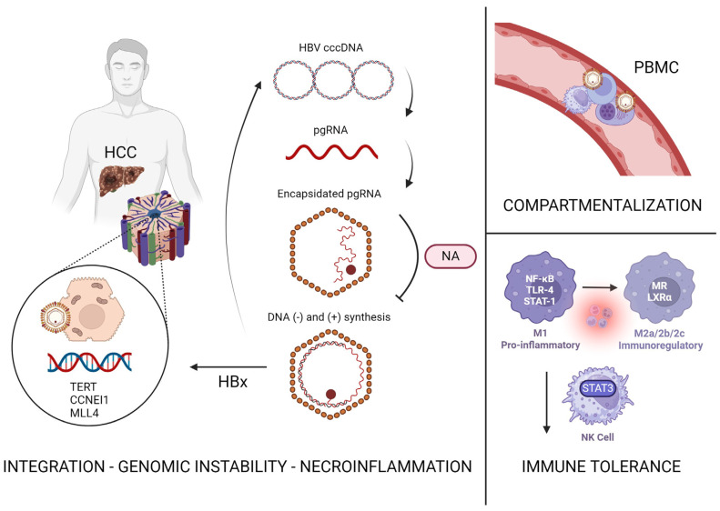
It has been discovered that HBV’s covalently closed circular DNA (cccDNA) integrates into the hepatocytes upon initial infection and can be detected even after antiviral treatment. This integration may occur at critical HCC driver genes which may give rise to malignant HBV-infected hepatocytes. Li et al. found that the common location of integration included the TERT, CCNEI1, and MLL4 genes, and that virus–host chimera DNA circulate in 97.7% of CHB patients with HCC [89,90]. Interestingly, the group demonstrated how virus–host chimera DNA could be used as a biomarker for detecting tumor load in this patient population, and how it may aid in monitoring recurrence after tumor resection [89]. In the first detailed molecular evaluation completed by Coffin et al., despite undetectable HBV DNA in plasma via clinical assays, patients treated with oral anti-HBV therapy undergoing liver transplant were found to have low levels of HBV genome in the liver, circulating lymphoid cells, as well as plasma using ultrasensitive PCR/nucleic acid hybridization assay. Their data promote the idea of HBV compartmentalization, where despite adequate suppression, the liver supports replication and extrahepatic HBV can be detected, particularly in peripheral blood mononuclear cells (PBMCs) that may serve as a sanctuary for wild-type viruses during antiviral therapy [91].
Other possible mechanisms that have been postulated include progression of cirrhosis, necroinflammation caused by HBV infection, and the oncogenic effect of HBV genome integration into the hepatocyte chromosomes. HBV DNA may remain detectable in the serum or liver cells, which is referred to as occult HBV infection [92]. Wong et al. found that 69% of 90 HBsAg-negative patients with HCC had occult infection and that of these patients, 47% had detectable cccDNA in hepatocytes and 53% had integration of HBV DNA near hepatic oncogenes [93]. In fact, the HBV genome has been detected in tumor tissue of HBsAg negative patients with HCC in a prevalence ranging from 30% to 80% [94,95]. Therefore, these patients continue to undergo surveillance per the AASLD guidelines [96]. Michalak et al. showed that traces of the HBV genome persist for years following recovery from hepatitis B (characterized by seronegativity) and that they can lead to HCC development in a woodchuck model [97].
HBV infection has an inhibitory effect on the innate and adaptive immune cells which may aggravate chronic inflammation and lead to carcinogenesis. These effects are thought to persist despite antiviral treatment as the microenvironment is altered upon initial infection. Data suggest that this immune-tolerant phase of CHB infection is when hepatocarcinogenesis begins, with integration of HBV DNA into the genome of hepatocytes [98]. A retrospective cohort study by Kim et al. demonstrated how untreated patients in an immune-tolerant phase had 2–3 times higher risk of HCC, liver transplantation, or death compared with patients in immune clearance phase who were initiated on NA therapy [99]. The immune-tolerant phase was defined as HBeAg-positive, HBV DNA level greater than or equal to 20,000 IU/mL and normal ALT for one year. It has been shown that the virus may decrease the expression of STAT3 in NK cells, ultimately inhibiting their ability to clear HBV [100]. Studies demonstrate that HBV may promote polarization of cells from M1 to M2 and inhibit secretion of antiviral cytokines by macrophages [101]. The idea that there are frequent integration events and detectable HBV viral load during the immune-tolerant phase suggests that there may be a role of early introduction of NA therapy to decrease risk of HCC and fibrosis progression [102,103]. This would ultimately mean initiation of therapy in young adults with immune-tolerant profile. However, the low rates of response, the costs of long-term treatment, and the lack of established benefits in prevention of HCC are obstacles to be considered [104,105]. Further research on the immune-tolerant population is necessary to fully understand this dynamic. Immune tolerance to HBV infection plays a key role in the acceleration of progression of HBV to HCC.
Most reported cases of HBV-HCC in the literature are within the first five years of NA therapy, but data on long-term risk of HCC while on treatment are lacking. The Liver Disease Prevention Center, Division of Gastroenterology and Hepatology at Thomas Jefferson University Hospital, has observed several HBV positive patients develop HCC even after 10 or more years of successful viral suppression [106]. Specifically, in our observational cohort, 17 patients developed HCC despite having noted undetectable HBV DNA for up to 12 years and on treatment for up to 19 years (Table 1) [107]. Currently, this is among the longest known follow-up studies on the development of HCC in patients on HBV treatment.
Table 1.
Development of HCC in patients with cirrhosis on long-term antiviral therapy. Sourced from Boortalary et al. [107].
| Pt | Date StartTx | Change in Child Class on Tx | Date HCC Dx |
Yrs on Anti-HBV Tx at HCC Dx | Yrs with HBV DNA (-) * | Age (Yr) at HCC Dx | Tumor Size at Dx | HBVDNA at HCC Dx | Anti-HBV Tx | Status |
|---|---|---|---|---|---|---|---|---|---|---|
| 1 | April 1998 | B → A | July 2007 | 9 | 3 | 53 | 1.1 Junction | UD | LAM + TDF | Alive |
| 2 | January 1998 | B → A | March 2008 | 10 | 8 | 68 | 2.8 Rt | UD | LAM + TDF | Dead |
| 3 | May 1998 | A →A | February 2008 | 10 | 7 | 76 | 1.8 Lt | UD | LAM + TDF | Alive |
| 4 | July 2001 | B → B | September 2010 | 9 | 4 | 54 | 2.8 Rt | UD | LAM + TDF | Dead |
| 5 | August 2004 | B → B | November 2010 | 16 | 4 | 53 | 3.9 Rt | UD | LAM + TDF | Alive |
| 6 | July 2001 | B → B | January 2011 | 10 | 5 | 55 | 2.8 Rt | UD | LAM + TDF | Dead |
| 7 | February 2004 | A → A | June 2013 | 9 | 8 | 57 | 2.5 Lt med | UD | TDF | Dead |
| 8 | February 1996 | A → A | July 2013 | 17 | 10 | 73 | 1.6 Rt | UD | TDF | Dead |
| 9 | August 1997 | A → A | June 2014 | 17 | 6 | 54 | 2.2 Lt lat | UD | ETV | Alive |
| 10 | March 2004 | B → B | June 2013 | 9 | 7 | 57 | 2.5 Lt | UD | TDF | Dead |
| 11 | July 2001 | A → A | June 2014 | 13 | 7 | 54 | 2.2 Lt | UD | TDF | Alive |
| 12 | May 1996 | A → A | October 2014 | 18 | 10 | 74 | 3.4 Rt | UD | LAM + TDF | Dead |
| 13 | February 2000 | A → A | October 2014 | 14 | 12 | 62 | 3.8 Rt | UD | ETV + TDF | Alive |
| 14 | February 2000 | A → A | April 2015 | 15 | 12 | 62 | 3.4 Rt | UD | TDF | Alive |
| 15 | February 2000 | B → A | May 2015 | 15 | 12 | 65 | 3.8 Rt | UD | TDF | Alive |
| 16 | December 1998 | A → A | August 2017 | 19 | 8 | 64 | 2.0 Rt | UD | LAM + TDF | Alive |
| 17 | February 2008 | A → A | June 2019 | 11 | 10 | 57 | 2.2 Rt | UD | ETV + TDF | Alive |
Dx: Diagnosis, ETV: Entecavir, LAM: Lamivudine, Lt: Left, Pt: Patient, Rt: Right, TDF: Tenofovir Disoproxil Fumarate, Tx: Treatment, UD: Undetectable, * Years with HBV DNA (-) at diagnosis of HCC.
Our center has observed six patients who developed new or recurrent HCC after undergoing curative therapy for HCC and achieving adequate HBV suppression for 5–11 years on antivirals [106]. Unfortunately, recent studies have shown that the patients who develop HCC after years of viral suppression, in fact, carry worse prognoses than those who develop HCC without prior treatment of HBV [108,109]. A case series of 26 patients, 13 antiviral-naive and 13 antiviral-treated, assessed clinical outcomes after HCC diagnosis. Garrido et al. showed that after the first HBV-HCC event, death was observed more frequently among the antiviral-treated cohort at 46.2% compared to 15.4% in the antiviral-naive cohort [108]. Li et al. compared patients who received NA therapy after hepatectomy for HCC and those who did not receive treatment and discovered that there was no difference in the recurrence rate of HCC [110]. The suggested mechanism by which this occurs is that halting HBV replication at the reverse transcription phase for a prolonged time may prime the infected hepatocytes to increasingly turnover HBV DNA integration, which can cause insertional mutagenesis and genomic instability, in the setting of HBV compartmentalization and immune-tolerant environment, as previously described (Figure 3) [111]. This additionally results in the accumulation of incompletely transcribed mRNAs which may lead to further integration.
Figure 3.
Proposed mechanisms behind persistent risk of HBV-associated HCC. cccDNA—covalently closed circular DNA; pgRNA—pre-genomic RNA; NA—nucleos(t)ide analog; HBx—HBV virus X protein; PBMC—peripheral blood mononuclear cells; NK—natural killer. Created with BioRender.com (accessed 24 January 2024).
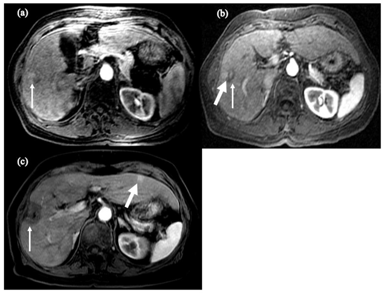
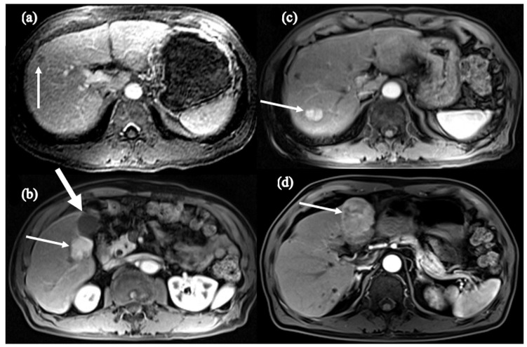
It is believed that HCC inoculation in this setting of disorganized products and instability may also cause cancerous cells to be primed to behave more aggressively. Chowdhury et al. described two cases of HBV-treatment-naive HCC where despite radiofrequency tumor ablation (RFA) and subsequent undetectable HBV viral load on NA therapy, the patients developed recurrent, more aggressive cancer [109]. The first case described an elderly female who underwent trans-arterial embolization (TACE) of a 5 × 2.1 cm mass suggestive of HCC. She started NA therapy and became HBV-DNA-negative. However, she developed recurrence 5 years later and required repeated treatment of TACE and RFA. Ten years after the recurrence, MRI demonstrated another 5 mm LI-RADS 4 lesion in segment 2 which was monitored with evidence of regression. Unfortunately, this tumor had grown to 1.2 cm two years later, which marked two decades after the development of her initial HCC (Figure 4) [109]. The second case described a male who was found to have cirrhosis with 1.0 cm HCC at age 68. He underwent NA therapy with undetectable HBV DNA and RFA; however, at age 79, he was found to have two new lesions on an abdominal MRI (3 × 2.8 cm and 2 × 1.7 cm) in segment 5, adjacent to the gallbladder, and segment 7/8, respectively. He received a cholecystectomy and tumor ablation but four years later was found to have a new 4.8 cm lesion in segment 4 on MRI (Figure 5) [109]. This illustrates the complexity of the relationship between HBV and HCC, as there may be accelerated progression and recurrence of tumor in patients after becoming treatment-experienced. Notably, this is a preliminary observation that requires larger studies to characterize, as random integration of HBV DNA and anatomical location of initial HCC lesion also play key roles in the development of further HCC events. Nonetheless, these cases place emphasis on the value of adhering to scheduled screening and monitoring of HCC recurrence years after initial diagnosis.
Figure 4.
Axial fat-suppressed, T1-weighted postcontrast images of (a) the initial mass concerning for HCC (arrow), (b) post-TACE lesion (thin arrow) with adjacent focal hyperenhancement (thick arrow), and (c) post-TACE and RFA lesion in segment 5 (thin arrow) with new hyper-enhancing lesion in segment 3 (thick arrow). Sourced from Chowdhury et al. [109].
Figure 5.
Axial fat-suppressed, T1-weighted postcontrast images of (a) the initial segment 8 mass concerning for HCC (arrow), (b) post-RFA and NA therapy 3 cm segment 5 hyper-enhancing HCC (thin arrow) adjacent to gallbladder (thick arrow), (c) second hyper-enhancing segment 7 HCC (arrow), and (d) post-cholecystectomy and tumor ablation large segment 4 HCC (arrow). Sourced from Chowdhury et al. [109].
Numerous studies have been conducted investigating the role of HBV infection in resistance to sorafenib therapy in patients with HBV-associated HCC. Newer systemic chemotherapies have been approved for the treatment of HCC, such as atezolizumab/bevacizumab, durvalumab/tremelimumab, and lenvatinib; however, for more than 10 years, sorafenib has been considered to be the standard of care for patients with advanced unresectable HCC. Sorafenib inhibits activity of many tyrosine kinases involved in tumor angiogenesis and progression including vascular endothelial growth factor receptor, platelet-derived growth factor receptor, Flt3 and c-Kit, and many Raf kinases involved in the MAPK/ERK pathway [112]. These pathways ultimately lead to proliferation, migration, survival, and metastasis of tumor cells. Liu et al. has shown that there is overexpression of variant 1 (tv1) of proliferating cell nuclear antigen clamp-associated factor (PCLAF) particularly in HBV-related HCC than in non-virus-related HCC [113]. In their biochemical studies, HBV promoted splicing of PCLAF tv1 through downregulating serine/arginine-rich splicing factor 2 (SRSF2). Through suppression of ferroptosis and regulation of abnormal splicing of the SRSF2/PCLAF tv1 axis, HBV was noted to induce sorafenib resistance. In a separate study by Liu et al., immunohistochemistry staining of HBC-associated HCC tissue demonstrated overexpression of WNT7B compared to nontumor liver tissues [114]. This signaling molecule along with its receptor frizzled-4 (FZD4) were noted to be upregulated in response to large hepatitis B surface antigens (L-HBs), which increased WNT signaling in HCC cells, leading to proliferation and metastasis. By this mechanism, L-HBs suppressed sorafenib-induced mitophagy, diminishing the therapeutic effect of chemotherapy. Zhang et al. demonstrated in mouse models that cellular inhibitor of apoptosis protein 2 (cIAP2) plays a key role in promotion of cell survival of carcinogenic hepatocytes in HBV-associated HCC [115]. Cell lines rich in cIAP2 had reduced caspase levels and increased cell viability when treated with sorafenib. However, when these cells were treated with lamivudine, cIAP2 expression decreased and sorafenib sensitivity was partially restored.
Other studies have investigated potential target strategies to overcome this resistance including regulatory T cells (Treg). CCR4+ Tregs are the predominant type of Tregs that are recruited to HBV-associated HCC and directly correlate with HBV titers, exhibiting increased immunosuppressive cytokines such as IL-10 and IL-35 [116]. It was shown in a mouse model study by Gao et al. that treatment with CCR4 antagonist could block Treg accumulation to overcome sorafenib resistance, and potentially serve as a therapeutic target in HCC immunotherapy [116]. This research group expanded on this finding by suggesting that blocking macrophage-derived chemokine (CCL22), which controls migration of Tregs via its receptor CCR4, may also be an immunotherapeutic strategy for HCC through a similar mechanism [117]. Notably, there is an increased risk of severe autoimmunity without adequate regulation by Tregs. Other mechanisms to sensitize HBV-associated HCC to sorafenib-induced apoptosis include modulation of miRNA such as miR-193b and miR-3677-3p, in addition to inhibiting pro-oncogenic Mitogen-Activated Protein Kinase 14 (pMAPK14) [118,119,120].
NAs have been critical in decreasing HBV-associated mortality worldwide, However, we demonstrate in this review that they do not eradicate the risk of HCC development. There is persistent risk of hepatocarcinogenesis despite adequate treatment marked by undetectable HBV DNA. Thus, there remains a public health threat as currently there is no cure for HBV infection. Achievement of a cure would require therapies which eliminate cccDNA and integrated HBV DNA as well as help restore the impaired innate and adaptive immune responses against HBV.
6. The Road to HBV Cure
The AASLD defines two different types of “cures” of HBV. First, it defines “immunological cure” as HBsAg loss and sustained HBV DNA suppression. Second, it defines “virological cure” as eradication of the virus, including cccDNA, which is not achievable with current therapies.
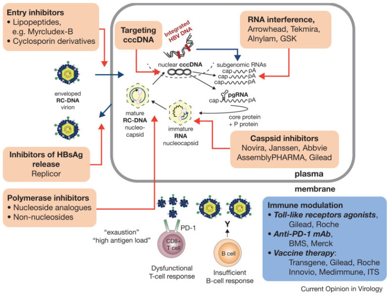
The next generation of drugs in development to treat hepatitis B can be divided into the following classes: drugs targeting the life cycle of the hepatitis B virus; drugs aiming to modulate the host’s immune response to CHB infection; monoclonal antibodies aiming to directly inhibit translation of HBV RNA; and therapeutic vaccines (Figure 6) [121]. A detailed discussion of the progress of these drugs’ development is beyond the scope of this paper; for further reading, the excellent review by Yardeni et al. is highly recommended [122].
Figure 6.
Therapeutic drug targets of hepatitis B virus replication. Sourced from Levrero et al. [121]. Used with permission from original copyright holder.
Drugs targeting the life cycle of the hepatitis B virus can be classified into entry inhibitors, capsid assembly modulators, post-transcriptional control inhibitors, and hepatitis B surface antigen release inhibitors.
Novel drugs aiming to modulate the host’s immune response to the HBV are comprised of innate immune activators—specifically, Toll-like receptor (TLR) agonists, RIG-1 agonists, and checkpoint inhibitors.
Monoclonal antibodies designed to inhibit the life cycle of HBV have been investigated. One such neutralizing monoclonal antibody, VIR-3434, enhances HBsAg uptake by dendritic cells to promote its presentation to naive T cells thereby stimulating an HBV-specific immune response [123].
Finally, there has been interest in using therapeutic vaccines in effort to stimulate the host’s immune response against CHB infection. However, early studies in humans showed only modest declines in HBsAg [124]. With that said, several new therapeutic vaccines are in development which may be tested in combination with drugs of other mechanisms of action, including siRNAs [125]. However, these therapies have not yet been tested in humans.
In summary, the emerging landscape for novel CHB treatments is composed of vast and varied mechanisms of action, but further clinical testing is still required to establish superiority in efficacy compared to the current standard of care.
7. Conclusions and Future Directions
HBV is a global health threat worldwide as infection can lead to HCC. Available treatment options do not fully eradicate the risk of hepatocarcinogenesis as initial integration of cccDNA into the host genome is not prevented. Continued transcription creates a milieu of genomic instability which can make recurrence of HCC even more aggressive. The phenomenon of worse prognosis in treatment-experienced HCC versus treatment-naive HCC is observed. While there remains a persistent risk of HBV-associated HCC despite viral suppression, recent studies show promising novel approaches to HBV cure.
Author Contributions
Conceptualization, H.-W.H.; Resources, H.-W.H., N.V., A.M. and S.N.; Writing—N.V., A.M. and S.N.; Original Draft Preparation, N.V., A.M. and S.N.; Writing—Review and Editing, H.-W.H., D.H.-D., N.V., A.M., S.N. and T.B.; Visualization, H.-W.H., D.H.-D., N.V. and T.B.; Supervision, H.-W.H., D.H.-D. and T.B. All authors have read and agreed to the published version of the manuscript.
Data Availability Statement
No new data were created or analyzed in this study. Data sharing does not apply to this article.
Conflicts of Interest
The authors declare no conflicts of interest.
Funding Statement
This research received no external funding.
Footnotes
Disclaimer/Publisher’s Note: The statements, opinions and data contained in all publications are solely those of the individual author(s) and contributor(s) and not of MDPI and/or the editor(s). MDPI and/or the editor(s) disclaim responsibility for any injury to people or property resulting from any ideas, methods, instructions or products referred to in the content.
References
- 1.Hou J., Liu Z., Gu F. Epidemiology and Prevention of Hepatitis B Virus Infection. Int. J. Med. Sci. 2005;2:50–57. doi: 10.7150/ijms.2.50. [DOI] [PMC free article] [PubMed] [Google Scholar]
- 2.Bashir Hamidu R., Hann R.R., Hann H.-W. Chronicles of HBV and the Road to HBV Cure. Livers. 2023;3:232–239. doi: 10.3390/livers3020015. [DOI] [Google Scholar]
- 3.Hepatitis B. [(accessed on 21 December 2023)]. Available online: https://www.who.int/news-room/fact-sheets/detail/hepatitis-b.
- 4.Pithawa A.K. Sleisenger and Fordtran’s Gastrointestinal and Liver Disease: Pathophysiology, diagnosis, management. Med. J. Armed Forces India. 2007;63:205. doi: 10.1016/S0377-1237(07)80085-2. [DOI] [Google Scholar]
- 5.Kao J.H., Chen P.J., Lai M.Y., Chen D.S. Hepatitis B genotypes correlate with clinical outcomes in patients with chronic hepatitis B. Gastroenterology. 2000;118:554–559. doi: 10.1016/S0016-5085(00)70261-7. [DOI] [PubMed] [Google Scholar]
- 6.Sunbul M. Hepatitis B virus genotypes: Global distribution and clinical importance. World J. Gastroenterol. 2014;20:5427–5434. doi: 10.3748/wjg.v20.i18.5427. [DOI] [PMC free article] [PubMed] [Google Scholar]
- 7.Guidotti L.G., Chisari F.V. Immunobiology and pathogenesis of viral hepatitis. Annu. Rev. Pathol. 2006;1:23–61. doi: 10.1146/annurev.pathol.1.110304.100230. [DOI] [PubMed] [Google Scholar]
- 8.Lok A.S. Chronic hepatitis B. N. Engl. J. Med. 2002;346:1682–1683. doi: 10.1056/NEJM200205303462202. [DOI] [PubMed] [Google Scholar]
- 9.Parkin D.M., Pisani P., Ferlay J. Global cancer statistics. CA Cancer J. Clin. 1999;49:33–64. doi: 10.3322/canjclin.49.1.33. [DOI] [PubMed] [Google Scholar]
- 10.Fattovich G. Natural history and prognosis of hepatitis B. Semin. Liver Dis. 2003;23:47–58. doi: 10.1055/s-2003-37590. [DOI] [PubMed] [Google Scholar]
- 11.El-Serag H.B., Rudolph K.L. Hepatocellular carcinoma: Epidemiology and molecular carcinogenesis. Gastroenterology. 2007;132:2557–2576. doi: 10.1053/j.gastro.2007.04.061. [DOI] [PubMed] [Google Scholar]
- 12.MacLachlan J.H., Cowie B.C. Hepatitis B virus epidemiology. Cold Spring Harb. Perspect. Med. 2015;5:a021410. doi: 10.1101/cshperspect.a021410. [DOI] [PMC free article] [PubMed] [Google Scholar]
- 13.Beasley R.P., Hwang L.Y., Lin C.C., Leu M.L., Stevens C.E., Szmuness W., Chen K.P. Incidence of hepatitis B virus infections in preschool children in Taiwan. J. Infect. Dis. 1982;146:198–204. doi: 10.1093/infdis/146.2.198. [DOI] [PubMed] [Google Scholar]
- 14.Alter M.J., Hadler S.C., Margolis H.S., Alexander W.J., Hu P.Y., Judson F.N., Mares A., Miller J.K., Moyer L.A. The changing epidemiology of hepatitis B in the United States. Need for alternative vaccination strategies. JAMA. 1990;263:1218–1222. doi: 10.1001/jama.1990.03440090052025. [DOI] [PubMed] [Google Scholar]
- 15.Chuang Y.C., Tsai K.N., Ou J.J. Pathogenicity and virulence of Hepatitis B virus. Virulence. 2022;13:258–296. doi: 10.1080/21505594.2022.2028483. [DOI] [PMC free article] [PubMed] [Google Scholar]
- 16.Ganem D., Prince A.M. Hepatitis B virus infection–Natural history and clinical consequences. N. Engl. J. Med. 2004;350:1118–1129. doi: 10.1056/NEJMra031087. [DOI] [PubMed] [Google Scholar]
- 17.Khanam A., Chua J.V., Kottilil S. Immunopathology of Chronic Hepatitis B Infection: Role of Innate and Adaptive Immune Response in Disease Progression. Int. J. Mol. Sci. 2021;22:5497. doi: 10.3390/ijms22115497. [DOI] [PMC free article] [PubMed] [Google Scholar]
- 18.Noverati N., Bashir-Hamidu R., Halegoua-DeMarzio D., Hann H.W. Hepatitis B Virus-Associated Hepatocellular Carcinoma and Chronic Stress. Int. J. Mol. Sci. 2022;23:3917. doi: 10.3390/ijms23073917. [DOI] [PMC free article] [PubMed] [Google Scholar]
- 19.Liaw Y.F., Tsai S.L., Sheen I.S., Chao M., Yeh C.T., Hsieh S.Y., Chu C.M. Clinical and virological course of chronic hepatitis B virus infection with hepatitis C and D virus markers. Am. J. Gastroenterol. 1998;93:354–359. doi: 10.1111/j.1572-0241.1998.00354.x. [DOI] [PubMed] [Google Scholar]
- 20.Wieland S., Thimme R., Purcell R.H., Chisari F.V. Genomic analysis of the host response to hepatitis B virus infection. Proc. Natl. Acad. Sci. USA. 2004;101:6669–6674. doi: 10.1073/pnas.0401771101. [DOI] [PMC free article] [PubMed] [Google Scholar]
- 21.Chisari F.V., Isogawa M., Wieland S.F. Pathogenesis of hepatitis B virus infection. Pathol. Biol. 2010;58:258–266. doi: 10.1016/j.patbio.2009.11.001. [DOI] [PMC free article] [PubMed] [Google Scholar]
- 22.Tripathi N., Mousa O.Y. StatPearls. Statpearls Publishing; Treasure Island, FL, USA: 2023. Hepatitis B. [PubMed] [Google Scholar]
- 23.Croagh C.M., Lubel J.S. Natural history of chronic hepatitis B: Phases in a complex relationship. World J. Gastroenterol. 2014;20:10395–10404. doi: 10.3748/wjg.v20.i30.10395. [DOI] [PMC free article] [PubMed] [Google Scholar]
- 24.Takashima H., Araki K., Miyazaki J., Yamamura K., Kimoto M. Characterization of T-cell tolerance to hepatitis B virus (HBV) antigen in transgenic mice. Immunology. 1992;75:398–405. [PMC free article] [PubMed] [Google Scholar]
- 25.Shi Y.H., Shi C.H. Molecular characteristics and stages of chronic hepatitis B virus infection. World J. Gastroenterol. 2009;15:3099–3105. doi: 10.3748/wjg.15.3099. [DOI] [PMC free article] [PubMed] [Google Scholar]
- 26.Wong G.L. Management of chronic hepatitis B patients in immunetolerant phase: What latest guidelines recommend. Clin. Mol. Hepatol. 2018;24:108–113. doi: 10.3350/cmh.2017.0068. [DOI] [PMC free article] [PubMed] [Google Scholar]
- 27.Hui C.K., Leung N., Yuen S.T., Zhang H.Y., Leung K.W., Lu L., Cheung S.K., Wong W.M., Lau G.K., Hong Kong Liver Fibrosis Study G. Natural history and disease progression in Chinese chronic hepatitis B patients in immune-tolerant phase. Hepatology. 2007;46:395–401. doi: 10.1002/hep.21724. [DOI] [PubMed] [Google Scholar]
- 28.McMahon B.J. The natural history of chronic hepatitis B virus infection. Hepatology. 2009;49:S45–S55. doi: 10.1002/hep.22898. [DOI] [PubMed] [Google Scholar]
- 29.Thimme R., Wieland S., Steiger C., Ghrayeb J., Reimann K.A., Purcell R.H., Chisari F.V. CD8(+) T cells mediate viral clearance and disease pathogenesis during acute hepatitis B virus infection. J. Virol. 2003;77:68–76. doi: 10.1128/JVI.77.1.68-76.2003. [DOI] [PMC free article] [PubMed] [Google Scholar]
- 30.Jeng W.J., Sheen I.S., Liaw Y.F. Hepatitis B virus DNA level predicts hepatic decompensation in patients with acute exacerbation of chronic hepatitis B. Clin. Gastroenterol. Hepatol. 2010;8:541–545. doi: 10.1016/j.cgh.2010.02.023. [DOI] [PubMed] [Google Scholar]
- 31.Chen Y.C., Chu C.M., Liaw Y.F. Age-specific prognosis following spontaneous hepatitis B e antigen seroconversion in chronic hepatitis B. Hepatology. 2010;51:435–444. doi: 10.1002/hep.23348. [DOI] [PubMed] [Google Scholar]
- 32.Yang H.I., Lu S.N., Liaw Y.F., You S.L., Sun C.A., Wang L.Y., Hsiao C.K., Chen P.J., Chen D.S., Chen C.J., et al. Hepatitis B e antigen and the risk of hepatocellular carcinoma. N. Engl. J. Med. 2002;347:168–174. doi: 10.1056/NEJMoa013215. [DOI] [PubMed] [Google Scholar]
- 33.Bonino F., Rosina F., Rizzetto M., Rizzi R., Chiaberge E., Tardanico R., Callea F., Verme G. Chronic hepatitis in HBsAg carriers with serum HBV-DNA and anti-HBe. Gastroenterology. 1986;90:1268–1273. doi: 10.1016/0016-5085(86)90395-1. [DOI] [PubMed] [Google Scholar]
- 34.Lok A.S., Hadziyannis S.J., Weller I.V., Karvountzis M.G., Monjardino J., Karayiannis P., Montano L., Thomas H.C. Contribution of low level HBV replication to continuing inflammatory activity in patients with anti-HBe positive chronic hepatitis B virus infection. Gut. 1984;25:1283–1287. doi: 10.1136/gut.25.11.1283. [DOI] [PMC free article] [PubMed] [Google Scholar]
- 35.Bixler D., Zhong Y., Ly K.N., Moorman A.C., Spradling P.R., Teshale E.H., Rupp L.B., Gordon S.C., Boscarino J.A., Schmidt M.A., et al. Mortality Among Patients With Chronic Hepatitis B Infection: The Chronic Hepatitis Cohort Study (CHeCS) Clin. Infect. Dis. 2019;68:956–963. doi: 10.1093/cid/ciy598. [DOI] [PMC free article] [PubMed] [Google Scholar]
- 36.Brechot C., Pourcel C., Louise A., Rain B., Tiollais P. Presence of integrated hepatitis B virus DNA sequences in cellular DNA of human hepatocellular carcinoma. Nature. 1980;286:533–535. doi: 10.1038/286533a0. [DOI] [PubMed] [Google Scholar]
- 37.Chemin I., Zoulim F. Hepatitis B virus induced hepatocellular carcinoma. Cancer Lett. 2009;286:52–59. doi: 10.1016/j.canlet.2008.12.003. [DOI] [PubMed] [Google Scholar]
- 38.De Mitri M.S., Cassini R., Bernardi M. Hepatitis B virus-related hepatocarcinogenesis: Molecular oncogenic potential of clear or occult infections. Eur. J. Cancer. 2010;46:2178–2186. doi: 10.1016/j.ejca.2010.03.034. [DOI] [PubMed] [Google Scholar]
- 39.Neuveut C., Wei Y., Buendia M.A. Mechanisms of HBV-related hepatocarcinogenesis. J. Hepatol. 2010;52:594–604. doi: 10.1016/j.jhep.2009.10.033. [DOI] [PubMed] [Google Scholar]
- 40.Block P.D., Shinn B., Kim J.H., Hann H.W. Hepatitis B-related hepatocellular carcinoma and stress: Untangling the host immune response from clinical outcomes. Hepat. Oncol. 2020;8:HEP35. doi: 10.2217/hep-2020-0028. [DOI] [PMC free article] [PubMed] [Google Scholar]
- 41.Li S., Hong M., Tan H.Y., Wang N., Feng Y. Insights into the Role and Interdependence of Oxidative Stress and Inflammation in Liver Diseases. Oxidative Med. Cell. Longev. 2016;2016:4234061. doi: 10.1155/2016/4234061. [DOI] [PMC free article] [PubMed] [Google Scholar]
- 42.Peneau C., Zucman-Rossi J., Nault J.C. Genomics of Viral Hepatitis-Associated Liver Tumors. J. Clin. Med. 2021;10:1827. doi: 10.3390/jcm10091827. [DOI] [PMC free article] [PubMed] [Google Scholar]
- 43.Wong G.L., Chan H.L., Yiu K.K., Lai J.W., Chan V.K., Cheung K.K., Wong E.W., Wong V.W. Meta-analysis: The association of hepatitis B virus genotypes and hepatocellular carcinoma. Aliment. Pharmacol. Ther. 2013;37:517–526. doi: 10.1111/apt.12207. [DOI] [PubMed] [Google Scholar]
- 44.Yang H.I., Yeh S.H., Chen P.J., Iloeje U.H., Jen C.L., Su J., Wang L.Y., Lu S.N., You S.L., Chen D.S., et al. Associations between hepatitis B virus genotype and mutants and the risk of hepatocellular carcinoma. J. Natl. Cancer Inst. 2008;100:1134–1143. doi: 10.1093/jnci/djn243. [DOI] [PMC free article] [PubMed] [Google Scholar]
- 45.Kao J.H., Chen P.J., Lai M.Y., Chen D.S. Clinical and virological aspects of blood donors infected with hepatitis B virus genotypes B and C. J. Clin. Microbiol. 2002;40:22–25. doi: 10.1128/JCM.40.1.22-25.2002. [DOI] [PMC free article] [PubMed] [Google Scholar]
- 46.Papatheodoridis G.V., Lampertico P., Manolakopoulos S., Lok A. Incidence of hepatocellular carcinoma in chronic hepatitis B patients receiving nucleos(t)ide therapy: A systematic review. J. Hepatol. 2010;53:348–356. doi: 10.1016/j.jhep.2010.02.035. [DOI] [PubMed] [Google Scholar]
- 47.Chen C.L., Yang H.I., Yang W.S., Liu C.J., Chen P.J., You S.L., Wang L.Y., Sun C.A., Lu S.N., Chen D.S., et al. Metabolic factors and risk of hepatocellular carcinoma by chronic hepatitis B/C infection: A follow-up study in Taiwan. Gastroenterology. 2008;135:111–121. doi: 10.1053/j.gastro.2008.03.073. [DOI] [PubMed] [Google Scholar]
- 48.Cheuk-Fung Yip T., Wai-Sun Wong V., Lik-Yuen Chan H., Tse Y.K., Pik-Shan Kong A., Long-Yan Lam K., Chung-Yan Lui G., Lai-Hung Wong G. Effects of Diabetes and Glycemic Control on Risk of Hepatocellular Carcinoma After Seroclearance of Hepatitis B Surface Antigen. Clin. Gastroenterol. Hepatol. 2018;16:765–773.e762. doi: 10.1016/j.cgh.2017.12.009. [DOI] [PubMed] [Google Scholar]
- 49.Orito E., Hasebe C., Kurosaki M., Osaki Y., Joko K., Watanabe H., Kimura H., Nishijima N., Kusakabe A., Izumi N., et al. Risk of hepatocellular carcinoma in cirrhotic hepatitis B virus patients during nucleoside/nucleotide analog therapy. Hepatol. Res. 2015;45:872–879. doi: 10.1111/hepr.12427. [DOI] [PubMed] [Google Scholar]
- 50.Song C., Zhu J., Ge Z., Yu C., Tian T., Wang H., Han J., Shen H., Dai J., Lu J., et al. Spontaneous Seroclearance of Hepatitis B Surface Antigen and Risk of Hepatocellular Carcinoma. Clin. Gastroenterol. Hepatol. 2019;17:1204–1206. doi: 10.1016/j.cgh.2018.08.019. [DOI] [PubMed] [Google Scholar]
- 51.Tan Y., Zhang X., Zhang W., Tang L., Yang H., Yan K., Jiang L., Yang J., Li C., Yang J., et al. The Influence of Metabolic Syndrome on the Risk of Hepatocellular Carcinoma in Patients with Chronic Hepatitis B Infection in Mainland China. Cancer Epidemiol. Biomarkers Prev. 2019;28:2038–2046. doi: 10.1158/1055-9965.EPI-19-0303. [DOI] [PubMed] [Google Scholar]
- 52.Motta B.M., Masarone M., Torre P., Persico M. From Non-Alcoholic Steatohepatitis (NASH) to Hepatocellular Carcinoma (HCC): Epidemiology, Incidence, Predictions, Risk Factors, and Prevention. Cancers. 2023;15:5458. doi: 10.3390/cancers15225458. [DOI] [PMC free article] [PubMed] [Google Scholar]
- 53.Ramai D., Singh J., Lester J., Khan S.R., Chandan S., Tartaglia N., Ambrosi A., Serviddio G., Facciorusso A. Systematic review with meta-analysis: Bariatric surgery reduces the incidence of hepatocellular carcinoma. Aliment. Pharmacol. Ther. 2021;53:977–984. doi: 10.1111/apt.16335. [DOI] [PubMed] [Google Scholar]
- 54.Yeung P., Wong D.K., Lai C.L., Fung J., Seto W.K., Yuen M.F. Association of hepatitis B virus pre-S deletions with the development of hepatocellular carcinoma in chronic hepatitis B. J. Infect. Dis. 2011;203:646–654. doi: 10.1093/infdis/jiq096. [DOI] [PMC free article] [PubMed] [Google Scholar]
- 55.Yuen M.F., Tanaka Y., Shinkai N., Poon R.T., But D.Y., Fong D.Y., Fung J., Wong D.K., Yuen J.C., Mizokami M., et al. Risk for hepatocellular carcinoma with respect to hepatitis B virus genotypes B/C, specific mutations of enhancer II/core promoter/precore regions and HBV DNA levels. Gut. 2008;57:98–102. doi: 10.1136/gut.2007.119859. [DOI] [PubMed] [Google Scholar]
- 56.Keng V.W., Largaespada D.A., Villanueva A. Why men are at higher risk for hepatocellular carcinoma? J. Hepatol. 2012;57:453–454. doi: 10.1016/j.jhep.2012.03.004. [DOI] [PMC free article] [PubMed] [Google Scholar]
- 57.Jang T.Y., Wei Y.J., Liu T.W., Yeh M.L., Liu S.F., Hsu C.T., Hsu P.Y., Lin Y.H., Liang P.C., Hsieh M.H., et al. Role of hepatitis D virus infection in development of hepatocellular carcinoma among chronic hepatitis B patients treated with nucleotide/nucleoside analogues. Sci. Rep. 2021;11:8184. doi: 10.1038/s41598-021-87679-w. [DOI] [PMC free article] [PubMed] [Google Scholar]
- 58.Hu J., Liu K., Luo J. HIV-HBV and HIV-HCV Coinfection and Liver Cancer Development. Cancer Treat. Res. 2019;177:231–250. doi: 10.1007/978-3-030-03502-0_9. [DOI] [PubMed] [Google Scholar]
- 59.Yang W.T., Wu L.W., Tseng T.C., Chen C.L., Yang H.C., Su T.H., Wang C.C., Kuo S.F., Liu C.H., Chen P.J., et al. Hepatitis B Surface Antigen Loss and Hepatocellular Carcinoma Development in Patients With Dual Hepatitis B and C Infection. Medicine. 2016;95:e2995. doi: 10.1097/MD.0000000000002995. [DOI] [PMC free article] [PubMed] [Google Scholar]
- 60.Clinical Practice Guidelines for Hepatocellular Carcinoma-The Japan Society of Hepatology 2009 update. Hepatol. Res. 2010;40((Suppl. S1)):2–144. doi: 10.1111/j.1872-034X.2010.00650.x. [DOI] [PubMed] [Google Scholar]
- 61.European Association for the Study of the Liver EASL Clinical Practice Guidelines: Management of hepatocellular carcinoma. J. Hepatol. 2018;69:182–236. doi: 10.1016/j.jhep.2018.03.019. [DOI] [PubMed] [Google Scholar]
- 62.Kanwal F., Singal A.G. Surveillance for Hepatocellular Carcinoma: Current Best Practice and Future Direction. Gastroenterology. 2019;157:54–64. doi: 10.1053/j.gastro.2019.02.049. [DOI] [PMC free article] [PubMed] [Google Scholar]
- 63.Marrero J.A., Kulik L.M., Sirlin C.B., Zhu A.X., Finn R.S., Abecassis M.M., Roberts L.R., Heimbach J.K. Diagnosis, Staging, and Management of Hepatocellular Carcinoma: 2018 Practice Guidance by the American Association for the Study of Liver Diseases. Hepatology. 2018;68:723–750. doi: 10.1002/hep.29913. [DOI] [PubMed] [Google Scholar]
- 64.Block P., Shinn B., Roth C., Needleman L., Rosato E., Hann H.-W. Vagaries of the Host Response in the Development of Hepatitis B-related Hepatocellular Carcinoma: A Case Series. Curr. Cancer Ther. Rev. 2020;16:253–258. doi: 10.2174/1573394716666200129121051. [DOI] [Google Scholar]
- 65.Noverati N., Nguyen A., Chalikonda D., Halegoua-DeMarzio D., Hann H.W. The Role of Host in the Spectrum of Outcomes in Family Clusters of Hepatitis Infection: From Asymptomatic to Hepatocellular Carcinoma. Case Rep. Gastroenterol. 2023;17:104–110. doi: 10.1159/000529153. [DOI] [PMC free article] [PubMed] [Google Scholar]
- 66.Akcay I.M., Katrinli S., Ozdil K., Doganay G.D., Doganay L. Host genetic factors affecting hepatitis B infection outcomes: Insights from genome-wide association studies. World J. Gastroenterol. 2018;24:3347–3360. doi: 10.3748/wjg.v24.i30.3347. [DOI] [PMC free article] [PubMed] [Google Scholar]
- 67.Russ T.C., Kivimäki M., Morling J.R., Starr J.M., Stamatakis E., Batty G.D. Association between Psychological Distress and Liver Disease Mortality: A Meta-analysis of Individual Study Participants. Gastroenterology. 2015;148:958–966.e4. doi: 10.1053/j.gastro.2015.02.004. [DOI] [PubMed] [Google Scholar]
- 68.Joung J.Y., Cho J.H., Kim Y.H., Choi S.H., Son C.G. A literature review for the mechanisms of stress-induced liver injury. Brain Behav. 2019;9:e01235. doi: 10.1002/brb3.1235. [DOI] [PMC free article] [PubMed] [Google Scholar]
- 69.He Y., Gao H., Li X., Zhao Y. Psychological stress exerts effects on pathogenesis of hepatitis B via type-1/type-2 cytokines shift toward type-2 cytokine response. PLoS ONE. 2014;9:e105530. doi: 10.1371/journal.pone.0105530. [DOI] [PMC free article] [PubMed] [Google Scholar]
- 70.Granito A., Muratori L., Lalanne C., Quarneti C., Ferri S., Guidi M., Lenzi M., Muratori P. Hepatocellular carcinoma in viral and autoimmune liver diseases: Role of CD4+ CD25+ Foxp3+ regulatory T cells in the immune microenvironment. World J. Gastroenterol. 2021;27:2994–3009. doi: 10.3748/wjg.v27.i22.2994. [DOI] [PMC free article] [PubMed] [Google Scholar]
- 71.Han Y.F., Zhao J., Ma L.Y., Yin J.H., Chang W.J., Zhang H.W., Cao G.W. Factors predicting occurrence and prognosis of hepatitis-B-virus-related hepatocellular carcinoma. World J. Gastroenterol. 2011;17:4258–4270. doi: 10.3748/wjg.v17.i38.4258. [DOI] [PMC free article] [PubMed] [Google Scholar]
- 72.Hosaka T., Suzuki F., Kobayashi M., Seko Y., Kawamura Y., Sezaki H., Akuta N., Suzuki Y., Saitoh S., Arase Y., et al. Long-term entecavir treatment reduces hepatocellular carcinoma incidence in patients with hepatitis B virus infection. Hepatology. 2013;58:98–107. doi: 10.1002/hep.26180. [DOI] [PubMed] [Google Scholar]
- 73.Kim W.R., Loomba R., Berg T., Aguilar Schall R.E., Yee L.J., Dinh P.V., Flaherty J.F., Martins E.B., Therneau T.M., Jacobson I., et al. Impact of long-term tenofovir disoproxil fumarate on incidence of hepatocellular carcinoma in patients with chronic hepatitis B. Cancer. 2015;121:3631–3638. doi: 10.1002/cncr.29537. [DOI] [PubMed] [Google Scholar]
- 74.Papatheodoridis G.V., Chan H.L., Hansen B.E., Janssen H.L., Lampertico P. Risk of hepatocellular carcinoma in chronic hepatitis B: Assessment and modification with current antiviral therapy. J. Hepatol. 2015;62:956–967. doi: 10.1016/j.jhep.2015.01.002. [DOI] [PubMed] [Google Scholar]
- 75.Singal A.K., Salameh H., Kuo Y.F., Fontana R.J. Meta-analysis: The impact of oral anti-viral agents on the incidence of hepatocellular carcinoma in chronic hepatitis B. Aliment. Pharmacol. Ther. 2013;38:98–106. doi: 10.1111/apt.12344. [DOI] [PubMed] [Google Scholar]
- 76.Lim Y.S., Chan H.L.Y., Ahn S.H., Seto W.K., Ning Q., Agarwal K., Janssen H.L.A., Pan C.Q., Chuang W.L., Izumi N., et al. Tenofovir alafenamide and tenofovir disoproxil fumarate reduce incidence of hepatocellular carcinoma in patients with chronic hepatitis B. JHEP Rep. 2023;5:100847. doi: 10.1016/j.jhepr.2023.100847. [DOI] [PMC free article] [PubMed] [Google Scholar]
- 77.Papatheodoridis G.V., Manolakopoulos S., Touloumi G., Nikolopoulou G., Raptopoulou-Gigi M., Gogos C., Vafiadis-Zouboulis I., Karamanolis D., Chouta A., Ilias A., et al. Hepatocellular carcinoma risk in HBeAg-negative chronic hepatitis B patients with or without cirrhosis treated with entecavir: HepNet.Greece cohort. J. Viral Hepat. 2015;22:120–127. doi: 10.1111/jvh.12283. [DOI] [PubMed] [Google Scholar]
- 78.Kim J.H., Sinn D.H., Kim K., Kim H., Gwak G.Y., Paik Y.H., Choi M.S., Lee J.H., Koh K.C., Paik S.W. Lamivudine versus Entecavir for Newly Diagnosed Hepatitis B Virus-Related Hepatocellular Carcinoma. Gut Liver. 2016;10:939–947. doi: 10.5009/gnl15527. [DOI] [PMC free article] [PubMed] [Google Scholar]
- 79.Yang S.C., Lee C.M., Hu T.H., Wang J.H., Lu S.N., Hung C.H., Changchien C.S., Chen C.H. Virological response to entecavir reduces the risk of liver disease progression in nucleos(t)ide analogue-experienced HBV-infected patients with prior resistant mutants. J. Antimicrob. Chemother. 2013;68:2154–2163. doi: 10.1093/jac/dkt147. [DOI] [PubMed] [Google Scholar]
- 80.Kim S.S., Ahn S.J., Park S.Y., Song G.W., Cheong J.Y., Cho S.W. 652 virological response to entecavir is associated with low probability of developing hepatocellular carcinoma in chronic Hepatitis B patients with cirrhosis. J. Hepatol. 2013;58:S265. doi: 10.1016/S0168-8278(13)60654-6. [DOI] [Google Scholar]
- 81.Kim J.H., Sinn D.H., Kang W., Gwak G.Y., Paik Y.H., Choi M.S., Lee J.H., Koh K.C., Paik S.W. Low-level viremia and the increased risk of hepatocellular carcinoma in patients receiving entecavir treatment. Hepatology. 2017;66:335–343. doi: 10.1002/hep.28916. [DOI] [PubMed] [Google Scholar]
- 82.Dargan A., Wong S.Y., Coben R., Conn M., Dimarino A.J., Hann H.W. Persistent risk for hepatocellular carcinoma after more than a decade of successful hepatitis B virus suppression. Minerva Gastroenterol. Dietol. 2017;63:74–76. doi: 10.23736/S1121-421X.16.02350-3. [DOI] [PubMed] [Google Scholar]
- 83.Papatheodoridis G.V., Manolakopoulos S., Touloumi G., Vourli G., Raptopoulou-Gigi M., Vafiadis-Zoumbouli I., Vasiliadis T., Mimidis K., Gogos C., Ketikoglou I., et al. Virological suppression does not prevent the development of hepatocellular carcinoma in HBeAg-negative chronic hepatitis B patients with cirrhosis receiving oral antiviral(s) starting with lamivudine monotherapy: Results of the nationwide HEPNET. Greece cohort study. Gut. 2011;60:1109–1116. doi: 10.1136/gut.2010.221846. [DOI] [PubMed] [Google Scholar]
- 84.Shinn B.J., Martin A., Coben R.M., Conn M.I., Prieto J., Kroop H., DiMarino A.J., Hann H.W. Persistent risk for new, subsequent new and recurrent hepatocellular carcinoma despite successful anti-hepatitis B virus therapy and tumor ablation: The need for hepatitis B virus cure. World J. Hepatol. 2019;11:65–73. doi: 10.4254/wjh.v11.i1.65. [DOI] [PMC free article] [PubMed] [Google Scholar]
- 85.Vlachogiannakos J., Papatheodoridis G. Hepatocellular carcinoma in chronic hepatitis B patients under antiviral therapy. World J. Gastroenterol. 2013;19:8822–8830. doi: 10.3748/wjg.v19.i47.8822. [DOI] [PMC free article] [PubMed] [Google Scholar]
- 86.Yoo J., Hann H.W., Coben R., Conn M., DiMarino A.J. Update Treatment for HBV Infection and Persistent Risk for Hepatocellular Carcinoma: Prospect for an HBV Cure. Diseases. 2018;6:27. doi: 10.3390/diseases6020027. [DOI] [PMC free article] [PubMed] [Google Scholar]
- 87.Ahn S.H., Park Y.N., Park J.Y., Chang H.Y., Lee J.M., Shin J.E., Han K.H., Park C., Moon Y.M., Chon C.Y. Long-term clinical and histological outcomes in patients with spontaneous hepatitis B surface antigen seroclearance. J. Hepatol. 2005;42:188–194. doi: 10.1016/j.jhep.2004.10.026. [DOI] [PubMed] [Google Scholar]
- 88.Gounder P.P., Bulkow L.R., Snowball M., Negus S., Spradling P.R., Simons B.C., McMahon B.J. Nested case-control study: Hepatocellular carcinoma risk after hepatitis B surface antigen seroclearance. Aliment. Pharmacol. Ther. 2016;43:1197–1207. doi: 10.1111/apt.13621. [DOI] [PMC free article] [PubMed] [Google Scholar]
- 89.Li C.L., Ho M.C., Lin Y.Y., Tzeng S.T., Chen Y.J., Pai H.Y., Wang Y.C., Chen C.L., Lee Y.H., Chen D.S., et al. Cell-Free Virus-Host Chimera DNA From Hepatitis B Virus Integration Sites as a Circulating Biomarker of Hepatocellular Cancer. Hepatology. 2020;72:2063–2076. doi: 10.1002/hep.31230. [DOI] [PubMed] [Google Scholar]
- 90.Lin C.L., Kao J.H. Development of hepatocellular carcinoma in treated and untreated patients with chronic hepatitis B virus infection. Clin. Mol. Hepatol. 2023;29:605–622. doi: 10.3350/cmh.2022.0342. [DOI] [PMC free article] [PubMed] [Google Scholar]
- 91.Coffin C.S., Mulrooney-Cousins P.M., Peters M.G., van Marle G., Roberts J.P., Michalak T.I., Terrault N.A. Molecular characterization of intrahepatic and extrahepatic hepatitis B virus (HBV) reservoirs in patients on suppressive antiviral therapy. J. Viral Hepat. 2011;18:415–423. doi: 10.1111/j.1365-2893.2010.01321.x. [DOI] [PMC free article] [PubMed] [Google Scholar]
- 92.Mak L.Y., Wong D.K., Pollicino T., Raimondo G., Hollinger F.B., Yuen M.F. Occult hepatitis B infection and hepatocellular carcinoma: Epidemiology, virology, hepatocarcinogenesis and clinical significance. J. Hepatol. 2020;73:952–964. doi: 10.1016/j.jhep.2020.05.042. [DOI] [PubMed] [Google Scholar]
- 93.Wong D.K., Cheng S.C.Y., Mak L.L., To E.W., Lo R.C., Cheung T.T., Seto W.K., Fung J., Man K., Lai C.L., et al. Among Patients with Undetectable Hepatitis B Surface Antigen and Hepatocellular Carcinoma, a High Proportion Has Integration of HBV DNA into Hepatocyte DNA and No Cirrhosis. Clin. Gastroenterol. Hepatol. 2020;18:449–456. doi: 10.1016/j.cgh.2019.06.029. [DOI] [PubMed] [Google Scholar]
- 94.Paterlini P., Driss F., Nalpas B., Pisi E., Franco D., Berthelot P., Brechot C. Persistence of hepatitis B and hepatitis C viral genomes in primary liver cancers from HBsAg-negative patients: A study of a low-endemic area. Hepatology. 1993;17:20–29. doi: 10.1002/hep.1840170106. [DOI] [PubMed] [Google Scholar]
- 95.Pollicino T., Squadrito G., Cerenzia G., Cacciola I., Raffa G., Craxi A., Farinati F., Missale G., Smedile A., Tiribelli C., et al. Hepatitis B virus maintains its pro-oncogenic properties in the case of occult HBV infection. Gastroenterology. 2004;126:102–110. doi: 10.1053/j.gastro.2003.10.048. [DOI] [PubMed] [Google Scholar]
- 96.Terrault N.A., Lok A.S.F., McMahon B.J., Chang K.M., Hwang J.P., Jonas M.M., Brown R.S., Jr., Bzowej N.H., Wong J.B. Update on prevention, diagnosis, and treatment of chronic hepatitis B: AASLD 2018 hepatitis B guidance. Hepatology. 2018;67:1560–1599. doi: 10.1002/hep.29800. [DOI] [PMC free article] [PubMed] [Google Scholar]
- 97.Michalak T.I., Pardoe I.U., Coffin C.S., Churchill N.D., Freake D.S., Smith P., Trelegan C.L. Occult lifelong persistence of infectious hepadnavirus and residual liver inflammation in woodchucks convalescent from acute viral hepatitis. Hepatology. 1999;29:928–938. doi: 10.1002/hep.510290329. [DOI] [PubMed] [Google Scholar]
- 98.Mason W.S., Gill U.S., Litwin S., Zhou Y., Peri S., Pop O., Hong M.L., Naik S., Quaglia A., Bertoletti A., et al. HBV DNA Integration and Clonal Hepatocyte Expansion in Chronic Hepatitis B Patients Considered Immune Tolerant. Gastroenterology. 2016;151:986–998.e4. doi: 10.1053/j.gastro.2016.07.012. [DOI] [PMC free article] [PubMed] [Google Scholar]
- 99.Kim G.A., Lim Y.S., Han S., Choi J., Shim J.H., Kim K.M., Lee H.C., Lee Y.S. High risk of hepatocellular carcinoma and death in patients with immune-tolerant-phase chronic hepatitis B. Gut. 2018;67:945–952. doi: 10.1136/gutjnl-2017-314904. [DOI] [PubMed] [Google Scholar]
- 100.Zheng B., Yang Y., Han Q., Yin C., Pan Z., Zhang J. STAT3 directly regulates NKp46 transcription in NK cells of HBeAg-negative CHB patients. J. Leukoc. Biol. 2019;106:987–996. doi: 10.1002/JLB.2A1118-421R. [DOI] [PubMed] [Google Scholar]
- 101.Faure-Dupuy S., Delphin M., Aillot L., Dimier L., Lebosse F., Fresquet J., Parent R., Matter M.S., Rivoire M., Bendriss-Vermare N., et al. Hepatitis B virus-induced modulation of liver macrophage function promotes hepatocyte infection. J. Hepatol. 2019;71:1086–1098. doi: 10.1016/j.jhep.2019.06.032. [DOI] [PubMed] [Google Scholar]
- 102.Zhou K., Terrault N. Immune tolerant HBV and HCC: Time to revise our tolerance levels for therapy? AME Med. J. 2018;3:27. doi: 10.21037/amj.2018.02.01. [DOI] [PMC free article] [PubMed] [Google Scholar]
- 103.Howell J., Chan H.L.Y., Feld J.J., Hellard M.E., Thompson A.J. Closing the Stable Door After the Horse Has Bolted: Should We Be Treating People With Immune-Tolerant Chronic Hepatitis B to Prevent Hepatocellular Carcinoma? Gastroenterology. 2020;158:2028–2032. doi: 10.1053/j.gastro.2020.02.027. [DOI] [PubMed] [Google Scholar]
- 104.Chan H.L., Chan C.K., Hui A.J., Chan S., Poordad F., Chang T.T., Mathurin P., Flaherty J.F., Lin L., Corsa A., et al. Effects of tenofovir disoproxil fumarate in hepatitis B e antigen-positive patients with normal levels of alanine aminotransferase and high levels of hepatitis B virus DNA. Gastroenterology. 2014;146:1240–1248. doi: 10.1053/j.gastro.2014.01.044. [DOI] [PubMed] [Google Scholar]
- 105.Jonas M.M., Block J.M., Haber B.A., Karpen S.J., London W.T., Murray K.F., Narkewicz M.R., Rosenthal P., Schwarz K.B., McMahon B.J., et al. Treatment of children with chronic hepatitis B virus infection in the United States: Patient selection and therapeutic options. Hepatology. 2010;52:2192–2205. doi: 10.1002/hep.23934. [DOI] [PubMed] [Google Scholar]
- 106.Boortalary T., Shinn B., Coben R.M., Conn M.I., Prieto J., Kroop H., Dimarino A., Hann H. Are We Close to Achieving a HBV Cure? Risk for Hepatocellular Carcinoma Persists Despite Long-term HBV Suppression: An Update on Our Experience. Arch. Gastroenterol. Res. 2020;1:105–110. [Google Scholar]
- 107.Boortalary T., Shinn B., Halegoua-DeMarzio D., Hann H.W. Achieving a Cure: The Next Frontier in Hepatitis B Treatment. In: Sergi C.M., editor. Liver Cancer [Internet] Exon Publications; Brisbane, Australia: 2021. [(accessed on 26 December 2023)]. Chapter 6. Available online: https://www.ncbi.nlm.nih.gov/books/NBK569795/ [PubMed] [Google Scholar]
- 108.Garrido D., Block P., Lin S., Halegoua-DeMarzio D., Hann H.W. Survival Disparity Between Antiviral-Treated and Antiviral-Naïve Patients Who Develop Their First HBV-Associated Hepatocellular Carcinoma. Arch. Gastroenterol. Res. 2021;2:86–94. [Google Scholar]
- 109.Chowdhury S., Garrido D., Halegoua-DeMarzio D., Roth C., Hann H.W. Poor Prognosis in HBV-associated Hepatocellular Carcinoma After Successful Viral Suppression: A Case Series Highlighting a Need for a Cure. J. Immunol. Sci. 2023;7:1–8. doi: 10.29245/2578-3009/2023/1.1242. [DOI] [Google Scholar]
- 110.Li N., Lai E.C., Shi J., Guo W.X., Xue J., Huang B., Lau W.Y., Wu M.C., Cheng S.Q. A comparative study of antiviral therapy after resection of hepatocellular carcinoma in the immune-active phase of hepatitis B virus infection. Ann. Surg. Oncol. 2010;17:179–185. doi: 10.1245/s10434-009-0694-z. [DOI] [PubMed] [Google Scholar]
- 111.Lin S.Y., Zhang A., Lian J., Wang J., Chang T.T., Lin Y.J., Song W., Su Y.H. Recurrent HBV Integration Targets as Potential Drivers in Hepatocellular Carcinoma. Cells. 2021;10:1294. doi: 10.3390/cells10061294. [DOI] [PMC free article] [PubMed] [Google Scholar]
- 112.Marisi G., Cucchetti A., Ulivi P., Canale M., Cabibbo G., Solaini L., Foschi F.G., De Matteis S., Ercolani G., Valgiusti M., et al. Ten years of sorafenib in hepatocellular carcinoma: Are there any predictive and/or prognostic markers? World J. Gastroenterol. 2018;24:4152–4163. doi: 10.3748/wjg.v24.i36.4152. [DOI] [PMC free article] [PubMed] [Google Scholar]
- 113.Liu L., Lv Z., Wang M., Zhang D., Liu D., Zhu F. HBV Enhances Sorafenib Resistance in Hepatocellular Carcinoma by Reducing Ferroptosis via SRSF2-Mediated Abnormal PCLAF Splicing. Int. J. Mol. Sci. 2023;24:3263. doi: 10.3390/ijms24043263. [DOI] [PMC free article] [PubMed] [Google Scholar]
- 114.Liu L.J., Lv Z., Xue X., Xing Z.Y., Zhu F. Canonical WNT Signaling Activated by WNT7B Contributes to L-HBs-Mediated Sorafenib Resistance in Hepatocellular Carcinoma by Inhibiting Mitophagy. Cancers. 2022;14:5781. doi: 10.3390/cancers14235781. [DOI] [PMC free article] [PubMed] [Google Scholar]
- 115.Zhang S., Li N., Sheng Y., Chen W., Ma Q., Yu X., Lian J., Zeng J., Yang Y., Yan J. Hepatitis B virus induces sorafenib resistance in liver cancer via upregulation of cIAP2 expression. Infect. Agent. Cancer. 2021;16:20. doi: 10.1186/s13027-021-00359-2. [DOI] [PMC free article] [PubMed] [Google Scholar]
- 116.Gao Y., You M., Fu J., Tian M., Zhong X., Du C., Hong Z., Zhu Z., Liu J., Markowitz G.J., et al. Intratumoral stem-like CCR4+ regulatory T cells orchestrate the immunosuppressive microenvironment in HCC associated with hepatitis B. J. Hepatol. 2022;76:148–159. doi: 10.1016/j.jhep.2021.08.029. [DOI] [PubMed] [Google Scholar]
- 117.Gao Y., Fan X., Li N., Du C., Yang B., Qin W., Fu J., Markowitz G.J., Wang H., Ma J., et al. CCL22 signaling contributes to sorafenib resistance in hepatitis B virus-associated hepatocellular carcinoma. Pharmacol. Res. 2020;157:104800. doi: 10.1016/j.phrs.2020.104800. [DOI] [PubMed] [Google Scholar]
- 118.Mao K., Zhang J., He C., Xu K., Liu J., Sun J., Wu G., Tan C., Zeng Y., Wang J., et al. Restoration of miR-193b sensitizes Hepatitis B virus-associated hepatocellular carcinoma to sorafenib. Cancer Lett. 2014;352:245–252. doi: 10.1016/j.canlet.2014.07.004. [DOI] [PubMed] [Google Scholar]
- 119.He H., Zhou J., Cheng F., Li H., Quan Y. MiR-3677-3p promotes development and sorafenib resistance of hepatitis B-related hepatocellular carcinoma by inhibiting FOXM1 ubiquitination. Hum. Cell. 2023;36:1773–1789. doi: 10.1007/s13577-023-00945-z. [DOI] [PubMed] [Google Scholar]
- 120.Witt-Kehati D., Fridkin A., Alaluf M.B., Zemel R., Shlomai A. Inhibition of pMAPK14 Overcomes Resistance to Sorafenib in Hepatoma Cells with Hepatitis B Virus. Transl. Oncol. 2018;11:511–517. doi: 10.1016/j.tranon.2018.02.015. [DOI] [PMC free article] [PubMed] [Google Scholar]
- 121.Levrero M., Testoni B., Zoulim F. HBV cure: Why, how, when? Curr. Opin. Virol. 2016;18:135–143. doi: 10.1016/j.coviro.2016.06.003. [DOI] [PubMed] [Google Scholar]
- 122.Yardeni D., Chang K.M., Ghany M.G. Current Best Practice in Hepatitis B Management and Understanding Long-term Prospects for Cure. Gastroenterology. 2023;164:42–60.e6. doi: 10.1053/j.gastro.2022.10.008. [DOI] [PMC free article] [PubMed] [Google Scholar]
- 123.Feld J.J., Lok A.S., Zoulim F. New Perspectives on Development of Curative Strategies for Chronic Hepatitis B. Clin. Gastroenterol. Hepatol. 2023;21:2040–2050. doi: 10.1016/j.cgh.2023.02.032. [DOI] [PubMed] [Google Scholar]
- 124.Lok A.S., Pan C.Q., Han S.H., Trinh H.N., Fessel W.J., Rodell T., Massetto B., Lin L., Gaggar A., Subramanian G.M., et al. Randomized phase II study of GS-4774 as a therapeutic vaccine in virally suppressed patients with chronic hepatitis B. J. Hepatol. 2016;65:509–516. doi: 10.1016/j.jhep.2016.05.016. [DOI] [PubMed] [Google Scholar]
- 125.Michler T., Kosinska A.D., Festag J., Bunse T., Su J., Ringelhan M., Imhof H., Grimm D., Steiger K., Mogler C., et al. Knockdown of Virus Antigen Expression Increases Therapeutic Vaccine Efficacy in High-Titer Hepatitis B Virus Carrier Mice. Gastroenterology. 2020;158:1762–1775.e9. doi: 10.1053/j.gastro.2020.01.032. [DOI] [PubMed] [Google Scholar]
Associated Data
This section collects any data citations, data availability statements, or supplementary materials included in this article.
Data Availability Statement
No new data were created or analyzed in this study. Data sharing does not apply to this article.


