Abstract
Sox11, a member of the SoxC family of transcription factors, has distinct functions at different times in neural development. Studies in mouse, frog, chick, and zebrafish show that Sox11 promotes neural fate, neural differentiation, and neuron maturation in the central nervous system. These diverse roles are controlled in part by spatial and temporal-specific protein interactions. However, the partner proteins and Sox11-interaction domains underlying these diverse functions are not well defined. Here, we identify partner proteins and the domains of Xenopus laevis Sox11 required for protein interaction and function during neurogenesis. Our data show that Sox11 co-localizes and interacts with Pou3f2 and Neurog2 in the anterior neural plate and in early neurons, respectively. We also demonstrate that Sox11 does not interact with Neurog1, a high-affinity partner of Sox11 in the mouse cortex, suggesting that Sox11 has species-specific partner proteins. Additionally, we determined that the N-terminus including the HMG domain of Sox11 is necessary for interaction with Pou3f2 and Neurog2, and we established a novel role for the N-terminal 46 amino acids in the specification of placodal progenitors. This is the first identification of partner proteins for Sox11 and of domains required for partner-protein interactions and distinct roles in neurogenesis.
Keywords: cell differentiation, neurogenesis, Xenopus laevis, transcription factor, protein interaction
1. Introduction
The Sry-related HMG box (Sox) family of transcription factors plays critical regulatory roles in the development of metazoans [1,2]. All Sox proteins contain a high mobility group (HMG) domain that binds both partner proteins and DNA in order to alter expression of downstream target genes [3,4]. Sox transcription factors are grouped into eight subfamilies (A–H) based on sequence homology and functional similarity. The SoxB and SoxC subfamily proteins play essential roles in orchestrating neurogenesis in the central nervous system (CNS). SoxB1 proteins drive neural progenitor specification and maintenance, and SoxC proteins promote differentiation and neuron maturation [5,6,7]. Both SoxB and SoxC proteins have multiple and seemingly opposite roles during neurogenesis. For example, the SoxB2 protein Sox21 expands the neural progenitor population when overexpressed but is also required for differentiation [8]. Similarly, the SoxB1 protein Sox2 is implicated in not only embryonic stem cell specification and maintenance but also neuron maintenance [9,10,11]. Thus, functional studies in numerous organisms demonstrate that Sox proteins perform unique functions in different neural cell types and at various times during neurogenesis.
Across subfamilies, the HMG domain of Sox proteins is more than 50% identical, resulting in all Sox proteins binding a similar DNA motif with low affinity [12]. Thus, interactions with partner proteins are required to facilitate specific and high-affinity binding of Sox proteins to DNA regulatory regions. The most commonly identified partners of Sox proteins in neurogenesis are Pou and E-Box proteins [13,14,15]. For example, Sox2 complexes with Pou5f1, Pou3f2, and Neurog2 at various stages in development and drives the expression of different genes in multiple developmental processes [8,16,17,18]. Sox2 cooperates with Pou5f1 (Oct3/4) to control embryonic stem cell differentiation and later complexes with Pou3f2 (Oct7) to promote neural specification [19,20,21]. Sox proteins can also form homo- and hetero-dimers; Sox2 interacts with Sox21 to promote ectodermal cell fate in stem cells [17]. Additionally, Sox2 and Sox21 each bind Neurog2 to promote or inhibit neural differentiation, respectively [8,22]. These data indicate that identifying and characterizing partner-protein interactions is essential to understanding the regulation of Sox protein function.
Sox proteins use various domains to complex with partner proteins. While many transcriptional partner proteins interact with the Sox HMG domain [15,20,23,24,25,26], some interact with multiple domains or a domain outside of the HMG. Sall4 interacts with the C-terminus, N-terminus, and HMG domain of Sox2 in embryonic stem cells. Others, like HDAC1, interact with only the C-terminus of Sox2 [27]. It has been proposed that protein interactions with domains outside of the HMG serve to stabilize binding of other transcriptional partner proteins [15,28]. The reliance on partner proteins for specificity allows two levels of regulation of Sox protein function: availability of partner proteins within a given cell or tissue type and the relative affinity of partner-protein interactions. Through identification of both Sox partner proteins and interaction domains, we can understand how Sox proteins precisely control gene networks during neurogenesis.
In this study, we identify partner proteins as well as the interaction and functional domains of the SoxC protein Sox11 during neurogenesis. Work from our lab and others has shown that Sox11 is involved in neural induction and in neuron differentiation and maturation [29,30,31,32]. We recently characterized Sox11 expression and function in the Xenopus laevis (Xl) neural plate and mouse cortex [33]. Sox11 expression is found throughout the developing neural plate and cortex and promotes neuronal maturation in both mouse cortical development and Xl neurogenesis. Surprisingly, we discovered that Sox11 is not functionally interchangeable between these two species, in contrast to many other Sox transcription factors [33,34]. This suggests that Sox11 is essential for neuron formation across species, but the molecular mechanism underlying Sox11 function is not conserved.
To investigate this and to better understand how Xl Sox11 drives neurogenesis, we establish that Xl Sox11 interacts with Neurog2 and Pou3f2/Brn2 but not Neurog1, all known partners of Sox11 in mouse cortex [30]. We also identify the domains of Sox11 needed for both partner-protein binding and formation of neural progenitors and neurons. Collectively, our data show that the first 46 amino acids of Sox11 are required for a strong interaction with Neurog2 and Pou3f2 and formation of the posterior placodes. Furthermore, the HMG domain of Sox11 is required for both partner-protein binding and Sox11 function. Additionally, the C-terminus and transcription activation domain of Sox11 are necessary for Sox11 to induce the formation of neurons in the developing neural plate. These data suggest that Sox11 engages species-specific partner proteins to drive neuron formation, and in Xenopus, it interacts with different partners to promote various functions in different regions of the developing neural plate. This study represents the first identification of partner proteins for Xl Sox11 and the first characterization of Sox11 protein interaction domains and their contribution to neurogenesis.
2. Materials and Methods
2.1. Plasmids
Mouse NEUROG2-MYC (gift from Qiang Lu), Pou3f2-HA, Neurog1-HA (generated by GeneWiz, Burlington, MA, USA), Neurog2-MYC (gift from Sally Moody), mouse Sox11-FLAG (gift from Maria Donoghue), and Sox11.S-FLAG were used. Sox11-FLAG mutants were created by site-directed mutagenesis (Agilent, Santa Clara, CA, USA). Each plasmid expressed the correct-sized protein as determined by immunoprecipitation (IP) and western blot (WB) analysis.
2.2. Identifying Cells Using Xenopus Time Series Database
Single-cell transcriptome measurements were taken from the Xenopus Jamboree database (kleintools.hms.harvard.edu/tools/spring.html, accessed on 2 May 2020). The threshold for sox11, neurog, and/or pou3f2 expression was set to 0.50 to include cells expressing low levels of all three genes.
2.3. Xenopus Animal Usage and Embryo Manipulation
All frog use and care were in accordance with federal and institutional guidelines. Frog embryos were obtained using standard methods [35,36] and staged according to Nieuwkoop & Faber.
2.4. Whole-Mount In Situ Hybridization
Whole-mount in situ hybridization was performed as previously described [37,38].
2.5. Frog Microinjection
mRNAs used for injection were made in vitro using the mMESSAGE mMACHINE® Transcription Kit (Life Technologies, Carlsbad, CA, USA). Embryos were injected with mRNA (1.2 ng) and 1:5 Dextran (tracer, ThermoFisher, Waltham, MA, USA) in one cell of two-cell stage embryos to over-express sox11 or deletion constructs. Embryos were cultured until stage 14 in 1/3 MMR at 18 °C and fixed in Bouin’s fixative. Embryos were visualized under 488 nm wavelength fluorescence to identify the injected side.
2.6. In Vitro Translation (IVT) and Co-Immunoprecipitation (co-IP)
The TNT® SP6 High-Yield Wheat Germ Protein Expression System (Promega, Madison, WI, USA) was used to confirm translation of mRNA and determine relative protein quantity. Protein products were denatured at 65 °C for 10 min and separated on an SDS-PAGE gel. Antibodies were anti-FLAG-HRP (Sigma-Aldrich, St. Louis, MO, USA, A8592, mouse monoclonal 1:2500), anti-HA-HRP (Roche, Basel, Switzerland, 12013819001, rat monoclonal 1:2500), and anti-cMyc-HRP (Abcam, Waltham, MA, USA, ab19312, rabbit polyclonal 1:2500).
For co-IP, IVT reactions were carried out with 500 ng of mRNA; 66% of the IVT reaction was subjected to co-IP, and 33% was saved as input; co-IP was performed as previously described [8] using 2 μg/mL of anti-FLAG (Sigma A8592, mouse monoclonal), anti-HA (Cell Signaling, Danvers, MA, USA, C29F4, rabbit monoclonal), or anti-MYC (Cell Signaling, D84C12, rabbit monoclonal). Samples were separated via SDS-PAGE on 12% pre-cast gels (Bio-rad, Hercules, CA, USA).
2.7. Western Blotting
Western blotting was performed using the Bio-Rad Mini TransBlot Transfer System, with the Bio-Rad PVDF Transfer Kit. Blots were incubated with anti-FLAG-HRP, anti-HA-HRP, anti-myc-HRP, and anti-β actin (Sigma, A2228, mouse monoclonal, 1:5000) primary antibodies and Pierce ECL Plus chemiluminescent substrate (ThermoFisher), imaged using the ImaqeQuant LAS-4000 mini digital imager (GE Healthcare, Salt Lake City, UT, USA), and visualized for a maximum of 5 min. Input blots were run as a control for each experiment. Relative band density co-immunoprecipitation was determined as previously described [39,40].
3. Results and Discussion
Our previous overexpression studies demonstrated that even though Xl Sox11 and mouse SOX11 (mSOX11) promote neural differentiation in their respective species, neither promotes differentiation in the other species [33]. To determine if Xl Sox11 and mSOX11 utilize different partner proteins and, therefore, different mechanisms to promote differentiation in the Xenopus neural plate and mouse cortex, we asked if Xl Sox11 interacts with three proteins that bind to mSOX11 in the mouse cortex: Neurog1, Neurog2, and Pou3f2/Brn2 [30].
3.1. Sox11, Neurog, and Pou3f2 Are Co-Expressed in Distinct Cell Types of the Neural Plate
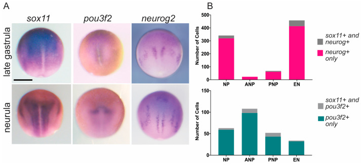
Whole mount in situ hybridization (WISH) results for sox11, pou3f2, neurog1, and neurog2 have been previously published and are available from the online Xenopus database Xenbase [41]. To illustrate the overlapping expression of pou3f2, neurog2, and sox11 in the neural plate and developing neural tube, we show expression in late gastrula and neurula embryos [41] (Figure 1A). Sox11 is expressed throughout the neural plate, whereas the proneural protein genes neurog1 and neurog2 are both expressed in three stripes of cells fated to become the motor, inter, and sensory neurons from medial to lateral. Notably, pou3f2 expression is undetectable at stage 12 by WISH due to low transcript levels, but it is visible at stage 14 in the anterior neural plate [42].
Figure 1.
Sox11, neurog2, and pou3f2 are co-expressed in distinct cell types of the neural plate. (A) WISH of sox11, pou3f2, and neurog2 at stage 12.5 (late gastrula) and stage 14 (early neurula). Embryos are dorsal view with anterior to the top. Sox11 is expressed throughout the neural plate and later in placodes. Pou3f2 expression is not detectable until the neurula stage and is strongest in the anterior neural plate. Neurog2 is expressed in the stripes of the neuronal progenitors. The scale bar is 500 microns. (B) Total number of cells across developmental stages that are sox11− and neurog+ (dark gray), sox11+ and neurog+ (magenta) (top) or sox11− and pou3f2+ (light gray), and sox11+ and pou3f2+ (teal) (bottom) within the neural plate (NP) at stage 12 and within the anterior neural plate (ANP), posterior neural plate (PNP), and early neurons (EN) at stage 13/14.
An interesting observation from single-cell transcriptome measurements in the open access database Jamboree [43] is that the majority of neurog+ and pou3f2+ cells also express sox11 in neurula stage 13/14 embryos (Figure 1B). However, due to a low sequencing depth, we could not distinguish between neurog1, neurog2, or neurog3, as neurog+ cells are grouped together. Most striking is in early neurons (EN), where 90% of neurog+ cells (413/458 cells) are sox11+, and in the neural plate, 88% of pou3f2+ cells are sox11+ (141/160 cells; Figure 1B). However, very few cells (<25) are positive for all three markers: sox11, neurog, and pou3f2. These findings suggest that sox11 is co-expressed in distinct cell types with neurog and pou3f2. Whereas the majority of early neurons co-express sox11 and neurog (413/585), the majority of the pou3f2+/sox11+ cells are located in the anterior neural plate, where there are very few sox11+/neurog+ cells. These single-cell data suggest that there are two populations of sox11-expressing cells: one that co-expresses neurog and another that is pou3f2+.
3.2. Sox11 Partners with Neurog2 and Pou3f2 but Not Neurog1
To examine the interactions of Sox11 with potential partner proteins, we conducted co-immunoprecipitation experiments using in vitro translated, epitope-tagged proteins: Sox11-FLAG, Pou3f2-HA, Neurog1-HA, and Neurog2-MYC. Our results indicate that Sox11-FLAG interacts with both Pou3f2-HA and Neurog2-MYC (Figure 2A,B) but not with Neurog1-HA (Figure 2C).
Figure 2.
Xl Sox11 interacts with Pou3f2 and Neurog2, but not Neurog1. (A–C). Immunoprecipitation (IP) of Xl Sox11-FLAG and Pou3f2-HA (A), Neurog2-MYC (B), or Neurog1-HA (C) from in vitro translated proteins. Proteins were immunoprecipitated using either FLAG (red), HA (green), or MYC (blue) antibodies. Samples were analyzed by western blotting (WB) indicated on the right with FLAG-HRP, MYC-HRP, or HA-HRP. Input represents the total protein lysate.
In summary, Xl Sox11 interacts with Neurog2 and Pou3f2 but not with Neurog1. This differs from mouse SOX11, which interacts with all three proteins. Even though Xl Sox11 and Neurog1 did not interact in our in vitro system, they may require post-translational modification (PTM) or a co-factor to interact. There is precedent for PTMs of mouse Sox11 in developing mouse retina and hippocampus [44,45], although to our knowledge, no studies have investigated PTMs of Sox11 or Neurog1 during neurogenesis. Furthermore, Neurog1 complexes with CBP/p300, which facilitates an interaction with Smad1 to inhibit glial cell differentiation and promote neurogenesis [46]. These modifications and interactions are absent in the in vitro system. As antibodies for Xenopus proteins become available, Sox11 protein interactions can be tested at different stages in the developing frog embryo.
These data support the idea that Sox11 has a different array of partner proteins in the mouse and frog during neurogenesis, especially considering that overexpression of mouse SOX11 has no effect on frog embryos and therefore does not recapitulate the overexpression phenotype of Xl Sox11 [33]. Since the DNA-binding domains of Sox proteins are highly homologous across species (100% amino acid identity of Sox11 DNA-binding domains in mammals, chicks, and zebrafish), it was proposed that the differential function of mouse and frog Sox11 was due to a single amino acid change from K to N in the frog DNA-binding domain at position 91 of the protein and position 43 of the HMG DNA-binding domain [33]. Based on currently available sequences, we have found that this amino acid difference is unique to anura amphibians (frogs). However, it is also worth noting that mouse and frog Neurogenins share only 36.3% similarity with eight amino acids divergent in the 57aa DNA-binding domain. Domain swap experiments will be used to determine if the amino acid changes in the DNA-binding domains lead to differences in protein interaction and function.
3.3. Sox11 N-Terminus and HMG Domain Are Necessary for Protein–Protein Interactions
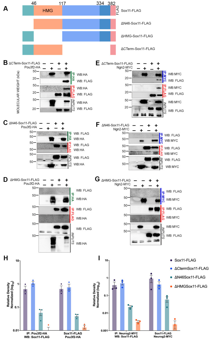
To investigate which domains of Sox11 are necessary for partner-protein binding, we generated three deletion constructs that express modified Sox11-FLAG protein: ΔN46-Sox11-FLAG (lacking the first 46 amino acids upstream of the HMG domain), ΔHMG-Sox11-FLAG (lacking the 72 amino acid HMG domain), and ΔCterm-Sox11-FLAG (containing only the N-terminus and HMG of Sox11 after removal of 265 C-terminal amino acids) (Figure 3A). We performed co-IP analysis of these modified Sox11 proteins with Neurog2 and Pou3f2, using FLAG, HA, or MYC antibodies, and examined them through Western blotting.
Figure 3.
Sox11 N-terminus is essential for protein–protein interactions. (A) Schematic of Sox11 and deletion constructs with referenced domains marked. ΔN46-Sox11-FLAG lacks the 46 amino acids (teal) upstream of the HMG domain. ΔHMG-Sox11-FLAG lacks the 72 amino acid HMG domain (orange), and ΔCterm-Sox11-FLAG lacks 265 amino acids (blue) and consists of the N-terminus and HMG domain. (C–G) Immunoprecipitation (IP) of ΔCterm-Sox11-FLAG, ΔN46-Sox11-FLAG, or ΔHMG-Sox11-FLAG with Pou3f2-HA or Neurog2-MYC (Ngn2-MYC). Proteins were generated via in vitro translation and immunoprecipitated using either FLAG (red), HA (green), or MYC (blue) antibodies. Samples were analyzed by WB with anti-FLAG-HRP, anti-HA-HRP, or anti-MYC-HRP. Inputs demonstrate each protein in the extract. (H) Graphical representation of protein co-immunoprecipitated (Sox11 variants and Pou3f2) from three replicates, one of which is represented in (B–D). (I) Graphical representation of protein co-immunoprecipitated (Sox11 variants and Neurog2) from three replicates, one of which is represented in (E–G). Co-expression bands were normalized to the input in each sample. Data represent the mean ± sem on a log10 scale with percent of pulldown across 3 experimental replicates.
Interestingly, the absence of the Sox11 C-terminus does not impact binding with Pou3f2-HA or Neurog2-MYC (Figure 3B,E). This suggests that the C-terminus is not necessary for the interaction of Sox11 with these two proteins. In contrast, the removal of the N46 domain decreases the interaction with both partner proteins (Figure 3C,F), and loss of the HMG domain results in no detectable interaction with these proteins (Figure 3D,G). We further normalized co-expression bands to the input of three replicates and quantified the relative band density [39,40]. The relative level of proteins of three replicates are graphically represented in Figure 3H,I. The graph reveals a reduction in the amount of protein recovered with immunoprecipitation of Neurgo2-MYC or Pou3f2-HA with ΔNterm-Sox11-FLAG or ΔHMG-Sox11-FLAG (Figure 3H,I, green and orange bars, respectively). These findings indicate that the 46 amino acid N-terminus of Sox11 is required for strong partner-protein interactions with Pou3f2 and Neurog2, while the HMG domain is essential for partner-protein binding.
The HMG domain’s significance for protein interaction is not unique to Sox11 but extends to other Sox proteins. For instance, SoxE proteins (Sox8, Sox9, and Sox10) require the C-terminal tail of the HMG domain to complex with partners [23,47]. However, in other cases, domains outside of the HMG are required for partner-protein interactions. For example, the B-homology domain and C-terminus of Sox2 are required for partner interactions with stem cells [27], and Sox18 binds to MEF2C in endothelial cells through its C-terminal domain [48]. Thus, similar to other Sox proteins, Sox11 utilizes domains beyond the HMG to complex with partners, likely contributing to its unique binding specificity [15].
3.4. Sox11 C-Terminus Is Required for Neuron Formation
Our prior research demonstrated that Sox11 plays a critical role in primary neurogenesis, with overexpression increasing both neural progenitors and neurons and MO knockdown decreasing neurons [33]. Together, these data establish Sox11 as a critical protein during early neurogenesis.
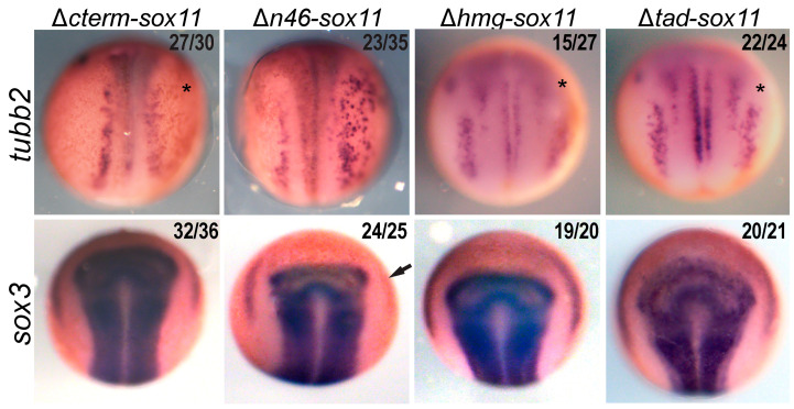
To determine the functional significance of the Sox11 protein domains, we analyzed the effect of the Sox11 deletion proteins on neurogenesis (Figure 4). For this analysis, we injected each mRNA into one cell of a two-cell blastomere embryo and examined changes in neurula embryos using WISH for tubb2b, a marker of neurons, and for sox3, a marker of neural progenitors. If a domain is necessary for Sox11 function, misexpression will not alter tubb2b expression. Conversely, the loss of a domain that is not essential for Sox11 function would drive an increase in tubb2 and/or sox3 consistent with our previous gain-of-function findings [33].
Figure 4.
Identification of Sox11 domains required for placode and neuron formation. WISH of neurula (stage 14/15) embryos injected in one of two cells (dorsal view, anterior to the top) with Dextran as a tracer and either Δcterm-Sox11, Δn46-Sox11, Δhmg-Sox11, or Δtad-Sox11 mRNA. The right side is the injected side, and the left side serves as control expression. Embryos were analyzed for expression of tubb2 for neurons or sox3 for neural progenitors. An arrow marks the reduction in sox3 expression in placodal progenitors, and an asterisk marks the loss of expression in the trigeminal placode. Numbers in the upper right of each image denote the number of embryos with the phenotype over the total analyzed. A one-sample proportion test reveals that the null hypothesis that the overexpression of the mRNA has no effect on tubb2 expression can be rejected in all cases except for the loss of the trigeminal placode for Δhmg-Sox11 p-value = 0.564.
Our data reveal that overexpression of Δcterm-Sox11 does not impact sox3 expression but slightly reduces tubb3 expression. This suggests that ΔCterm-Sox11 functions as a dominant negative protein and, like the morpholino knockdown, reduces neuron formation [33]. On the other hand, overexpression of Δn46-Sox11 leads to ectopic expression of tubb2b in the neural plate resembling the effect of full-length Sox11, albeit to a lesser extent. In addition, sox3 expression is lost in the placodes (Figure 4, asterisk). These results indicate that the 46 N-terminal amino acids are not only required for full activity in the neural plate but also crucial for placode development. It is possible that ΔN46-Sox11 functions as a dominant negative in the placode, which necessitates these 46 aa for proper placode development. One hypothesis is that ΔN46-Sox11 fails to effectively bind a partner protein essential for placode formation and instead interferes with the function of endogenous Sox11.
Next, we explored the function of two internal domains—the HMG DNA-binding domain and the serine-rich transactivation domain (TAD) in the C-terminus. Numerous studies have shown that the HMG domain of many Sox proteins is essential to both DNA- and partner-protein binding [49,50]. Therefore, as expected, overexpression of Δhmg-Sox11 did not mimic full-length Sox11 and increase expression of either tubb2b or sox3 but did lead to a decrease in expression of tubb2b in the trigeminal ganglion in 15/27 embryos (asterisk). Additionally, we explored the role of the 48 amino acid TAD [13,51,52]. We found that in the absence of the TAD, overexpression of Sox11 decreased tubb2b in the trigeminal ganglia in 56% of the embryos (asterisk) but did not affect sox3 expression or tubb2b expression in the neural plate. This aligns with prior studies showing TAD’s essential role in Sox11 function in vitro. Sox11 is known as the most potent transactivator of the SoxC family and has even been shown to be more potent than Sox2 [52].
Our functional analysis of Sox11 domains has uncovered embryo phenotypes that support the involvement of protein domains in protein interactions outside of the HMG DNA-binding domain. Overexpression of ΔCterm-Sox11 replicates the knockdown phenotype, resulting in reduced neuron formation (Figure 4). This effect could be due to the sequestration of partner proteins in the neural plate, hindering endogenous Sox11 from interacting with these proteins. Since ΔCterm-Sox11 lacks the TAD, these complexes are unable to activate target genes.
Additionally, we show that excess ΔN46-Sox11 functions similarly to Sox11 overexpression in embryos, leading to increased neurons, albeit less effectively. This aligns with the weak interaction of ΔN46-Sox11 and Neurog2. We also show that overexpression of ΔN46-Sox11 decreases neural progenitors as marked by sox3 in the placodes (Figure 4, arrow). To further investigate this result, we used the single-cell sequencing database [43]. We identified posterior placodal cells as those expressing pax8, six1, and sox9 and found that 487 out of 618 placodal cells co-expressed sox11, supporting the role for Sox11 in placodal development as previously described [53,54]. We confirmed that neither pou3f2 nor neurog is expressed in the posterior placode. Thus, Sox11 likely interacts with an unknown placodal partner protein to regulate posterior placodal development. These findings underscore the essential role of the C-terminus of Sox11 in neuron formation, emphasizing the need for further research to elucidate Sox11 partner proteins in placodal progenitors.
In total, these functional deletion studies represent the first in vivo characterization of Sox11 domains to determine their contribution to neurogenesis. Together, our data suggest that the HMG domain is necessary for Sox11 function during neuron formation, the N46 domain is necessary for development of neural progenitors in the placodes, and the C-terminus of Sox11, including the TAD, is necessary for neuron formation.
4. Conclusions
Sox transcription factors cooperate with region-specific partner proteins to regulate downstream targets and orchestrate neurogenesis. To identify and characterize Sox11 partner-protein interactions essential to neurogenesis, we tested whether Sox11 partner proteins are conserved between mouse and Xenopus and identified the domains of Sox11 necessary for protein interaction and function. In conclusion, here, we uncover several critical features of Sox11, a protein necessary for neurogenesis. First, Sox11 is co-expressed with Pou3f2 and Neurog2 in the anterior neural plate and early neurons, respectively. Second, Sox11 partner proteins are not conserved across species leading to the enticing possibility that changes in SoxC proteins evolved to enable expansion of the cortex in mammals. Third, the HMG domain and the first 46 amino acids in the N-terminus are necessary for strong partner-protein interactions, whereas the C-terminus plays no role in the binding of Pou3f2 and Neurog2. Lastly, we show that the C-terminus of Sox11, and specifically, the TAD, is required for promoting neuron formation and that the N46 domain of Sox11 is essential in posterior placodal development. To our knowledge, these are the first partner proteins identified for Xenopus Sox11 and the first identification of Sox11 domains essential for protein interaction and neuron formation in the developing neural plate.
Acknowledgments
We thank members of the Silva lab for scientific editing of the manuscript.
Author Contributions
K.S.S. performed co-immunoprecipitations and single-cell seq analysis. P.S.-R. performed the injections and in situ hybridization. Conceptualization, formal analysis, writing—original draft and revision, figure generation, supervision, and funding acquisition were performed by E.M.S. Data analysis, manuscript revision, and figure generation were performed by K.S.S. and D.D.C. All authors have read and agreed to the published version of the manuscript.
Institutional Review Board Statement
The animal study protocol was approved by the Georgetown University Institutional Animal Care and Use Committee (protocol code 2016-1114 and approval 29 October 2021).
Informed Consent Statement
Not applicable.
Data Availability Statement
The data are contained within the article.
Conflicts of Interest
The authors declare no conflicts of interest.
Funding Statement
This work was supported in part by an NIH grant NS078741. K.S.S. is supported by the Center of Neural Injury and Plasticity (5T32NS041218) and an NINDS Pre-doctoral to postdoctoral advancement in neuroscience grant (F99NS108539). P.S.-R. received support from the Fulbright Foundation.
Footnotes
Disclaimer/Publisher’s Note: The statements, opinions and data contained in all publications are solely those of the individual author(s) and contributor(s) and not of MDPI and/or the editor(s). MDPI and/or the editor(s) disclaim responsibility for any injury to people or property resulting from any ideas, methods, instructions or products referred to in the content.
References
- 1.Sarkar A., Hochedlinger K. The Sox family of transcription factors: Versatile regulators of stem and progenitor cell fate. Cell Stem Cell. 2013;12:15–30. doi: 10.1016/j.stem.2012.12.007. [DOI] [PMC free article] [PubMed] [Google Scholar]
- 2.Phochanukul N., Russell S. No backbone but lots of Sox: Invertebrate Sox genes. Int. J. Biochem. Cell Biol. 2010;42:453–464. doi: 10.1016/j.biocel.2009.06.013. [DOI] [PubMed] [Google Scholar]
- 3.Kondoh H., Kamachi Y. SOX-partner code for cell specification: Regulatory target selection and underlying molecular mechanisms. Int. J. Biochem. Cell Biol. 2010;42:391–399. doi: 10.1016/j.biocel.2009.09.003. [DOI] [PubMed] [Google Scholar]
- 4.Kiefer J.C. Back to basics: Sox genes. Dev. Dyn. 2007;236:2356–2366. doi: 10.1002/dvdy.21218. [DOI] [PubMed] [Google Scholar]
- 5.Bergsland M., Ramsköld D., Zaouter C., Klum S., Sandberg R., Muhr J., Ramskold D., Zaouter C., Klum S., Sandberg R., et al. Sequentially acting Sox transcription factors in neural lineage development. Genes Dev. 2011;25:2453–2464. doi: 10.1101/gad.176008.111. [DOI] [PMC free article] [PubMed] [Google Scholar]
- 6.Reiprich S., Wegner M. From CNS stem cells to neurons and glia: Sox for everyone. Cell Tissue Res. 2014;359:111–124. doi: 10.1007/s00441-014-1909-6. [DOI] [PubMed] [Google Scholar]
- 7.Hyodo-Miura J., Urushiyama S., Nagai S., Nishita M., Ueno N., Shibuya H. Involvement of NLK and Sox11 in neural induction in Xenopus development. Genes Cells. 2002;7:487–496. doi: 10.1046/j.1365-2443.2002.00536.x. [DOI] [PubMed] [Google Scholar]
- 8.Whittington N., Cunningham D., Le T.K., De Maria D., Silva E.M. Sox21 regulates the progression of neuronal differentiation in a dose-dependent manner. Dev. Biol. 2015;397:237–247. doi: 10.1016/j.ydbio.2014.11.012. [DOI] [PMC free article] [PubMed] [Google Scholar]
- 9.Kopp J.L., Ormsbee B.D., Desler M., Rizzino A. Small Increases in the Level of Sox2 Trigger the Differentiation of Mouse Embryonic Stem Cells. Stem Cells. 2008;26:903–911. doi: 10.1634/stemcells.2007-0951. [DOI] [PubMed] [Google Scholar]
- 10.Taranova O.V., Magness S.T., Fagan B.M., Wu Y., Surzenko N., Hutton S.R., Pevny L.H. SOX2 is a dose-dependent regulator of retinal neural progenitor competence. Genes Dev. 2006;20:1187–1202. doi: 10.1101/gad.1407906. [DOI] [PMC free article] [PubMed] [Google Scholar]
- 11.Ferri A.L., Cavallaro M., Braida D., Di Cristofano A., Canta A., Vezzani A., Ottolenghi S., Pandolfi P.P., Sala M., DeBiasi S., et al. Sox2 deficiency causes neurodegeneration and impaired neurogenesis in the adult mouse brain. Development. 2004;131:3805–3819. doi: 10.1242/dev.01204. [DOI] [PubMed] [Google Scholar]
- 12.Wegner M. From head to toes: The multiple facets of Sox proteins. Nucleic Acids Res. 1999;27:1409–1420. doi: 10.1093/nar/27.6.1409. [DOI] [PMC free article] [PubMed] [Google Scholar]
- 13.Kuhlbrodt K., Herbarth B., Sock E., Enderich J., Hermans-Borgmeyer I., Wegner M. Cooperative function of POU proteins and SOX proteins in glial cells. J. Biol. Chem. 1998;273:16050–16057. doi: 10.1074/jbc.273.26.16050. [DOI] [PubMed] [Google Scholar]
- 14.Kamachi Y., Uchikawa M., Kondoh H. Pairing SOX off: With partners in the regulation of embryonic development. Trends Genet. 2000;16:182–187. doi: 10.1016/S0168-9525(99)01955-1. [DOI] [PubMed] [Google Scholar]
- 15.Wilson M., Koopman P. Matching SOX: Partner proteins and co-factors of the SOX family of transcriptional regulators. Curr. Opin. Genet. Dev. 2002;12:441–446. doi: 10.1016/S0959-437X(02)00323-4. [DOI] [PubMed] [Google Scholar]
- 16.Chew J.-L., Loh Y.-H., Zhang W., Chen X., Tam W.-L., Yeap L.-S., Li P., Ang Y.-S., Lim B., Robson P., et al. Reciprocal Transcriptional Regulation of Pou5f1 and Sox2 via the Oct4/Sox2 Complex in Embryonic Stem Cells. Mol. Cell. Biol. 2005;25:6031–6046. doi: 10.1128/MCB.25.14.6031-6046.2005. [DOI] [PMC free article] [PubMed] [Google Scholar]
- 17.Mallanna S.K., Ormsbee B.D., Iacovino M., Gilmore J.M., Cox J.L., Kyba M., Washburn M.P., Rizzino A. Proteomic analysis of Sox2-associated proteins during early stages of mouse embryonic stem cell differentiation identifies Sox21 as a novel regulator of stem cell fate. Stem Cells. 2010;28:1715–1727. doi: 10.1002/stem.494. [DOI] [PMC free article] [PubMed] [Google Scholar]
- 18.Wegner M., Stolt C.C. From stem cells to neurons and glia: A Soxist’s view of neural development. Trends Neurosci. 2005;28:583–588. doi: 10.1016/j.tins.2005.08.008. [DOI] [PubMed] [Google Scholar]
- 19.Chew L.J., Gallo V. The Yin and Yang of Sox proteins: Activation and repression in development and disease. J. Neurosci. Res. 2009;87:3277–3287. doi: 10.1002/jnr.22128. [DOI] [PMC free article] [PubMed] [Google Scholar]
- 20.Yuan H., Corbi N., Basilico C., Dailey L. Developmental-specific activity of the FGF-4 enhancer requires the synergistic action of Sox2 and Oct-3. Genes Dev. 1995;9:2635–2645. doi: 10.1101/gad.9.21.2635. [DOI] [PubMed] [Google Scholar]
- 21.Tanaka S., Kamachi Y., Tanouchi A., Hamada H., Jing N., Kondoh H. Interplay of SOX and POU factors in regulation of the Nestin gene in neural primordial cells. Mol. Cell. Biol. 2004;24:8834–8846. doi: 10.1128/MCB.24.20.8834-8846.2004. [DOI] [PMC free article] [PubMed] [Google Scholar]
- 22.Zhao P., Zhu T., Lu X., Zhu J., Li L. Neurogenin 2 enhances the generation of patient-specific induced neuronal cells. Brain Res. 2015;1615:51–60. doi: 10.1016/j.brainres.2015.04.027. [DOI] [PubMed] [Google Scholar]
- 23.Wissmüller S., Kosian T., Wolf M., Finzsch M., Wegner M., Wißmü S., Kosian T., Wolf M., Finzsch M., Wegner M. The high-mobility-group domain of Sox proteins interacts with DNA-binding domains of many transcription factors. Nucleic Acids Res. 2006;34:1735–1744. doi: 10.1093/nar/gkl105. [DOI] [PMC free article] [PubMed] [Google Scholar]
- 24.Yuan X., Lu M.L., Li T., Balk S.P. SRY Interacts with and Negatively Regulates Androgen Receptor Transcriptional Activity. J. Biol. Chem. 2001;276:46647–46654. doi: 10.1074/jbc.M108404200. [DOI] [PubMed] [Google Scholar]
- 25.De Santa Barbara P., Bonneaud N., Boizet B., Desclozeaux M., Moniot B., Sudbeck P., Scherer G., Poulat F., Berta P. Direct Interaction of SRY-Related Protein SOX9 and Steroidogenic Factor 1 Regulates Transcription of the Human Anti-Müllerian Hormone Gene. Mol. Cell. Biol. 1998;18:6653–6665. doi: 10.1128/MCB.18.11.6653. [DOI] [PMC free article] [PubMed] [Google Scholar]
- 26.Botquin V., Hess H., Fuhrmann G., Anastassiadis C., Gross M.K., Vriend G., Schöler H.R. New POU dimer configuration mediates antagonistic control of an osteopontin preimplantation enhancer by Oct-4 and Sox-2. Genes Dev. 1998;12:2073–2090. doi: 10.1101/gad.12.13.2073. [DOI] [PMC free article] [PubMed] [Google Scholar]
- 27.Cox J.L., Mallanna S.K., Luo X., Rizzino A. Sox2 uses multiple domains to associate with proteins present in Sox2-protein complexes. PLoS ONE. 2010;5:e15486. doi: 10.1371/journal.pone.0015486. [DOI] [PMC free article] [PubMed] [Google Scholar]
- 28.Bowles J., Schepers G., Koopman P. Phylogeny of the SOX family of developmental transcription factors based on sequence and structural indicators. Dev. Biol. 2000;227:239–255. doi: 10.1006/dbio.2000.9883. [DOI] [PubMed] [Google Scholar]
- 29.Uy B.R., Simoes-Costa M., Koo D.E.S., Sauka-Spengler T., Bronner M.E. Evolutionarily conserved role for SoxC genes in neural crest specification and neuronal differentiation. Dev. Biol. 2015;397:282–292. doi: 10.1016/j.ydbio.2014.09.022. [DOI] [PMC free article] [PubMed] [Google Scholar]
- 30.Chen C., Lee G.A., Pourmorady A., Sock E., Donoghue M.J. Orchestration of Neuronal Differentiation and Progenitor Pool Expansion in the Developing Cortex by SoxC Genes. J. Neurosci. 2015;35:10629–10642. doi: 10.1523/JNEUROSCI.1663-15.2015. [DOI] [PMC free article] [PubMed] [Google Scholar]
- 31.Bergsland M., Werme M., Malewicz M., Perlmann T., Muhr J. The establishment of neuronal properties is controlled by Sox4 and Sox11. Genes Dev. 2006;20:3475–3486. doi: 10.1101/gad.403406. [DOI] [PMC free article] [PubMed] [Google Scholar]
- 32.Jankowski M.P., Cornuet P.K., McIlwrath S., Koerber H.R., Albers K.M. SRY-box containing gene 11 (Sox11) transcription factor is required for neuron survival and neurite growth. Neuroscience. 2006;143:501–514. doi: 10.1016/j.neuroscience.2006.09.010. [DOI] [PMC free article] [PubMed] [Google Scholar]
- 33.Chen C., Jin J., Lee G.A., Silva E., Donoghue M. Cross-species functional analyses reveal shared and separate roles for Sox11 in frog primary neurogenesis and mouse cortical neuronal differentiation. Biol. Open. 2016;5:409–417. doi: 10.1242/bio.015404. [DOI] [PMC free article] [PubMed] [Google Scholar]
- 34.Carl S.H., Russell S. Common binding by redundant group B Sox proteins is evolutionarily conserved in Drosophila. BMC Genom. 2015;16:292. doi: 10.1186/s12864-015-1495-3. [DOI] [PMC free article] [PubMed] [Google Scholar]
- 35.Sive H.L., Grainger R.M., Harland R.M. Baskets for In Situ Hybridization and Immunohistochemistry. Cold Spring Harb. Protoc. 2007;2007:pdb.prot4777. doi: 10.1101/pdb.prot4777. [DOI] [PubMed] [Google Scholar]
- 36.Sive H.L., Grainger R.M., Harland R.M. Early Development of Xenopus laevis: A Laboratory Manual. Cold Spring Harbor Laboratory Press; Long Island, NY, USA: 2000. [Google Scholar]
- 37.Harland R.M. In situ hybridization: An improved whole-mount method for Xenopus embryos. Methods Cell Biol. 1991;36:685–695. doi: 10.1016/s0091-679x(08)60307-6. [DOI] [PubMed] [Google Scholar]
- 38.Hemmati-Brivanlou A., Frank D., Bolce M.E., Brown B.D., Sive H.L., Harland R.M. Localization of specific mRNAs in Xenopus embryos by whole-mount in situ hybridization. Development. 1990;110:325–330. doi: 10.1242/dev.110.2.325. [DOI] [PubMed] [Google Scholar]
- 39.Gassmann M., Grenacher B., Rohde B., Vogel J. Quantifying Western blots: Pitfalls of densitometry. Electrophoresis. 2009;30:1845–1855. doi: 10.1002/elps.200800720. [DOI] [PubMed] [Google Scholar]
- 40.Tan H.Y., Ng T.W. Accurate step wedge calibration for densitometry of electrophoresis gels. Opt. Commun. 2008;281:3013–3017. doi: 10.1016/j.optcom.2008.01.012. [DOI] [Google Scholar]
- 41.Bowes J.B., Snyder K.A., Segerdell E., Jarabek C.J., Azam K., Zorn A.M., Vize P.D. Xenbase: Gene expression and improved integration. Nucleic Acids Res. 2010;38:D607–D612. doi: 10.1093/nar/gkp953. [DOI] [PMC free article] [PubMed] [Google Scholar]
- 42.Cosse-Etchepare C., Gervi I., Buisson I., Formery L., Schubert M., Riou J.F., Umbhauer M., Le Bouffant R. Pou3f transcription factor expression during embryonic development highlights distinct pou3f3 and pou3f4 localization in the Xenopus laevis kidney. Int. J. Dev. Biol. 2018;62:325–334. doi: 10.1387/ijdb.170260RL. [DOI] [PubMed] [Google Scholar]
- 43.Briggs J.A., Weinreb C., Wagner D.E., Megason S., Peshkin L., Kirschner M.W., Klein A.M. The dynamics of gene expression in vertebrate embryogenesis at single-cell resolution. Science. 2018;360:eaar5780. doi: 10.1126/science.aar5780. [DOI] [PMC free article] [PubMed] [Google Scholar]
- 44.Balta E.A., Wittmann M.T., Jung M., Sock E., Haeberle B.M., Heim B., von Zweydorf F., Heppt J., von Wittgenstein J., Gloeckner C.J., et al. Phosphorylation modulates the subcellular localization of SOX11. Front. Mol. Neurosci. 2018;11:211. doi: 10.3389/fnmol.2018.00211. [DOI] [PMC free article] [PubMed] [Google Scholar]
- 45.Chang K.-C.C., Hertz J., Zhang X., Jin X.-L.L., Shaw P., Derosa B.A., Li J.Y., Venugopalan P., Valenzuela D.A., Patel R.D., et al. Novel Regulatory Mechanisms for the SoxC Transcriptional Network Required for Visual Pathway Development. J. Neurosci. 2017;37:4967–4981. doi: 10.1523/JNEUROSCI.3430-13.2017. [DOI] [PMC free article] [PubMed] [Google Scholar]
- 46.Sun Y., Nadal-Vicens M., Misono S., Lin M.Z., Zubiaga A., Hua X., Fan G., Greenberg M.E. Neurogenin promotes neurogenesis and inhibits glial differentiation by independent mechanisms. Cell. 2001;104:365–376. doi: 10.1016/S0092-8674(01)00224-0. [DOI] [PubMed] [Google Scholar]
- 47.Marshall O.J., Harley V.R. Identification of an interaction between SOX9 and HSP70. FEBS Lett. 2001;496:75–80. doi: 10.1016/S0014-5793(01)02407-3. [DOI] [PubMed] [Google Scholar]
- 48.Hosking B.M., Wang S.C.M., Chen S.L., Penning S., Koopman P., Muscat G.E.O. SOX18 directly interacts with MEF2C in endothelial cells. Biochem. Biophys. Res. Commun. 2001;287:493–500. doi: 10.1006/bbrc.2001.5589. [DOI] [PubMed] [Google Scholar]
- 49.Bernard P., Harley V.R. Acquisition of SOX transcription factor specificity through protein-protein interaction, modulation of Wnt signalling and post-translational modification. Int. J. Biochem. Cell Biol. 2010;42:400–410. doi: 10.1016/j.biocel.2009.10.017. [DOI] [PubMed] [Google Scholar]
- 50.Weiss M.A. Floppy SOX: Mutual induced fit in hmg (high-mobility group) box-DNA recognition. Mol. Endocrinol. 2001;15:353–362. doi: 10.1210/mend.15.3.0617. [DOI] [PubMed] [Google Scholar]
- 51.Dy P., Penzo-Méndez A., Wang H., Pedraza C.E., Macklin W.B., Lefebvre V., Penzo-Mendez A., Wang H., Pedraza C.E., Macklin W.B., et al. The three SoxC proteins–Sox4, Sox11 and Sox12–exhibit overlapping expression patterns and molecular properties. Nucleic Acids Res. 2008;36:3101–3117. doi: 10.1093/nar/gkn162. [DOI] [PMC free article] [PubMed] [Google Scholar]
- 52.Wiebe M.S., Nowling T.K., Rizzino A. Identification of novel domains within Sox-2 and Sox-11 involved in autoinhibition of DNA binding and partnership specificity. J. Biol. Chem. 2003;278:17901–17911. doi: 10.1074/jbc.M212211200. [DOI] [PubMed] [Google Scholar]
- 53.Moody S.A., Lamantia A.-S. Transcriptional regulation of cranial sensory placode development HHS Public Access. Curr. Top. Dev. Biol. 2015;111:301–350. doi: 10.1016/bs.ctdb.2014.11.009. [DOI] [PMC free article] [PubMed] [Google Scholar]
- 54.Saint-Jeannet J.P., Moody S.A. Establishing the pre-placodal region and breaking it into placodes with distinct identities. Dev. Biol. 2014;389:13–27. doi: 10.1016/j.ydbio.2014.02.011. [DOI] [PMC free article] [PubMed] [Google Scholar]
Associated Data
This section collects any data citations, data availability statements, or supplementary materials included in this article.
Data Availability Statement
The data are contained within the article.


