Abstract
Sporotrichosis, the cutaneous mycosis most commonly reported in Latin America, is caused by the Sporothrix clinical clade species, including Sporothrix brasiliensis and Sporothrix schenckii sensu stricto. In Brazil, S. brasiliensis represents a vital health threat to humans and domestic animals due to its zoonotic transmission. Itraconazole, terbinafine, and amphotericin B are the most used antifungals for treating sporotrichosis. However, many strains of S. brasiliensis and S. schenckii have shown resistance to these agents, highlighting the importance of finding new therapeutic options. Here, we demonstrate that milteforan, a commercial veterinary product against dog leishmaniasis whose active principle is miltefosine, is a possible therapeutic alternative for the treatment of sporotrichosis, as observed by its fungicidal activity in vitro against different strains of S. brasiliensis and S. schenckii, and by its antifungal activity when used to treat infected epithelial cells and macrophages. Our results suggest milteforan as a possible alternative to treat feline sporotrichosis.
Keywords: Sporothrix brasiliensis, Sporothrix schenckii, sporotrichosis, milteforan, miltefosine, antifungal agent
Introduction
Sporotrichosis, a chronic cutaneous and subcutaneous infection, is the most commonly reported mycosis in Latin America and Asia, with a high prevalence in tropical and subtropical areas, including Brazil, Mexico, Argentina, India, Japan, and China (1, 2). Since 1998, Brazil has experienced large outbreaks of sporotrichosis that have been expanding throughout the country, mainly in the southeastern regions, the reason for which Brazil is considered a hyperendemic area (3-5).
Until 2007, Sporothrix schenckii was assumed to be the unique etiological agent for sporotrichosis, but recent molecular analyses have revealed the existence of several cryptic species capable of causing infection (6). These species comprise the S. schenckii clinical/pathogenic clade, which includes S. schenckii sensu stricto, S. brasiliensis, Sporothrix globosa, and Sporothrix lurei (7, 8). These species are thermodimorphic fungi, with a mycelial phase that grows in decaying organic matter at 25°C (known as the infectious morphology) and a yeast phase that develops inside the host during infection (known as the parasitic morphology) (9, 10). The virulence profile varies among the species of the pathogenic clade being S. brasiliensis the most virulent, followed by S. schenckii, both with the capacity to cause severe infection even in immunocompetent individuals, while S. globosa and S. lurei are classified as low virulent species (11, 12).
Sporotrichosis can present different clinical manifestations, such as cutaneous (lymphocutaneous and fixed cutaneous), disseminated cutaneous, and extracutaneous (pulmonary, osteoarticular, ocular, meningeal, and visceral) (13). The development of one or other clinical forms depends on different factors, which include the host immune competence, site and depth of inoculation, amount of inoculum, and the etiological agent, all of which should be considered for proper patient management (14).
The transmission of the Sporothrix species is through traumatic implantation with contaminated material, the sapronosis, and the classical route. However, in hyperendemic zones, such as Brazil, zoonotic infection by S. brasiliensis is highly reported, transmitted mainly by cats through scratching, biting, and even through contact with fluids from infected animals. This zoonotic transmission is considered a severe health problem in Brazil, especially in the area of Rio de Janeiro, due to the rapid spread of S. brasiliensis, which is associated with severe clinical manifestations in both humans and cats (15-18). Besides cats, dogs, albeit to a lesser extent, have also been affected by sporotrichosis, making this infection a significant veterinarian problem. Five thousand hundred-thirteen cases of feline sporotrichosis (from 1988 to 2017) and 244 canine cases (from 1988 to 2014) have been reported by the Evandro Chagas National Institute of Infectious Diseases in Rio de Janeiro, Brazil. However, this number is likely underestimated because sporotrichosis incidence is a mandatory notification only in a few states of Brazil (18).
Identification of the sporotrichosis causative agent is essential for treatment since the Sporothrix species show different antifungal susceptibility profiles (19-21), but this is not always possible given that the identification of the species requires molecular tools (8). In general, for the treatment of the cutaneous forms, itraconazole (ITZ) is considered the gold standard for the cutaneous clinical forms, while amphotericin B (AMB) is the first-line antifungal therapy used for disseminated forms (22, 23). However, in the last few years, many S. brasiliensis clinical strains have been reported to show resistance to both azoles and AMB (24-26), which complicates sporotrichosis treatment.
Miltefosine (MFS), also known as hexadecyl phosphocholine, is a synthetic glycerol-free phospholipid analog initially used as an antineoplastic drug (27, 28). Nowadays, MFS is the only available oral drug used in the treatment of visceral and cutaneous leishmaniasis in dogs and humans due to its significant antiparasitic activity, in vitro and in vivo, against Leishmania species (29-32). MFS's action mechanism(s) has yet to be entirely understood. However, it has been demonstrated to act as a multi-target drug associated with the disruption of many vital pathways, such as (i) the inhibition of the biosynthesis of phosphatidylcholine, which causes low levels of this phospholipid (33, 34); (ii) the interference of the cell membrane calcium channels, which induces an increase of intracellular Ca2+ (35, 36); (iii) the inhibition of the sphingomyelin biosynthesis, which increases ceramide concentration (37), resulting in cell apoptosis; and (iv) the immune response, in which its immunomodulatory effects induce the activation of the Th1 response, mainly through the increased production of IFNγ and IL-12, which prevails over the Th2 response driven by Leishmania sp (38).
MFS has also been reported as an antifungal agent in vitro against some of the most clinically significant pathogenic and opportunistic fungi, such as Candida spp., Aspergillus spp., Fusarium spp., and Cryptococcus spp. (39-44). In addition, it was recently shown that MFS has in vitro fungicidal activity against Sporothrix spp., inhibiting the growth of the mycelial phase of S. brasiliensis, S. schenckii, and Sporothrix globosa (45), and the yeast phase of S. brasiliensis strains resistant to (ITZ) and AMB (46). It was also demonstrated that alone or in combination with potassium iodide, MFS inhibits the biofilm formation of S. brasiliensis, S. schenckii, and S. globosa (47, 48). All of this evidence suggests the potential of MFS for treating sporotrichosis. Repurposing orphan drugs, which are the application of existing drugs for different therapeutic purposes than the ones initially marketed for, is a good alternative for treating infections caused by susceptible or resistant microorganisms (49). Such is the case of MFS, which, besides being repurposed for treating leishmaniasis, has been recently designated for treating primary amebic meningoencephalitis and invasive candidiasis (50).
Here, we demonstrate that MFS has fungicidal in vitro activity against both morphologies (hyphae and yeast) of different S. brasiliensis and S. schenckii strains. We also showed that milteforan (ML), a commercial veterinary product against dog leshmaniasis whose active principle is miltefosine (Virbac), can inhibit and kill Sporothrix spp in vitro. ML treatment also increases the killing of S. brasiliensis yeast by the epithelial cells A549 and bone marrow-derived macrophages (BMDMs). Our results suggest ML as a possible veterinary alternative to treat feline sporotrichosis.
Results
ML and MFS have fungicidal activity against Sporothrix spp. in vitro
Several drugs' in vitro antifungal activity against six strains of S. schenckii and S. brasiliensis, three from each species, were assessed according to their MIC and MFC values for the mycelial and yeast phases (Table 1). From these drugs, ITZ has already been reported to show fungistatic activity against Sporothrix spp., while terbinafine (TRB), AMB, and MFS are fungicidal drugs (19, 23, 24). On the other hand, voriconazole (VCZ) was reported to show low activity in inhibiting Sporothrix growth, while caspofungin (CSP) does not exhibit antifungal activity in vitro (20). We also included brilacidin (BRI), a host defense peptide mimetic that synergizes CSP against several human pathogenic fungi (51), to assess its antifungal activity against Sporothrix species.
Table 1.
MIC and MFC values of several antifungals against S. schenckii and S. brasiliensis yeast and mycelial phases.
| CSP (4-0.06 μg/mL) |
VCZ (4-0.06 μg/mL) |
BRI (80-1.25 μM) |
ITZ (8-0.125 μg/mL) |
MFS (16-0.25 μg/mL) |
ML (16-0.25 μg/mL) |
TRB (4-0.06 μg/mL) |
AMB (8-0.125 μg/mL) |
|||
|---|---|---|---|---|---|---|---|---|---|---|
| Ss4820 | Y | MIC | >4 | >4 | 5 | 0.25 | 2 | 2 | 1 | 2 |
| MFC | >4 | >4 | 5 | 0.5 | 2 | 2 | 1 | 2 | ||
| M | MIC | >4 | >4 | >80 | 2 | 2 | 2 | 1 | ND | |
| MFC | >4 | >4 | >80 | >8 | 2 | 2 | 1 | ND | ||
| Ss4821 | Y | MIC | >4 | >4 | 5 | 0.5 | 2 | 2 | 1 | 2 |
| MFC | >4 | >4 | 5 | 2 | 2 | 2 | 1 | 2 | ||
| M | MIC | >4 | >4 | >80 | 1 | 2 | 2 | 1 | ND | |
| MFC | >4 | >4 | >80 | >8 | 2 | 2 | 1 | ND | ||
| Ss4822 | Y | MIC | >4 | >4 | 2.5 | 0.125 | 2 | 2 | 0.5 | 2 |
| MFC | >4 | >4 | 2.5 | 0.25 | 2 | 2 | 0.5 | 2 | ||
| M | MIC | >4 | >4 | >80 | 2 | 2 | 2 | 1 | ND | |
| MFC | >4 | >4 | >80 | >8 | 2 | 2 | 1 | ND | ||
| Sb4823 | Y | MIC | >4 | >4 | 2.5 | 2 | 2 | 2 | 0.5 | 2 |
| MFC | >4 | >4 | 2.5 | >8 | 2 | 2 | 0.5 | 2 | ||
| M | MIC | >4 | >4 | >80 | 1 | 2 | 2 | 1 | ND | |
| MFC | >4 | >4 | >80 | >8 | 2 | 2 | 1 | ND | ||
| Sb4824 | Y | MIC | >4 | >4 | 2.5 | 0.5 | 2 | 2 | 0.5 | >8 |
| MFC | >4 | >4 | 2.5 | 1 | 2 | 2 | 0.5 | >8 | ||
| M | MIC | >4 | >4 | >80 | 2 | 2 | 2 | 1 | ND | |
| MFC | >4 | >4 | >80 | >8 | 2 | 2 | 1 | ND | ||
| Sb4858 | Y | MIC | >4 | >4 | 2.5 | 0.125 | 2 | 2 | 0.125 | 2 |
| MFC | >4 | >4 | 2.5 | 2 | 2 | 2 | 0.125 | 2 | ||
| M | MIC | >4 | >4 | >80 | 1 | 2 | 2 | 1 | ND | |
| MFC | >4 | >4 | >80 | >8 | 2 | 2 | 1 | ND | ||
Ss: S. schenckii, Sb: S. brasiliensis, Y: yeast phase, M: mycelial phase; CSP: caspofungin, VCZ: voriconazole, BRI: brilacidin, ITZ: itraconazole, MFS: miltefosine, ML: milteforan, TRB: terbinafine, AMB: amphotericin B; ND: not determined.
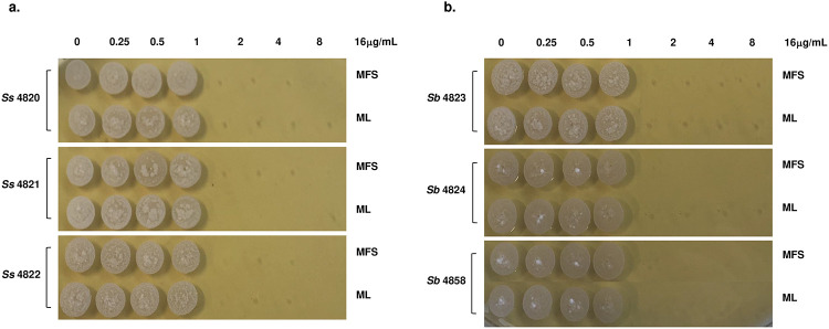
Similar to previous reports, we found that none of the Sporothrix strains, in either yeast or mycelium states, were inhibited by CSP or VCZ. At the same time, both morphologies from all the isolates were sensitive to low concentrations of TRB and AMB (MIC ≤ 2 μg/mL). For ITZ, all strains' conidia were highly resistant (MFC > 8 μg/mL). At the same time, the yeast phase was more sensitive with MIC and MFC values ≤ 2 μg/mL, except the S. brasiliensis clinical isolate 4823 yeast phase, which shows resistance to the drug (MFC > 8 μg/mL), as already reported (52). In the case of BRI, the yeast morphology from all of the Sporothrix strains was susceptible to low concentrations (MIC ≤ 5 μg/mL) of the drug, while conidia are highly resistant. TRB, AMB, and BRI present fungicidal activity against Sporothrix species, while ITZ is a fungistatic drug (Table 1). MFS and ML also have fungicidal activity in vitro against both morphologies from the S. schenckii and S. brasiliensis strains, with MIC and MFC values ≤ 2 μg/mL (Table 1 and Figure 1).
Figure 1. In vitro fungicidal activity of miltefosine and milteforan against the yeast morphology of S. schenckii and S. brasiliensis.
a) S. schenckii (strains 4820, 4821, and 4822) yeast were grown in liquid YDP pH 7.8 at 37°C in the presence of several concentrations of MFS or ML (16, 8, 4, 2, 1, 0.5, and 0.25μg/mL). After 4 days of incubation, the cells were plated in solid YPD pH 7.8 and incubated for 4 days at 37°C. b) S. brasiliensis (strains 4823, 4824, and 4858) yeast were grown in liquid YDP pH 7.8 at 37°C in the presence of several concentrations of MFS or ML (16, 8, 4, 2, 1, 0.5, and 0.25μg/mL). After 4 days of incubation, the cells were plated in solid YPD pH 7.8 and incubated for 4 days at 37°C. As control, yeast cells of each strain were grown without the drugs. Results represent the average of three independent experiments performed by duplicate.
Once we showed the antifungal activity of MFS and ML against Sporothrix spp., we evaluated their ability to interact with some of the drugs already being used for treating sporotrichosis. MIC and MFC values of CSP, VCZ, ITZ, TRB, BRI, and AMB in combination with half MIC of MFS or ML were determined for the yeast morphology of each Sporothrix strain (Table 2). No differences in the activity of CSP and VCZ were observed since neither of these drugs could inhibit S. schenckii or S. brasiliensis growth in the presence of MFS or ML. Combining BRI and MFS or ML does not increase BRI fungicidal activity, as the MIC and MFC values are the same as those of BRI alone. On the other hand, the interaction of MFS or ML with either ITZ, TRB, or AMB increases the antifungal activity against all of the Sporothrix strains tested, decreasing their MIC and MFC values.
Table 2.
MIC and MFC values of MFS and ML combination with several antifungals against S. schenckii and S. brasiliensis yeast phase.
| CSP (16-0.25μg/mL) |
VCZ (16-0.25μg/mL) |
ITZ (8-0.125μg/mL) |
TRB (4-0.06μg/mL) |
BRI (20-0.03μM) |
AMB (8μg-0.125μg/mL) |
|||
|---|---|---|---|---|---|---|---|---|
| Ss4820 | Y | MIC | >16 | >16 | 0.25 | 1 | 5 | 2 |
| MFC | >16 | >16 | 0.5 | 1 | 5 | 2 | ||
| ML | MIC | >16 | >16 | <0.125 | <0.06 | 5 | 1 | |
| MFC | >16 | >16 | 0.5 | <0.06 | 5 | 1 | ||
| MFS | MIC | >16 | >16 | <0.125 | <0.06 | 5 | 1 | |
| MFC | >16 | >16 | 0.5 | <0.06 | 5 | 1 | ||
| Ss4821 | Y | MIC | >16 | >16 | 0.5 | 0.5 | 5 | 2 |
| MFC | >16 | >16 | 2 | 0.5 | 5 | 2 | ||
| ML | MIC | >16 | >16 | <0.125 | 0.25 | 5 | 0.5 | |
| MFC | >16 | >16 | 0.5 | 0.25 | 5 | 0.5 | ||
| MFS | MIC | >16 | >16 | <0.125 | 0.25 | 5 | 0.5 | |
| MFC | >16 | >16 | 0.5 | 0.25 | 5 | 0.5 | ||
| Ss4822 | Y | MIC | >16 | >16 | 0.125 | 0.5 | 5 | 2 |
| MFC | >16 | >16 | 0.25 | 0.5 | 5 | 2 | ||
| ML | MIC | >16 | >16 | ND | ND | 5 | 1 | |
| MFC | >16 | >16 | ND | ND | 5 | 1 | ||
| MFS | MIC | >16 | >16 | ND | ND | 5 | 1 | |
| MFC | >16 | >16 | ND | ND | 5 | 1 | ||
| Sb4823 | Y | MIC | >16 | >16 | 2 | 0.5 | 2.5 | 2 |
| MFC | >16 | >16 | >8 | 0.5 | 2.5 | 2 | ||
| ML | MIC | >16 | >16 | 0.5 | 0.125 | 2.5 | 0.5 | |
| MFC | >16 | >16 | >8 | 0.125 | 2.5 | 0.5 | ||
| MFS | MIC | >16 | >16 | 0.5 | 0.125 | 2.5 | 0.5 | |
| MFC | >16 | >16 | >8 | 0.125 | 2.5 | 0.5 | ||
| Sb4824 | Y | MIC | >16 | >16 | 0.5 | 0.5 | 2.5 | >8 |
| MFC | >16 | >16 | 1 | 0.5 | 2.5 | >8 | ||
| ML | MIC | >16 | >16 | 0.25 | 0.25 | 2.5 | 8 | |
| MFC | >16 | >16 | 0.25 | 0.25 | 2.5 | 8 | ||
| MFS | MIC | >16 | >16 | 0.25 | 0.25 | 2.5 | 8 | |
| MFC | >16 | >16 | 0.25 | 0.25 | 2.5 | 8 | ||
| Sb4858 | Y | MIC | >16 | >16 | 0.125 | 0.125 | 5 | 2 |
| MFC | >16 | >16 | 2 | 0.125 | 5 | 2 | ||
| ML | MIC | >16 | >16 | ND | ND | 5 | 0.25 | |
| MFC | >16 | >16 | ND | ND | 5 | 0.25 | ||
| MFS | MIC | >16 | >16 | ND | ND | 5 | 0.25 | |
| MFC | >16 | >16 | ND | ND | 5 | 0.25 | ||
Ss: S. schenckii, Sb: S. brasiliensis; Y: untreated yeasts, ML: yeast treated with milteforan (1μg/mL), MFS: yeast treated with miltefosine (1μg/mL); CSP: caspofungin, VCZ: voriconazole, ITZ: itraconazole, TRB: terbinafine, BRI: brilacidin, AMB: amphotericin B; ND: Not determined.
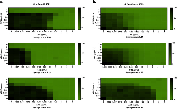
Next, in order to determine what kind of interaction MFS has with ITZ, TRB, and AMB, the drug combination responses were analyzed using checkerboard assays and the SynergyFinder software (53), which evaluates the potential synergy of 2 or more drugs. The dose-response data obtained for combining MFS with either TRB, ITZ, or AMB against S. brasiliensis and S. schenckii yeast cells shows a likely additive interaction (synergy score from −10 to 10) (Figure 2). As previously reported for ITZ (46), we found that MFS does not synergize with the drug against S. brasiliensis and S. schenckii.
Figure 2. MFS has an additive interaction with ITZ, TRB, and AMB against S. brasiliensis and S. schenckii yeast cells.
The synergy score for MFS x TRB, MFS x ITZ, and MFS x AMB against Sporothrix was determined by analyzing the SynergyFinder software's checkerboard data. a) S. schenckii and b) S. brasiliensis yeast were grown in liquid YDP pH 7.8 at 37°C in different concentrations of the selected drugs. After 4 days of incubation, the metabolic activity of the cells was assessed by the XTT reduction assay. Results are expressed as the % of metabolic activity and represent the average of three independent experiments.
MFS localizes to the Sporothrix cell membrane and mitochondria and causes cell death
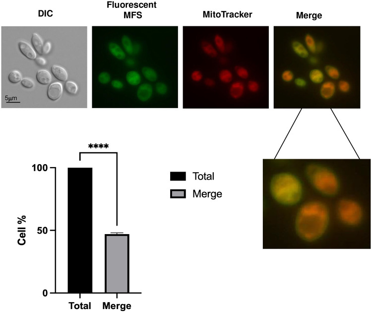
Although the antifungal effect of MFS against Sporothrix has been reported, the localization of the drug in the yeast is still unknown. In Leishmania (54) and A. fumigatus (43), MFS localizes in the cell membrane and the mitochondria, increasing mitochondrial fragmentation and damage. Here, we found that in S. brasiliensis, fluorescent MFS is also localized in the cell membrane and the mitochondria in 47% of the cells investigated (three repetitions of 100 cells each), as shown by MitoTracker colocalization (Figure 3).
Figure 3. MFS is localized in the mitochondria and cell surface of S. brasiliensis yeast.
S. brasiliensis yeast cells were exposed to fluorescent MFS (2μg/mL) for 1 h and then stained with MitoTracker Deep Red FM. The MFS and MitoTracker signals merge on the mitochondria, while the MFS signal is observed on the cell surface. Three independent experiments were performed, and 100 cells were counted for each to calculate a 47.06 ± 1.01 % of MFS and MitoTracker colocalization (merge).
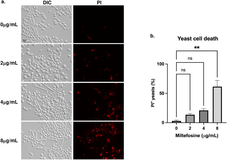
Subsequently, to evaluate the viability of the yeast in the presence of MFS, drug-treated cells were stained with propidium iodide (PI) and analyzed by fluorescence microscopy. Since PI only penetrates cells with damaged membranes, PI+ cells are considered to be going through late apoptosis or early necrosis (55). Treatment of S. brasiliensis yeasts with 2, 4, and 8μg/mL of MFS showss dose-dependent damage of the cells since the PI signal increased with the drug concentration (Figure 4), as early as 6 hours of exposure, confirming the MFS fungicidal activity against Sporothrix.
Figure 4. MFS causes dose-dependent death in S. brasiliensis yeast.
a) S. brasiliensis yeast were exposed to 0, 2, 4, and 8μg/mL of MFS for 6 hours, stained with PI, and analyzed by fluorescence microscopy. b) Quantification of PI+ yeast exposed to MFS, in which 100 yeast-like cells were counted for each condition. Results represent the average of two independent experiments. **p-value<0.001 when compared to untreated cells. ns: not significant.
ML decreases S. brasiliensis fungal burden in A549 pulmonary cells and bone marrow-derived macrophages (BMDM)
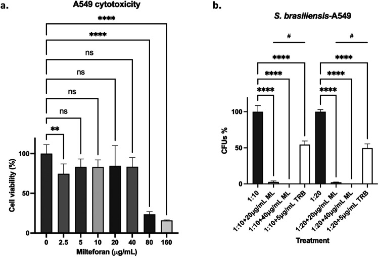
To determine the antifungal activity of ML against S. brasiliensis in the host tissues, two cell lines were used: lung A549 cells and Bone Marrow-Derived Macrophages (BMDMs). As shown in Figure 5a, ML concentrations of 40μg/mL and lower did not reduce A549 cell viability compared to the control. A549 cells were challenged with 1:10 and 1:20 ratios (A549-yeast), and we observed a significant reduction of more than 90 % in the fungal viability in both ML treatments, which contrasts with TRB treatment that shows about 50 % viability (Figure 5b).
Figure 5. Concentrations up to 40μg/mL of ML are not toxic to human cells and can significantly decrease S. brasiliensis survival in A549 epithelial cells.
a) A459 epithelial cells were treated with different ML concentrations, with a decrease of cell viability only at 80μg/mL or higher concentrations. b) A459 cells were challenged with S. brasiliensis yeast at a proportion of 1:10 and 1:20 and then treated with 20 and 40μg/mL of ML. The fungicidal drug TRB was included as a control. **p-value<0.01 when compared to untreated cells. ****p-value<0.0001 when compared to untreated cells. #p-value<0.0001 when compared to cells treated with TRB.
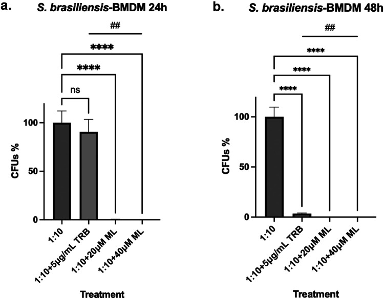
When we challenged BMDMs with S. brasiliensis at a 1:10 ratio (BMDMs-yeast) in the presence of 20 and 40μg/ml ML, we observed complete clearing of S. brasiliensis compared to TRB that showed about 80 and 40 % clearing, respectively, at 24 and 48 h (Figure 6). Our results strongly indicated that ML can help both A549 and BMDMs to clear S. brasiliensis infection.
Figure 6. Killing of S. brasiliensis yeast by BMDM is significantly increased in the presence of ML.
a) BMDM cells were infected with S. brasiliensis yeast and then treated with 20 and 40μg/mL for 24h, which decreased the fungal survival by almost 100% compared to untreated cells. b) BMDM cells were infected with S. brasiliensis yeast and were then treated with 20 and 40μg/mL for 48h, which decreased the fungal survival to 100% when compared to untreated cells. The fungicidal drug TRB was included as a control. *p-value<0.05 when compared to untreated cells. ***p-value<0.0005 when compared to untreated cells. ****p-value<0.0001 when compared to untreated cells. #p-value<0.01 when compared to cells treated with TRB. ##p-value<0.01.
We also assessed the ability of the BMDMs to produce cytokines after stimulation by S. brasiliensis and treatment with the drug. It has already been reported that S. brasiliensis yeast stimulates higher production of TNF-α, IL-6, IL-1β, and IL-10 in human monocyte-derived macrophages when compared to S. schenckii, and it is also more phagocytosed under these conditions (56), which might contribute to the higher virulence of this species.
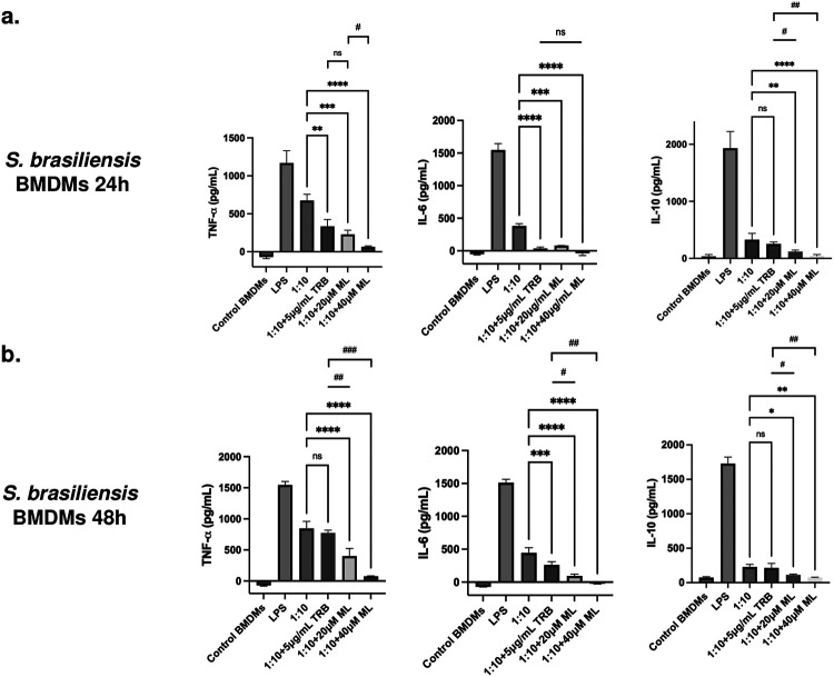
After infection of BMDMs and treatment during 24h, we observed a significant decrease in the stimulation of TNF-α and IL-6 when the yeast cells were treated with TRB and 20 and 40μg/mL of ML, when compared to untreated cells (1:10) (Figure 7a). However, when compared to TRB treatment, a significant decrease was observed in the stimulation of TNF-α only at 40μg/mL of ML. In contrast, no difference was observed in the case of IL-6 with both ML concentrations compared to TRB. Finally, for the secretion of IL-10, a significant decrease was only observed when the yeast cells were treated with both ML concentrations. However, no difference was found with the TRB treatment compared to untreated cells (Figure 7a).
Figure 7. Cytokine secretion by BMDM infected with S. brasiliensis and treated with ML.
a) BMDM cells were infected with S. brasiliensis yeast and treated with ML 20 and 40μg/mL for 24h. The interaction supernatant was collected and the cytokines TNF-α (ns: not significant; **p-value<0.005 when compared to untreated cells; ***p-value<0.0005 when compared to untreated cells; ****p-value<0.0001 when compared to untreated cells; #p-value<0.01 when compared to TRB treatment), IL-6 (ns: not significant; ***p-value<0.0005 when compared to untreated cells; ****p-value<0.0001 when compared to untreated cells), and IL-10 (ns: not significant; **p-value<0.005 when compared to untreated cells; ****p-value<0.0001 when compared to untreated cells; #p-value<0.0005 when compared to TRB treatment; ##p-value<0.0001 when compared to TRB treatment) were measured. b) BMDM cells were infected with S. brasiliensis yeast and treated with 20 and 40μg/mL for 48h. The interaction supernatant was collected and the cytokines TNF-α (ns: not significant; ****p-value<0.0001 when compared to untreated cells; #p-value<0.0005 when compared to TRB treatment; ##p-value<0.0001 when compared to TRB treatment), IL-6 (***p-value<0.001 when compared to untreated cells; (****p-value<0.0001 when compared to untreated cells; #p-value<0.005 when compared to TRB treatment; ##p-value<0.0001 when compared to TRB treatment), and IL-10 (ns: not significant; *p-value<0.05 when compared to untreated cells; **p-value<0.01 when compared to untreated cells; #p-value<0.05 when compared to TRB treatment; ##p-value<0.01 when compared to TRB treatment) were measured.
After 48h of infection, treatment with TRB did not cause a significant decrease in the TNF-α production, while both ML concentrations did when compared to untreated cells and TRB treatment (Figure 7b). In the case of the IL-6 secretion, the same trend as that of 24h was observed, with the only exception that treatment with 20 and 40μg/mL of ML results in a significant decrease compared to TRB (Figure 7b). The secretion of IL-10 did not decrease with the TRB treatment, while significantly decreased in macrophages infected and uninfected treated with ML, confirming the participation of this drug in the immune response modulation (Figure 7b).
Discussion
Although there are several therapeutic options for the treatment of sporotrichosis, fungal resistance and cytotoxicity of the drugs to the host are essential obstacles that hinder the efficient recovery of the patient. ITZ is considered the first-line treatment, an azole known for its fungistatic activity against Sporothrix species (22, 24), which has increased the development of resistance in some isolates, mainly from S. brasiliensis (46, 57, 58). TRB, a drug with fungicidal activity against Sporothrix, has been reported to be effective in treating the cutaneous forms but not for the disseminated infections for which AMB is used. AMB is considered a second-line treatment and is commonly used to treat the invasive and disseminated forms, with the disadvantage that it is very toxic in the doses and time needed to eradicate the infection, in addition to recent reports of isolates resistant to this antifungal agent (22, 46).
In Brazil, cat-transmitted sporotrichosis, caused by S. brasiliensis, is a vital health treat that has been spreading since 1998 (5, 8) across the country, affecting domestic animals and humans, another reason for which is of great importance to find new drugs for the treatment and control of this mycosis. For this objective, drug repurposing is an excellent alternative to finding new treatments since these drugs already approved to be used in humans and animals, initially developed to treat other diseases, can help treat infections caused by different pathogens (59, 60). Such is the case of commercial MFS, which was initially used as an antineoplastic drug (27, 28) that is now the only available oral treatment for leishmaniasis in dogs and humans (29-32), and was recently proven to be effective for the treatment of infections caused by Candida species (39, 40). As previously demonstrated (45, 46, 48), MFS also has in vitro fungicidal activity against Sporothrix species by inhibiting the growth of both fungal morphologies. S. brasiliensis and S. schenckii strains are sensitive to low concentrations of this drug, with an antifungal activity of 2μg/mL for both hyphae and yeast cells. Unlike ITZ, we found no strain resistant to MFS or ML.
We also assessed the ability of MFS to synergize with other drugs used for the treatment of sporotrichosis, including TRB, ITZ, and AMB, and as previously reported for ITZ (45), MFS does not synergize the activity of other antifungals. However, it has instead an additive effect, which suggest they do not interact, or act on independent pathways (61). Similarly to A. fumigatus (43), MFS is directed to the mitochondria of S. brasiliensis yeast, staying also on the cell surface and causing cell death, suggesting that this drug might be affecting the mitochondria and membrane integrity, which might be related to its mechanism of action.
This drug has been reported to be toxic in high doses in mice, with high mortality in concentrations higher than 25mg/kg (62, 63), with maximum concentrations in the kidney and liver, probably due to its amphiphilic nature (64, 65). We assessed ML cytotoxicity in A549 human pulmonary cells and observed a significant viability reduction at 80μg/mL. When we tested the ability of ML to decrease the fungal burden in A549 cells and BMDMs, at 24h and 24 and 48h, respectively, we observed that ML could significantly decrease the CFUs more efficiently than the fungicidal drug TRB in both cell types, with an almost complete clearing of the yeast cells as early as 24 h of treatment.
One of the proposed mechanisms of action for MFS is its immunomodulatory ability, which is essential for the treatment of leishmaniasis since the drug induces the Th1 response and suppresses the Th2, by increasing the production of proinflammatory cytokines such as IFNγ, TNFα, and IL-12 for the clearance of intracellular pathogens, while relapses of leishmaniasis have been related with an increase of the Th2 response and the production of IL-10 (32, 38). We observed that ML decreases the fungal burden and the production of TNFα, IL-6, and IL-10, secreted by the infected BMDMs. We propose three non-excluded hypotheses to explain it: (i) The cytokines reduction might be related to the fact that the drug is killing the yeast cells before being phagocytosed, where there is the death of the yeast cells as early as 6 hours of MFS treatment; (ii) since the drug is localized to the cell surface, MFS could act as an opsonizing agent helping in the macrophage recognition and further phagocytosis: and (iii) MFS could bind to essential virulence factors, such as adhesins, or immunogenic components, such a β-glucans, in a way that is attenuating S. brasiliensis ability to infect and generate an immune response. All three options would reduce the fungal load, tissue damage, and inflammation, making this veterinary drug a suitable treatment alternative for feline sporotrichosis.
Materials and Methods
Fungal strains and culture conditions
In this study, three Sporothrix schenckii (ATCC-MYA 4820, ATCC-MYA 4821, and ATCC-MYA 4822) and three S. brasiliensis strains (ATCC-MYA 4823, ATCC-MYA 4824, and ATCC-MYA 4858) were used for the in vitro antifungal susceptibility assays; S. schenckii ATCC-MYA 4821 and S. brasiliensis ATCC-MYA 4823 were used for the checkerboard assays; and S. brasiliensis ATCC-MYA 4823, a highly virulent clinical isolate obtained from feline sporotrichosis (66), was used for the infection assays.
The mycelial phase from Sporothrix spp. was obtained and maintained on solid YPD pH 4.5 (yeast extract 1% (w/v), gelatin peptone 2% (w/v), and dextrose 3% (w/v)) at 28°C for four days. In contrast, the yeast morphology was grown in liquid YPD pH 7.8, at 37°C under orbital agitation for four days, as previously reported (67). Each phase was confirmed by observing the cells with light microscopy.
Antifungal drugs
For the in vitro assays, voriconazole (VCZ, Sigma-Aldrich), itraconazole (ITZ, Sigma-Aldrich), amphotericin B (AMB, Sigma-Aldrich), terbinafine (TRB, Sigma-Aldrich), and brilacidin (BRI, supplied by Innovation Pharmaceuticals) were diluted in dimethyl sulfoxide (DMOS); while miltefosine (MFS, Sigma-Aldrich), the milteforan active compound, was diluted in ethanol; and caspofungin (CSP, Sigma-Aldrich) was diluted in distilled water. Milteforan (miltefosine 2%) was purchased from Virbac as an oral solution.
In vitro antifungal susceptibility testing
The minimum inhibitory concentrations (MICs) were determined by the broth microdilution method adapted from protocols published by the Clinical Laboratory Standard Institute for the mycelial and yeast phases (24, 68). Briefly, serial twofold dilutions of the antifungal drugs were performed in YPD pH 4.5 and 7.8, for mycelial and yeast, respectively, into 96-well microtiter plates to obtain concentrations of 4-0.06μg/mL for CSP, VCZ and TRB; 8-0.125μg/mL for ITZ and AMB; 16-0.25μg/mL for MFS and ML; and 80-1.25μM for BRI, with a final concentration of 2x10³ and 2x10⁴ conidia or yeast cells, respectively, in a volume of 100μL. The plates were incubated at 28°C (for conidia) or 37°C (for yeast) for four days, and the MIC was determined by visual inspection and defined as the lowest concentration that inhibits 90-100% of fungal growth about untreated cells. Finally, 5μL of conidia or yeast cells from each well were grown in drug-free solid YPD pH 4.5 and pH 7.8 at 28°C and 37°C, respectively, for four days. The minimum fungicidal concentration (MFC) value was the lowest concentration, showing no fungal growth. Three independent experiments were performed by duplicate.
Checkerboard assays and synergy testing
The drug combination effect was determined through the MIC and MFC values of the yeast phase, as described before. Briefly, serial twofold dilutions of the antifungal drugs were performed in liquid YPD pH 7.8 containing half MIC of MFS or ML (1μg/mL) in 96-well microtiter plates to obtain concentrations of 16-0.25μg/mL for CSP and VCZ; 8-0.125μg/mL for ITZ and AMB; 4-0.06μg/mL for TRB; and 80-1.25μM for BRI, with a final concentration of 2x10⁴ yeast, in a volume of 100μL. The plates were incubated at 37°C for four days, and the MIC was determined by visual inspection. It was defined as the lowest concentration inhibiting 90-100% of fungal growth in cells treated only with 1μg/mL of MFS or ML. After MIC determination, 5μL of yeast from each well were grown in drug-free solid YPD pH 7.8 at 37°C for four days. The MFC value was the lowest concentration, which showed no fungal growth.
Checkerboard assays were performed to quantify the interaction (synergistic, additive, or antagonistic) between MFS and ITZ, AMB, or TRB. Briefly, a stock solution of 2x10⁵ yeast/mL and each drug (8μg of MFS and 16μg/mL of ITZ, 16μg of AMB, or 8μg of TRB) were prepared in RMPI-1640. In 96-well microtiter plates, the first antibiotic (MFS) was diluted sequentially along the ordinate. In contrast, the second drug (ITZ, AMB, or TRB) was diluted along the abscissa to obtain a final volume of 100μL. The plates were incubated at 37°C for four days, and the metabolic activity was determined through the XTT reduction assay (47). Briefly, 50μL of a solution of XTT 1mg/mL and menadione 1mM resuspended in water were added to each well, mixed, and incubated in the dark at 37°C for three h. The supernatant of each well was transferred to a new plate and read in a spectrophotometer at 492nm. Results are expressed as means ± SD of three independent experiments.
To determine the type of drug interaction, the SynergyFinder software (53) was used, with the following parameters: detect outliners: yes; curve fitting: LL4; method: Bliss; correction: on. The summary synergy scores represent the average excess response due to drug interaction, in which a value less than −10 suggest an antagonistic interaction between two drugs; values from −10 to 10 suggest an additive interaction; and values larger that 10 suggest a synergistic interaction.
Yeast cells death
The effect of ML on the cell membrane potential was assessed by staining with propidium iodide (PI). Yeast cells grown for 4 days in liquid YPD pH 7.8 were treated with 0, 2, 4 and 8μg/mL of ML during 6 h, stained with PI 20mM for 30 minutes, and washed with PBS 1X three times. Fluorescence was analyzed at an excitation wavelength of 572/25nm and emission of 629/62nm with the Observer Z1 fluorescence microscope using a 100x oil immersion lens objective. Differential interference contrast (DIC) and fluorescent images were capture with an AxioCam camera (Carl Zeiss) and processed using AxioVision software (version 4.8). The experiment was performed twice, and for each treatment at least 100 cells were counted. The results were plotted using Graphpad Prism (GraphPad software, Inc.). A p-value<0.001 was considered significant.
Miltefosine localization
S. brasiliensis yeast cells cultured for 4 days in YPD pH 7.8 were washed 3 times with PBS 1X and then treated with the fluorescent MFS analogue MT-11 C-BDP (excitation wavelength 450-490nm and emission wavelength 500-550nm) for 6 hours, also in liquid YPD pH 7.8. The cells were washed 3 times and stained with 250nM of MitoTracker Deep Red FM (Invitrogen) (wavelength absorbance/emission 644/665nm) and washed again. The yeast cells were visualized in slides with the Observer Z1 fluorescent microscope using a 100x oil immersion lens objective. DIC and fluorescent images were capture with an AxioCam camera (Carl Ziess) and processed using AxioVision software (version 4.8). Two independent experiments were performed, and 100 cells were counted of each to calculate the merge %.
Cytotoxicity assay
The cytotoxicity of ML was determined in A549 human lung cancer cells using the XTT reduction assay. 2x10⁵ cells/well were seeded in 96-well tissue plates and incubated in Dulbecco’s Modified Eagle Medium (DMEM, ThermoFischer). After 24 h of incubation with CO2 5%, the cells were treated with different concentrations of ML (0, 2.5, 5, 10, 20, 40, 80 and 160μg/mL), and after 48 h of incubation, cell viability was assessed using the XTT assay. Briefly, 80μL of a solution of XTT 1mg/mL in DMEM, HEPES 1M, and menadione 8μg/mL were added to each well, and after 30 min, formazan formation was quantified spectrophotometrically at 450nm using a microplate reader. Each treatment was performed by triplicate and the results were plotted using Graphpad Prism (GraphPad Software, Inc.). A p-value<0.0001 was considered significant.
A549 and bone marrow derived macrophages (BMDMs) killing assays
The cell line A549 and BMDMs were cultured using DMEM supplemented with fetal bovine serum (FBS) 10% and penicillin-streptomycin 1% (Sigma-Aldrich), and seeded at a concentration of 1x10⁶ cells/mL in 24-well plates (Corning). The cells were challenged with S. brasiliensis yeasts at a multiplicity of infection of 1:10 and were then treated with ML 20 and 40μM. As control, we included untreated cells and cells treated with TRB 5μg/mL. For the BMDMs, cells treated with LPS were also included as control. The A549 were incubated during 24 h at 37°C with CO2 5%, while the BMDM were incubated for 24 and 48h under the same conditions. After incubation, the culture media was removed, each well was washed 3 times with PBS 1X, and 1mL of sterile cold water was added to recover and collect the cell monolayer. To assess the number of CFUs, 100μL of the cell suspensions were plated on YDP pH 4.5 and incubated at 28°C for 4 days. When necessary, the cell suspensions were diluted at 1:100 or 1:1000 and 100μL were plated. 50μL of the inoculum adjusted to 1x10³ cells/mL was also plated to correct the CFU count. Each treatment was performed by triplicate to calculate the CFU %, and the results were plotted using Graphpad Prism (GraphPad Software, Inc.). A p-value<0.0001 was considered significant.
Cytokines quantification
The Elisa-assay kits (R&D Systems) were used to evaluate the concentration of the proinflammatory cytokines TNFα and IL-6, and the anti-inflammatory cytokine IL-10 in the supernatants of the S. brasiliensis and BMDMs interaction for 24 and 48 h, according to the manufacturers instruction. The plates absorbance was read at 450nm and the cytokine concentration (pg/mL) was calculated according to the values obtained in the standard curve of each cytokine. The results were plotted using Graphpad Prism (GraphPad software, Inc.).
Statistical analyses
The GraphPad Prism 10 (GraphPad Software, Inc.) was used for the statistical analyses. The results are reported as the media ± SD from two or three independent experiments performed by duplicate and were analyzed using the Ordinary one-way ANOVA or the Unpaired T test. The statistical significance was considered with a p-value<0.05 or lower.
Acknowledgements
We thank the Fundação de Amparo à Pesquisa do Estado de São Paulo (FAPESP) grant numbers 2021/04977-5 (GHG) and 2022/08556-7 (LCGC) 2022/08796-8 (CD), 2022/09882-5 (LP), the Conselho Nacional de Desenvolvimento Científico e Tecnológico (CNPq), FAPESP and Fundação Coordenação de Aperfeiçoamento do Pessoal do Ensino Superior (CAPES) grant number 405934/2022-0 (The National Institute of Science and Technology INCT Funvir), and CNPq 301058/2019-9 from Brazil to GHG., both from Brazil, the National Institutes of Health/National Institute of Allergy and Infectious Diseases from the USA, grant R01AI153356 to GHG. This work was also funded by the Joint Canada-Israel Health Research Program, jointly supported by the Azrieli Foundation, Canada’s International Development Research Centre, Canadian Institutes of Health Research, and the Israel Science Foundation (GHG).
References
- 1.Rodrigues AM, Della Terra PP, Gremião ID, Pereira SA, Orofino-Costa R, de Camargo ZP. 2020. The threat of emerging and re-emerging pathogenic Sporothrix species. Mycopathologia 185(5):813–842. 10.1007/s11046-020-00425-0. [DOI] [PubMed] [Google Scholar]
- 2.Chakrabarti A, Bonifaz A, Gutierrez-Galhardo MC, Mochizuki T, Li S. 2014. Global epidemiology of sporotrichosis. Med Mycol 53(1):3–14. 10.1093/mmy/myu062. [DOI] [PubMed] [Google Scholar]
- 3.Barros MBL, Schubach AO, Schubach TMP, Wanke B, Lambert-Passos SR. 2008. An epidemic of sporotrichosis in Rio de Janeiro, Brazil: Epidemiological aspects of a series of cases. Epidemiol Infect 136(9):1192–1196. 10.1017/S0950268807009727. [DOI] [PMC free article] [PubMed] [Google Scholar]
- 4.Falcão EMM, Pires MCDS, Andrade HB, Gonçalves MLC, Almeida-Paes R, Do Valle ACF, Bastos FI, Gutierrez-Galhardo MC, Freitas DFS. 2020. Zoonotic sporotrichosis with greater severity in Rio de Janeiro, Brazil: 118 hospitalizations and 11 deaths in the last 2 decades in a reference institution. Med Mycol 58(1):141–143. 10.1093/mmy/myz024. [DOI] [PubMed] [Google Scholar]
- 5.Gremião IDF, Marques M, Oliveira E, de Miranda LHM, Freitas DFS, Pereira SA. 2020. Geographic expansion of sporotrichosis, Brazil. Emerg Infect Dis 26(3):621–624. 10.3201/eid2603.190803. [DOI] [PMC free article] [PubMed] [Google Scholar]
- 6.Marimon R, Cano J, Gené J, Sutton DA, Kawasaki M, Guarro J. 2007. Sporothrix brasiliensis, S. globosa, and S. mexicana, three new Sporothrix species of clinical interest. J Clin Microbiol 45(10):3198–3206. 10.1128/jcm.00808-07. [DOI] [PMC free article] [PubMed] [Google Scholar]
- 7.de Beer ZW, Duong TA, Wingfield MJ. 2016. The divorce of Sporothrix and Ophiostoma: Solution to a problematic relationship. Stud Mycol 83:165–191. 10.1016/j.simyco.2016.07.001. [DOI] [PMC free article] [PubMed] [Google Scholar]
- 8.Lopes-Bezerra LM, Mora-Montes HM, Zhang Y, Nino-Vega G, Rodrigues AM, de Camargo ZP, de Hoog S. 2018. Sporotrichosis between 1898 and 2017: The evolution of knowledge on a changeable disease and on emerging etiological agents. Med Mycol 56(S1):126–143. 10.1093/mmy/myx103. [DOI] [PubMed] [Google Scholar]
- 9.de Lima Barros MB, de Almeida Paes R, Schubach AO. 2011. Sporothrix schenckii and sporotrichosis. Clin Microbiol Rev 24(4):354–633. 10.1128/cmr.00007-11. [DOI] [PMC free article] [PubMed] [Google Scholar]
- 10.de Carolis E, Posteraro B, Sanguinetti M. 2022. Old and new insights into Sporothrix schenckii complex biology and identification. Pathogens 11(3), 297. 10.3390/pathogens11030297. [DOI] [PMC free article] [PubMed] [Google Scholar]
- 11.Arrillaga-Moncrieff I, Capilla J, Mayayo E, Marimon R, Mariné M, Gené J, Cano J, Guarro J. 2009. Different virulence levels of the species of Sporothrix in a murine model. Clinical Microbiology and Infection 15(7):651–655. 10.1111/j.1469-0691.2009.02824.x. [DOI] [PubMed] [Google Scholar]
- 12.Della Terra PP, Rodrigues AM, Fernandes GF, Nishikaku AS, Burger E, de Camargo ZP. 2017. Exploring virulence and immunogenicity in the emerging pathogen Sporothrix brasiliensis. PLoS Negl Trop Dis 11(8). 10.1371/journal.pntd.0005903. [DOI] [PMC free article] [PubMed] [Google Scholar]
- 13.Gutierrez-Galhardo MC, Freitas DFS, Do Valle ACF. 2015. Clinical forms of human sporotrichosis and host immunocompetence, 73-82. In: Zeppone Carlos I. (eds) Sporotrichosis. Springer, Cham. 10.1007/978-3-319-11912-0_5. [DOI] [Google Scholar]
- 14.García-Carnero LC, Lozoya-Pérez NE, González-Hernández SE, Martínez-Álvarez JA. 2018. Immunity and treatment of sporotrichosis. Journal of Fungi 4 (3),100. 10.3390/jof4030100. [DOI] [PMC free article] [PubMed] [Google Scholar]
- 15.Gremião IDF, Miranda LHM, Reis EG, Rodrigues AM, Pereira SA. 2017. Zoonotic epidemic of sporotrichosis: cat to human transmission. PLoS Pathog 13(1). 10.1371/journal.ppat.1006077. [DOI] [PMC free article] [PubMed] [Google Scholar]
- 16.de Oliveira Bento A, de Sena Costa AS, Lima SL, Do Monte Alves M, de Azevedo Melo AS, Rodrigues AM, da Silva-Rocha WP, Milan EP, Chaves GM. 2021. The spread of cat-transmitted sporotrichosis due to Sporothrix brasiliensis in Brazil towards the Northeast region. PLoS Negl Trop Dis 15 (8). 10.1371/journal.pntd.0009693. [DOI] [PMC free article] [PubMed] [Google Scholar]
- 17.Boechat JS, Oliveira MME, Gremião IDF, Almeida-Paes R, Machado AC de S, Zancopé-Oliveira RM, Oliveira R de VC, Morgado DS, Corrêa ML, Figueiredo ABF, Menezes RC, Pereira SA. 2022. Sporothrix brasiliensis and feline sporotrichosis in the metropolitan region of Rio de Janeiro, Brazil (1998-2018). Journal of Fungi 8(7),749. 10.3390/jof8070749. [DOI] [PMC free article] [PubMed] [Google Scholar]
- 18.Morgado DS, Castro R, Ribeiro-Alves M, Corrêa-Moreira D, Castro-Alves J, Pereira SA, Menezes RC, Oliveira MME. 2022. Global distribution of animal sporotrichosis: a systematic review of Sporothrix sp. identified using molecular tools. Curr Res Microb Sci 19(3). 10.1016/j.crmicr.2022.100140. [DOI] [PMC free article] [PubMed] [Google Scholar]
- 19.Marimon R, Serena C, Gené J, Cano J, Guarro J. 2008. In vitro antifungal susceptibilities of five species of Sporothrix. Antimicrob Agents Chemother 52(2):732–734. 10.1128/aac.01012-07. [DOI] [PMC free article] [PubMed] [Google Scholar]
- 20.Rodrigues AM, de Hoog GS, de Cássia Pires D, Brihante RSN, da Costa Sidrim JJ, Gadelha MF, Colombo AL, De Camargo ZP. 2014. Genetic diversity and antifungal susceptibility profiles in causative agents of sporotrichosis. BMC Infect Dis 14,219. 10.1186/1471-2334-14-219. [DOI] [PMC free article] [PubMed] [Google Scholar]
- 21.Flórez-Muñoz SV, Alzate JF, Mesa-Arango AC. 2019. Molecular identification and antifungal susceptibility of clinical isolates of Sporothrix schenckii complex in Medellin, Colombia. Mycopathologia 184:53–63. 10.1007/s11046-018-0310-5. [DOI] [PubMed] [Google Scholar]
- 22.Kauffman CA, Bustamante B, Chapman SW, Pappas PG. 2007. Clinical practice guidelines for the management of sporotrichosis: 2007 Update by the Infectious Diseases Society of America. Clinical Infectious Diseases 45(10):1255–1265. 10.1086/522765. [DOI] [PubMed] [Google Scholar]
- 23.Orofino-Costa R, Freitas DFS, Bernardes-Engemann AR, Rodrigues AM, Talhari C, Ferraz CE, Veasey JV, Quintella L, de Sousa MSLA, Vettorato R, de Almeida-Paes R, de Macedo PM. 2022. Human sporotrichosis: recommendations from the Brazilian Society of Dermatology for the clinical, diagnostic and therapeutic management. An Bras Dermatol 97(6):757–777. 10.1016/j.abd.2022.07.001. [DOI] [PMC free article] [PubMed] [Google Scholar]
- 24.Borba-Santos LP, Rodrigues AM, Gagini TB, Fernandes GF, Castro R, de Camargo ZP, Nucci M, Lopes-Bezerra LM, Ishida K, Rozental S. 2015. Susceptibility of Sporothrix brasiliensis isolates to amphotericin B, azoles, and terbinafine. Med Mycol 53(2):178–188. 10.1093/mmy/myu056. [DOI] [PubMed] [Google Scholar]
- 25.Waller SB, Dalla Lana DF, Quatrin PM, Ferreira MRA, Fuentefria AM, Mezzari A. 2021. Antifungal resistance on Sporothrix species: an overview. Brazilian Journal of Microbiology 52(1):73–80. 10.1007/s42770-020-00307-z. [DOI] [PMC free article] [PubMed] [Google Scholar]
- 26.Bombassaro A, Spruijtenburg B, Medeiros F, Favoreto de Souza Lima BJ, Ballardin LB, Rodrigues de Farias M, Vicente VA, de Queiroz-Telles F, Meis JF, de Groot T. 2023. Genotyping and antifungal susceptibility testing of Sporothrix brasiliensis isolates from Southern Brazil. Mycoses 66(7):585–593. 10.1111/myc.13584. [DOI] [PubMed] [Google Scholar]
- 27.Unger C, Damenz W, Fleer EAM, Kim DJ, Breiser A, Hilgard P, Engel J, Nagel G, Eibl H. 1989. Hexadecylphosphocholine, a new ether lipid analogue studies on the antineoplastic activity in vitro and in vivo. Acta Oncol (Madr) 28(2):213–217. 10.3109/02841868909111249. [DOI] [PubMed] [Google Scholar]
- 28.SpruB T, Bernhardt G, Schonenberger H, Engel J. 1993. Antitumour activity of miltefosine alone and after combination with platinum complexes on MXT mouse mammary carcinoma models. J Cancer Res Clin Oncol 119(3):142–149. 10.1007/BF01229528. [DOI] [PMC free article] [PubMed] [Google Scholar]
- 29.Croft SL, Snowdon D, Yardley V. 1996. The activities of four anticancer alkyllysophospholipids against Leishmania donovani, Trypanosoma cruzi and Trypanosoma brucei. Journal of Antimicrobial Chemotherapy 38(6):1041–1047. 10.1093/jac/38.6.1041. [DOI] [PubMed] [Google Scholar]
- 30.Sundar S, Rosenkaimer F, Makharia MK, Goyal AK, Mandal AK, Voss A, Hilgard P, Murray HW. 1998. Trial of oral miltefosine for visceral leishmaniasis. The Lancet 352:1821–1823. 10.1016/S0140-6736(98)04367-0. [DOI] [PubMed] [Google Scholar]
- 31.Croft SL, Engel J. 2006. Miltefosine - discovery of the antileishmanial activity of phospholipid derivatives. Trans R Soc Trop Med Hyg 100:S4–S8. 10.1016/j.trstmh.2006.03.009. [DOI] [PubMed] [Google Scholar]
- 32.Dorlo TPC, Balasegaram M, Beijnen JH, de vries PJ. 2012. Miltefosine: a review of its pharmacology and therapeutic efficacy in the treatment of leishmaniasis. Journal of Antimicrobial Chemotherapy 67(11):2576–2597. 10.1093/jac/dks275. [DOI] [PubMed] [Google Scholar]
- 33.Geilen CC, Wieder T, Reutter W. 1991. Hexadecylphosphocholine inhibits translocation of CTP:Choline-phosphate cytidylyltransferase in Madin-Darby canine kidney cells. J Biol Chem 267(10):6719–6724. [PubMed] [Google Scholar]
- 34.Cui Z, Houweling M, Chen MH, Record M, Chap H, Vance DE, Ois Tercé F. 1996. A genetic defect in phosphatidylcholine biosynthesis triggers apoptosis in chinese hamster ovary cells. J Biol Chem 271(25):14668–14671. 10.1074/jbc.271.25.14668. [DOI] [PubMed] [Google Scholar]
- 35.Paris C, Loiseau PM, Bories C, Bréard J. 2004. Miltefosine induces apoptosis-Like death in Leishmania donovani promastigotes. Antimicrob Agents Chemother 48(3):852–859. 10.1128/aac.48.3.852-859.2004. [DOI] [PMC free article] [PubMed] [Google Scholar]
- 36.Pinto-Martinez AK, Rodriguez-Durán J, Serrano-Martin X, Hernandez-Rodriguez V, Benaim G. 2018. Mechanism of action of miltefosine on Leishmania donovani involves the impairment of acidocalcisome function and the activation of the sphingosine-dependent plasma membrane Ca2+ channel. Antimicrob Agents Chemother 62(1). 10.1128/aac.01614-17. [DOI] [PMC free article] [PubMed] [Google Scholar]
- 37.Wieder T, Orfanos CE, Geilen CC. 1998. Induction of ceramide-mediated apoptosis by the anticancer phospholipid analog, hexadecylphosphocholine. J Biol Chem 273(18):11025–11031. 10.1074/jbc.273.18.11025. [DOI] [PubMed] [Google Scholar]
- 38.Palic S, Bhairosing P, Beijnen JH, Dorlo TPC. 2019. Systematic review of host-mediated activity of miltefosine in Leishmaniasis through immunomodulation. Antimicrob Agents Chemother 63(7). 10.1128/aac.02507-18. [DOI] [PMC free article] [PubMed] [Google Scholar]
- 39.Machado Vila TV, Sousa Quintanilha N, Rozental S. 2015. Miltefosine is effective against Candida albicans and Fusarium oxysporum nail biofilms in vitro. J Med Microbiol 64(11):1436–1449. 10.1099/jmm.0.000175. [DOI] [PubMed] [Google Scholar]
- 40.Vila T, Ishida K, Seabra SH, Rozental S. 2016. Miltefosine inhibits Candida albicans and non-albicans Candida spp. biofilms and impairs the dispersion of infectious cells. Int J Antimicrob Agents 48(5):512–520. 10.1016/j.ijantimicag.2016.07.022. [DOI] [PubMed] [Google Scholar]
- 41.de Castro Spadari C, Vila T, Rozental S, Ishida K. 2018. Miltefosine has a postantifungal effect and induces apoptosis in Cryptococcus yeasts. Antimicrob Agents Chemother 62(8). 10.1128/aac.00312-18. [DOI] [PMC free article] [PubMed] [Google Scholar]
- 42.Barreto TL, Rossato L, de Freitas ALD, Meis JF, Lopes LB, Colombo AL, Ishida K. 2020. Miltefosine as an alternative strategy in the treatment of the emerging fungus Candida auris. Int J Antimicrob Agents 56(2). 10.1016/j.ijantimicag.2020.106049. [DOI] [PubMed] [Google Scholar]
- 43.dos Reis TF, Crivelente Horta MA, Colabardini AC, Mota Fernandes C, Pereira Silva L, Wesley Bastos R, de Lazari Fonseca MV, Wang F, Martins C, Rodrigues ML, Silva Pereira C, Del Poeta M, Ho Wong K, Goldman GH. 2021. Screening of chemical libraries for new antifungal drugs against Aspergillus fumigatus reveals sphingolipids are involved in the mechanism of action of miltefosine. mBio 12(4). 10.1128/mbio.01458-21. [DOI] [PMC free article] [PubMed] [Google Scholar]
- 44.Nosratabadi M, Akhtari J, Faeli L, Haghani I, Aghili SR, Shokohi T, Hedayati MT, Zarrinfar H, Mohammadi R, Najafzadeh MJ, Khodavaisy S, Al-Harrasi A, Javan-Nikkhah M, Kachuei R, Salimi M, Fattahi M, Badali H, Al Hatmi AMS, Abastabar M. 2022. In vitro antifungal susceptibility profile of miltefosine against a collection of azole and echinocandins resistant Fusarium strains. Journal of Fungi 8(7), 709. 10.3390/jof8070709. [DOI] [PMC free article] [PubMed] [Google Scholar]
- 45.Nogueira Brilhante RS, Maia Malaquias ÂD, Pacheco Caetano É, de Souza Collares Maia Castelo-Branco D, Chaves De Lima RA, de Farias Marques FJ, Fechine Silva N, Pereira de Alencar L, Monteiro AJ, de Camargo ZP, de Jesus Pinheiro Gomes Bandeira T, Rodrigues AM, de Aguiar Cordeiro R, Bezerra Moreira JL, Costa Sidrim JJ, Gadelha Rocha MF. 2014. In vitro inhibitory effect of miltefosine against strains of Histoplasma capsulatum var. capsulatum and Sporothrix spp. Med Mycol 52(3):320–325. 10.1093/mmy/myt027. [DOI] [PubMed] [Google Scholar]
- 46.Borba-Santos LP, Gagini T, Ishida K, de Souza W, Rozental S. 2015. Miltefosine is active against Sporothrix brasiliensis isolates with in vitro low susceptibility to amphotericin B or itraconazole. J Med Microbiol 64(4):415–422. 10.1099/jmm.0.000041. [DOI] [PubMed] [Google Scholar]
- 47.Nogueira Brilhante RS, Queiroz Da Silva ML, Santos Pereira V, Sales de Oliveira J, Maciel JM, Goes da Silva IN, Silva Garcia LG, de Melo Guedes GM, de Aguiar Cordeiro R, de Aquino Pereira-Neto W, de Camargo ZP, Rodrigues AM, Costa Sidrim JJ, de Souza Collares Maia Castelo-Branco D, Gadelha Rocha MF. 2019. Potassium iodide and miltefosine inhibit biofilms of Sporothrix schenckii species complex in yeast and filamentous forms. Med Mycol 57(6):464–472. 10.1093/mmy/myy119. [DOI] [PubMed] [Google Scholar]
- 48.dos Santos GMP, Borba-Santos LP, Vila T, Gremião IDF, Pereira SA, de Souza W, Rozental S. 2022. Sporothrix spp. biofilms impact in the zoonotic transmission route: feline claws associated biofilms, itraconazole tolerance, and potential repurposing for miltefosine. Pathogens 11(2), 206. 10.3390/pathogens11020206. [DOI] [PMC free article] [PubMed] [Google Scholar]
- 49.Peyclit L, Yousfi H, Rolain JM, Bittar F. 2021. Drug repurposing in medical mycology: identification of compounds as potential antifungals to overcome the emergence of multidrug-resistant fungi. Pharmaceuticals 14(5), 488. 10.3390/ph14050488. [DOI] [PMC free article] [PubMed] [Google Scholar]
- 50.Profounda Pharmaceuticals. PROFOUNDA, INC. https://www.profounda.com. Accessed 13 August 2023. [Google Scholar]
- 51.dos Reis TF, de Castro PA, Bastos RW, Pinzan CF, Souza PFN, Ackloo S, Hossain MA, Drewry DH, Alkhazraji S, Ibrahim AS, Jo H, Lightfoot JD, Adams EM, Fuller KK, deGrado WF, Goldman GH. 2023. A host defense peptide mimetic, brilacidin, potentiates caspofungin antifungal activity against human pathogenic fungi. Nat Commun 14(1), 2052. 10.1038/s41467-023-37573-y [DOI] [PMC free article] [PubMed] [Google Scholar]
- 52.Ishida K, Castro RA, Torrado JJ, Serrano DR, Borba-Santos LP, Quintella LP, de Souza W, Rozental S, Lopes-Bezerra LM. 2018. Efficacy of a poly-aggregated formulation of amphotericin B in treating systemic sporotrichosis caused by Sporothrix brasiliensis. Med Mycol 56(3):288–296. 10.1093/mmy/myx040. [DOI] [PubMed] [Google Scholar]
- 53.Ianevski A, Giri KA, Aittokallio T. 2022. SynergyFinder 3.0: an interactive analysis and consensus interpretation of multi-drug synergies across multiple samples. Nucleic Acids Res 50(W1):W739–W743. 10.1093/nar/gkac382. [DOI] [PMC free article] [PubMed] [Google Scholar]
- 54.Santa-Rita RM, Henriques-Pons A, Barbosa HS, de Castro SL. 2004. Effect of the lysophospholipid analogues edelfosine, ilmofosine and miltefosine against Leishmania amazonensis. Journal of Antimicrobial Chemotherapy 54(4):704–710. 10.1093/jac/dkh380. [DOI] [PubMed] [Google Scholar]
- 55.Davey HM, Hexley P. 2011. Red but not dead? Membranes of stressed Saccharomyces cerevisiae are permeable to propidium iodide. Environ Microbiol 13(1):163–171. 10.1111/j.1462-2920.2010.02317.x. [DOI] [PubMed] [Google Scholar]
- 56.Gómez-Gaviria M, Martínez-Duncker I, García-Carnero LC, Mora-Montes HM. 2023. Differential recognition of Sporothrix schenckii, Sporothrix brasiliensis, and Sporothrix globosa by human monocyte-derived macrophages and dendritic cells. Infect Drug Resist 16:4817–4834. 10.2147/IDR.S419629. [DOI] [PMC free article] [PubMed] [Google Scholar]
- 57.Kohler LM, Soares BM, de Assis Santos D, da Silva Barros ME, Hamdan JS. 2006. In vitro susceptibility of isolates of Sporothrix schenckii to amphotericin B, itraconazole, and terbinafine: Comparison of yeast and mycelial forms. Can J Microbiol 52(9):843–847. 10.1139/w06-040. [DOI] [PubMed] [Google Scholar]
- 58.Alvarado-Ramírez E, Torres-Rodríguez JM. 2007. In vitro susceptibility of Sporothrix schenckii to six antifungal agents determined using three different methods. Antimicrob Agents Chemother 51(7):2420–2423. 10.1128/aac.01176-06. [DOI] [PMC free article] [PubMed] [Google Scholar]
- 59.Nosengo N. 2016. Can you teach old drugs new tricks? Nature 534:314–316. 10.1038/534314a [DOI] [PubMed] [Google Scholar]
- 60.Kaul G, Shukla M, Dasgupta A, Chopra S. 2019. Update on drug-repurposing: is it useful for tackling antimicrobial resistance? Future Microbiol 14(10). 10.2217/fmb-2019-0122. [DOI] [PubMed] [Google Scholar]
- 61.Roell KR, Reif DM, Motsinger-Reif AA. 2017. An introduction to terminology and methodology of chemical synergy-perspectives from across disciplines. Front Pharmacol 8. 10.3389/fphar.2017.00158. [DOI] [PMC free article] [PubMed] [Google Scholar]
- 62.Lima Prado Godinho J, Simas-Rodrigues C, Silva R, Ürmenyi TP, de Souza W, Fernandes Rodrigues JC. 2012. Efficacy of miltefosine treatment in Leishmania amazonensis-infected BALB/c mice. Int J Antimicrob Agents 39(4):326–331. 10.1016/j.ijantimicag.2011.11.008. [DOI] [PubMed] [Google Scholar]
- 63.García Bustos MF, Barrio A, Prieto GG, de Raspi EM, Cimino RO, Cardozo RM, Parada LA, Yeo M, Soto J, Uncos DA, Parodi C, Basombrío MA. 2014. In vivo antileishmanial efficacy of miltefosine against Leishmania (Leishmania) amazonensis. Journal of Parasitology 100(6):840–847. 10.1645/13-376.1. [DOI] [PubMed] [Google Scholar]
- 64.Kotting J, Berger MR, Unger C, Eibl H. 1992. Alkylphosphocholines: influence of structural variation on biodistribution at antineoplastically active concentrations. Cancer Chemother Pharmacol 30(2):105–112. 10.1007/BF00686401. [DOI] [PubMed] [Google Scholar]
- 65.Jiménez-Antón MD, García-Calvo E, Gutiérrez C, Escribano MD, Kayali N, Luque-García JL, Olías-Molero AI, Corral MJ, Costi MP, Torrado JJ, Alunda JM. 2018. Pharmacokinetics and disposition of miltefosine in healthy mice and hamsters experimentally infected with Leishmania infantum. European Journal of Pharmaceutical Sciences 121:281–286. 10.1016/j.ejps.2018.06.002. [DOI] [PubMed] [Google Scholar]
- 66.Castro RA, Kubitschek-Barreira PH, Teixeira PAC, Sanches GF, Teixeira MM, Quintella LP, Almeida SR, Costa RO, Camargo ZP, Felipe MSS, de Souza W, Lopes-Bezerra LM. 2013. Differences in cell morphometry, cell wall topography and Gp70 expression correlate with the virulence of Sporothrix brasiliensis clinical isolates. PLoS One 8(10). 10.1371/journal.pone.0075656. [DOI] [PMC free article] [PubMed] [Google Scholar]
- 67.Martínez-Álvarez JA, Pérez-García LA, Mellado-Mojica E, López MG, Martínez-Duncker I, Lópes-Bezerra LM, Mora-Montes HM. 2017. Sporothrix schenckii sensu stricto and Sporothrix brasiliensis are differentially recognized by human peripheral blood mononuclear cells. Front Microbiol 8. 10.3389/fmicb.2017.00843. [DOI] [PMC free article] [PubMed] [Google Scholar]
- 68.Wayne PA. 2017. Reference method for broth dilution antifungal susceptibility testing of yeasts. 4th ed. CLSI standard M27. Clinical and Laboratory Standard Institute. [Google Scholar]


