Abstract
Aims
Tumour necrosis factor α (TNF-α) represents a classical pro-inflammatory cytokine, and its increased levels positively correlate with the severity of many cardiovascular diseases. Surprisingly, some heart failure patients receiving high doses of anti-TNF-α antibodies showed serious health worsening. This work aimed to examine the role of TNF-α signalling on the development and progression of myocarditis and heart-specific autoimmunity.
Methods and results
Mice with genetic deletion of TNF-α (Tnf+/− and Tnf−/−) and littermate controls (Tnf+/+) were used to study myocarditis in the inducible and the transgenic T cell receptor (TCRM) models. Tnf+/− and Tnf−/− mice immunized with α-myosin heavy chain peptide (αMyHC) showed reduced myocarditis incidence, but the susceptible animals developed extensive inflammation in the heart. In the TCRM model, defective TNF-α production was associated with increased mortality at a young age due to cardiomyopathy and cardiac fibrosis. We could confirm that TNF-α as well as the secretome of antigen-activated heart-reactive effector CD4+ T (Teff) cells effectively activated the adhesive properties of cardiac microvascular endothelial cells (cMVECs). Our data suggested that TNF-α produced by endothelial in addition to Teff cells promoted leucocyte adhesion to activated cMVECs. Analysis of CD4+ T lymphocytes from both models of myocarditis showed a strongly increased fraction of Teff cells in hearts, spleens, and in the blood of Tnf+/− and Tnf−/− mice. Indeed, antigen-activated Tnf−/− Teff cells showed prolonged long-term survival and TNF-α cytokine-induced cell death of heart-reactive Teff.
Conclusion
TNF-α signalling promotes myocarditis development by activating cardiac endothelial cells. However, in the case of established disease, TNF-α protects from exacerbating cardiac inflammation by inducing activation-induced cell death of heart-reactive Teff. These data might explain the lack of success of standard anti-TNF-α therapy in heart failure patients and open perspectives for T cell–targeted approaches.
Keywords: TNF-alpha, Experimental autoimmune myocarditis, CD4+ T cells, Inflammation
Graphical Abstract
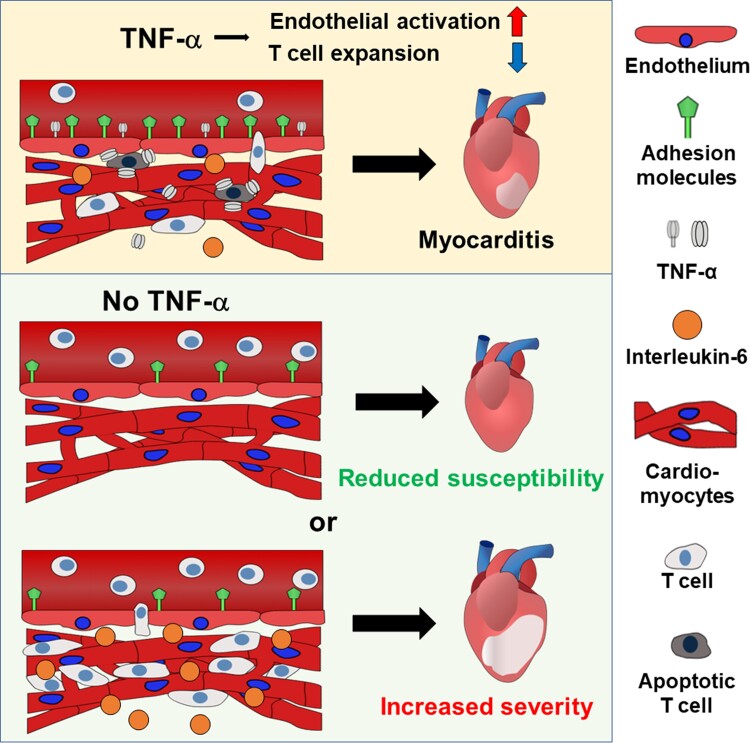
Graphical Abstract.
A dual role of TNF-α in autoimmune myocarditis. The top panel illustrates that TNF-α can have a pro-inflammatory effect by activating cardiac endothelial cells and an anti-inflammatory effect by inducing activation-induced cell death of heart-reactive T cells. The bottom panel depicts two alternative scenarios in case of the absence of TNF-α.
Time of primary review: 62 days
1. Introduction
Heart disease is a leading cause of death in the world affecting millions of people worldwide.1 Inflammation has been implicated in the development and progression of heart disease. Chronic systemic inflammation enhances the development of atherosclerosis and increases the risk of life-threatening ischaemic events, and active inflammatory processes in the heart can trigger tissue remodelling and profibrotic and hypertrophic changes in the cardiac muscle leading to systolic and diastolic dysfunctions and cardiac arrhythmias.2 Myocarditis refers to an inflammatory condition in the heart in the absence of ischaemic events and is characterized by a massive influx of inflammatory cells, mostly myeloid cells, and T lymphocytes, into the cardiac tissue.3 Cardiotropic infections, systemic autoimmune diseases, immune checkpoint inhibitor therapy, and most recently COVID-19 represent major causes of myocarditis.4,5 Diagnosed myocarditis cases are relatively infrequent, and its true prevalence is likely underestimated. This is primarily due to the lack of specific disease manifestation, the need for endomyocardial biopsies as a gold standard diagnostic, and the limitations of magnetic resonance imaging (MRI). Fulminant myocarditis may cause fatal arrhythmias and represents an important cause of unexpected and sudden death.5 Resolution of acute cardiac inflammation is often spontaneous and can be followed by full recovery; however, some myocarditis patients develop dilated cardiomyopathy phenotype characterized by left ventricular dilation and systolic dysfunction.
Tumour necrosis factor α (TNF-α) represents one of the most important cytokines in the immune response. Elevated TNF-α levels are observed during the course of cardiovascular pathologies and positively correlate with disease severity.6,7 TNF-α is synthesized as a transmembrane protein that can be cleaved and released in a soluble form.8 TNF-α is secreted mainly by activated myeloid cells and T lymphocytes but also by other cell types, and its production is often triggered by infections or injury. Both soluble and transmembrane TNF-α are biologically active, as they can bind to TNF-α receptor (TNFR)1 and TNFR2, respectively. Of note, transmembrane TNF-α acts also as a receptor for TNFR2 in reverse signalling.9 TNFR1 signalling triggers a pro-inflammatory response, immune activation, cell apoptosis, cytokine secretion, and endothelial activation, while TNFR2 activation promotes cell survival, and regeneration and modulates the immune response by counteracting effects mediated by TNFR1.7,10 Over 20 years ago, targeting TNF-α has been suggested as a promising anti-inflammatory treatment strategy across multiple diseases. Indeed, TNF-α inhibitors revolutionized the treatment of certain systemic inflammatory diseases, such as rheumatoid arthritis, psoriasis, or Crohn’s disease.11 Conversely, treatment of heart failure patients with etanercept showed no benefits, and high doses of infliximab adversely affected the clinical condition.12,13 Similarly, treatment with infliximab significantly increased cardiovascular mortality in immune checkpoint inhibitor–induced myocarditis patients.14 The reasons for opposing outcomes of anti-TNF-α therapies in the treatment of systemic inflammatory diseases and heart failure remain unclear.
Heart-specific autoimmunity plays an important role in the progression of myocarditis alpha-myosin heavy chain (α-MyHC) represents a major cardiac autoantigen in humans and mice. Unlike other cardiac proteins, α-MyHC is not expressed in the thymus, and therefore, α-MyHC-reactive T lymphocytes escape the negative selection process and circulate in the body.15 Release of α-MyHC from damaged cardiomyocytes during heart injury may activate circulating α-MyHC-reactive T cells and trigger a heart-specific immune response under various cardiac conditions.
Mouse models of experimental autoimmune myocarditis (EAM) use the ability of α-MyHC-reactive CD4+ T lymphocytes to induce heart-specific inflammation and reflect key aspects of the human disease.16 In the classical EAM model, immunization with α-MyHC peptide and complete Freund’s adjuvant (CFA) is used to induce myocarditis in susceptible strains. In this model, following the resolution of the inflammation, some mice show progressive accumulation of fibrotic tissue in the myocardium, ventricular dilatation, and impaired heart function.16 Published data demonstrated that mice deficient for TNFR1 were protected from the development of inducible EAM17; however, the effect of TNF-α cytokine itself has not been investigated yet. Mice with transgenic overexpression of T cell receptor (TCR) specific to α-MyHC (TCRM) represent an alternative EAM model. Due to the high number of circulating α-MyHC-reactive T cells, TCRM mice spontaneously develop progressive myocarditis-associated with ventricular wall thickening but without evident systolic dysfunction.18
2. Methods
2.1. Mice
To obtain Tnf+/− and Tnf−/− BALB/c mice, knockout of the Tnf allele was performed in one-cell-stage BALB/c embryos using CRISPR/Cas9-mediated non-homologous end-joining method. Generation of TCRM mice (on BALB/c background) was described previously.18 TCRM mice were crossed with Tnf−/− mice to obtain TCRM × Tnf+/− and TCRM × Tnf−/− mice. Mice reaching humane endpoints (defined as abnormal physical appearance, reduced mobility, and impaired behavioural response to external stimuli) were prematurely euthanized and classified as non-survivors. All experiments were performed in accordance with Polish law and were approved by local authorities (licence numbers 206/2017 and 234/2019 and 106/2021). Animal experiments followed the guidelines from Directive 2010/63/EU of the European Parliament on the protection of animals used for scientific purposes.
2.2. Experimental autoimmune myocarditis
EAM was induced in 6–8-week-old sex-matched BALB/c mice by subcutaneous injection of 200 µg of α-MyHC614–634 peptide (Ac-RSLKLMATLFSTYASADR-OH, Caslo, Denmark) emulsified 1:1 with CFA (BD Difco, USA) at Days 0 and 7. On Day 21, mice were euthanized by cervical dislocation under anaesthesia (intraperitoneal injection of 75 mg/kg ketamine). Myocarditis severity was evaluated using a blinded approach on haematoxylin/eosin stainings using a 0–5 scale (0, no leucocyte infiltrates; 1, small foci of leucocytes; 2, larger foci of >100 inflammatory cells; 3, >10% of a cross-section involved; 4, >30% of a cross-section involved, 5, >50% of a cross-section involved.
2.3. Cardiac function
Global heart function was assessed by MRI using a 9.4 T small animal MRI scanner (Bruker BioSpec, Ettlingen, Germany) equipped with a 1000 mT/m gradient coil with a maximum slew rate of 3500 T/m/s. During the experiment, mice were anaesthetized using isoflurane (1.7 vol%) in oxygen and air (1:2) mixture. Dobutamine hydrochloride (Sigma-Aldrich, Germany) was dissolved in 0.9% NaCl at a concentration of 0.5 mg/mL and injected i.p. at a dose of 2 mg/kg. Body temperature was monitored with an endorectal probe and maintained in the range of 35.5–36.5°C. The response to dobutamine stimulation was measured pre- and 6 min post-dobutamine (2 mg/kg i.p.) injections. A detailed description is provided in the Supplemental Methods section.
2.4. Cell cultures
To obtain CD4+ T lymphocytes and antigen-presenting cells (APCs), single-cell suspensions of splenocytes were sorted using BD FACSAria II (BD Biosciences, San Jose, CA, USA). CD4+ T cells and APCs were co-cultured in a 10:1 ratio. CD4+ T cells were activated with anti-CD3/CD28 beads (ThermoFisher, USA) or 10 ng/mL of α-MyHC (Caslo), and the conditioned medium was collected on Day 3 for further experiments. Experiments were performed on primary cardiac microvascular endothelial cells (cMVECs) isolated from hearts of 4-week-old Tnf+/+ and Tnf−/− mice as described previously.19 cMVECs were cultured in RPMI 1640 supplemented with 10% foetal bovine serum (FBS) and 2 ng/mL recombinant mouse epidermal growth factor (BioLegend, USA). cMVECs were activated with recombinant mouse TNF-α (5 ng/mL, BioLegend) or conditional medium. A detailed description is provided in the Supplemental Methods section.
2.5. Flow cytometry
Isolation of single-cell suspension from spleens and hearts was previously described.20 cMVECs were detached and collected using 0.25% trypsin with 0.02% ethylenediaminetetraacetic acid (EDTA) (Sigma). Cells were stained with appropriate fluorochrome-conjugated anti-mouse antibodies (see Supplemental Methods section for detailed description and gating strategies used in the experiments) for 30 min at 4°C and analysed with the FACSCanto 10 Flow Cytometer (BD Biosciences) and the FlowJo v10 software (Tree Star, Ashland, USA).
2.6. Adhesion assay
Adhesion of splenocytes to cMVECs was performed using the BioFlux 200 system (Fluxion, USA) as described previously.19 Briefly, mouse splenocytes (3 × 10⁵ cells/mL) labelled with CellTrace™ carboxyfluorescein diacetate succinimidyl ester (CFSE) (ThermoFisher) were passed through microcapillaries coated with cMVECs at 1 dyn/cm3 for 30 min. Cell adhesion was recorded with a fluorescence microscope LS720 (Etaluma, USA) and analysed using the ImageJ software (version 1.52a, NIH, Bethesda, USA).
2.7. Enzyme-linked immunosorbent assay
TNF-α, IL-6, IL-10, IFN-γ, and GM-CSF were measured using respective enzyme-linked immunosorbent assay (ELISA) MAX kits (BioLegend) according to manufacturer instructions and analysed on an M200 PRO plate reader (TECAN Instruments, Switzerland).
2.8. Histology
Hearts were fixed in 4% formalin and embedded in paraffin. Conventional haematoxylin/eosin and Masson’s trichrome staining were used to assess cardiac inflammation and fibrosis, respectively. Staining with the wheat germ agglutinin conjugated with Alexa Fluor 488 (ThermoFisher) was performed to assess cardiomyocyte size. The longitudinal cross-sectional area of >100 randomly selected cardiomyocytes with a visible nucleus from each heart was measured using ImageJ software.
2.9. Statistical analysis
For normally distributed data unpaired Student’s t-test or one-way ANOVA followed by Fisher’s least significant difference (LSD) tests was used. Mann–Whitney U test or Kruskal–Wallis followed by Dunn’s test was used for non-parametric data. Differences were considered statistically significant for P < 0.05. All analyses were performed with the Prism 8 software (GraphPad).
3. Results
3.1. TNF-α deficiency is associated with reduced incidence of myocarditis in an inducible model of EAM
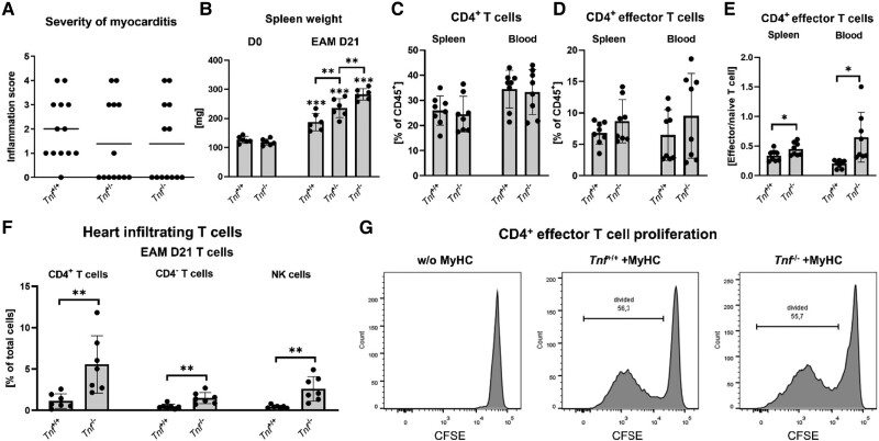
To address the role of TNF-α in myocarditis, we generated BALB/c mice with genetic deletion of the Tnf gene and used them in an inducible model of EAM. Histological analysis of hearts at Day 21 showed a reduced incidence of myocarditis in Tnf+/− (TNF-α haploinsufficient) and in Tnf−/− (TNF-α knockout) mice compared with Tnf+/+ (wild-type) littermates (Figure 1A). Although about half of Tnf+/− and Tnf−/− mice were completely resistant to immunization with α-MyHC and CFA, the other half of transgenic animals developed extensive inflammation in the cardiac muscle (Figure 1A). In this model, the immunization led to spleen enlargement, which was particularly enhanced in Tnf−/− mice. Of note, splenomegaly was observed in all immunized Tnf−/− mice regardless of myocarditis incidence and severity (Figure 1B). EAM is mediated by MyHC-specific effector CD4+ T (Teff) cells; therefore, in the next step, we analysed the development of CD44hiCD62Llow Teff in the immunized mice. While no difference in the total percentage of CD4+ T cells in spleens and blood was found (Figure 1C), we observed a trend towards increased Teff percentage (Figure 1D) and significantly increased Teff/Tn ratio in the spleen and blood of Tnf+/− and Tnf−/− mice at Day 21 of EAM (Figure 1E). In line with these data, increased prevalence of CD4+ T cells was observed in myocarditis-positive Tnf−/− hearts (Figure 1F, Supplementary material online, Figure S1). CD4+ T cells infiltrating the heart during myocarditis show nearly exclusively Teff phenotype20; therefore, an increased fraction of CD4+ T cells in the inflammatory compartment of TNF-α-deficient hearts was not surprising. In the next step, we sorted splenic CD44hiCD62Llow Teff from MyHC/CFA-immunized mice and challenged them with MyHC peptide in the presence of wild-type APCs. Both Tnf+/+ and Tnf−/− Teff showed a potent antigen-specific response (Figure 1G).
Figure 1.
Effect of TNF-α in experimental autoimmune myocarditis. (A) Severity of myocarditis in wild-type BALB/c (Tnf+/+), haploinsufficient (Tnf+/−), and mice deficient in TNF-α (Tnf−/−) at Day 21 after immunization, n = 12. (B) Average spleen weight of healthy (D0) and age-matched mice 21 days from disease induction (D21). (C) Total percentage of splenic and blood CD4+ T cells at D21, n = 8. (D) Percentage of effector CD4+ T cells. (E) Ratio of effector to naïve CD4+ T cells in spleens and blood at D21, n = 8. (F) Percentage of selected leucocyte populations in myocarditis-positive hearts (defined as >3% of CD45+ cells; see Supplementary material online, Figure S3) of wild-type and TNF-α-deficient mice 21 days from immunization, n = 7. (G) Representative proliferation of effector T cells isolated from the spleens of immunized mice in the presence (+MyHC) or absence (w/o MyHC) of α-MyHC peptide. P values calculated with Kruskal–Wallis followed by Dunn’s test (A), one-way ANOVA followed by multiple comparison using Fisher’s LSD test (B), or unpaired Student’s t-test (C). ns P > 0.05, *P < 0.05, **P < 0.01, ***P < 0.001.
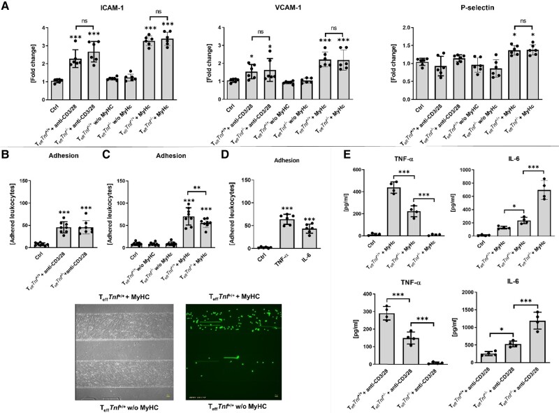
Local activation of cMVECs resulting in adhesion and transmigration of blood leucocytes through the endothelial barrier represents the first step in the development of myocarditis. We confirmed that TNF-α could potently activate adhesive molecules ICAM-1, VCAM-1, and P-selectin in cMVECs and effectively promote adhesion of leucocytes to cMVECs under shear flow conditions in time- and dose-dependent manners (see Supplementary material online, Figure S2). However, conditional medium obtained from Tnf−/− Teff activated with anti-CD3/CD28 showed similar up-regulation of ICAM-1 and VCAM-1 and activation of adhesive properties of cMVECs as a conditional medium from activated Tnf+/+ Teff (Figure 2A–C). Similarly, stimulation of cMVECs with conditioned medium obtained from MyHC-activated Teff isolated from MyHC/CFA-immunized Tnf+/+ and Tnf−/− mice resulted in potent activation of ICAM-1, VCAM-1, and P-selectin in cMVECs and adhesion of leucocytes to cMVECs (Figure 2A–C). Conditioned medium of Tnf−/− Teff (activated with MyHC in the presence of Tnf−/− APCs) was, however, slightly less effective in inducing adhesive properties of cMVECs (Figure 2C). Of note, stimulation with TNF-α or IL-6 alone was sufficient in activating cMVECs (Figure 2D). Analysis of TNF-α and IL-6 in secretomes of activated Teff showed over-production of pro-inflammatory IL-6 by Tnf−/− cells (Figure 2E), which might explain effective activation of cMVECs by Teff in the absence of TNF-α.
Figure 2.
Endothelial activation in inducible EAM model. Teff cells isolated from Tnf+/+ or Tnf−/− mice at D21 of EAM were stimulated with anti-CD3/28 magnetic beads (+anti-CD3/28) or with APCs in the presence (+MyHC) or absence (w/o MyHC) of α-MyHC peptide for 3 days. (A) Changes in surface expression of ICAM-1, VCAM-1, and P-selectin on cMVECs stimulated with the indicated supernatants for 16 h, n = 6. (B–D) Results of leucocyte–endothelial adhesion assay expressed as an average count of firmly adhered leucocytes per field of view after 30 min. (B, C) Adhesion to cMVECs stimulated with Teff supernatants (n = 8) and in (D) stimulated with 5 ng/mL of TNF-α or 60 ng/mL of IL-6 (n = 6) for 16 h. Brightfield microphotograph shows representative microcapillaries coated with cMVECs, and fluorescence image indicates CFSE-labelled splenocytes. (E) TNF-α and IL-6 levels measured in the indicated supernatants by ELISA (n = 4). P values calculated with one-way ANOVA followed by multiple comparison using Fisher’s LSD test. ns P > 0.05, *P < 0.05, **P < 0.01, ***P < 0.001.
3.2. TNF-α protects from cardiac death in TCRM mice
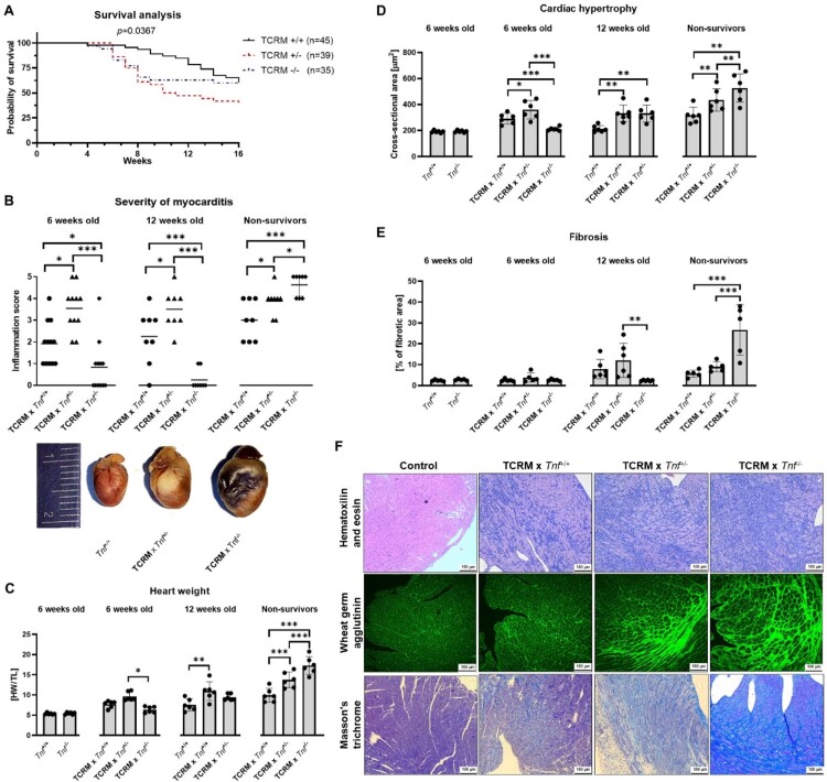
Next, we studied how TNF-α deficiency affected myocarditis and heart failure in transgenic TCRM mice. In the TCRM model, all T lymphocytes recognize the MyHC peptide and therefore the TCRM mice spontaneously develop myocarditis and cardiomyopathy leading in some cases to cardiac death.18 Strikingly, TCRM mice with reduced TNF-α production (TCRM × Tnf+/−) and complete TNF-α deficiency (TCRM × Tnf−/−) showed high mortality, particularly between Weeks 5 and 10 of age (Figure 3A). Analysis of 6-week-old and 12-week-old mice showed more severe myocarditis in TCRM × Tnf+/− animals (Figure 3B). In line with these data, increased size of the heart and cardiomyocytes were observed in TCRM × Tnf+/− mice (Figure 3C, D).
Figure 3.
Effects of TNF-α deficiency in the TCRM model. (A) Mortality of TCRM mice up to age of 16 weeks (Tnf+/+ n = 45, Tnf+/− n = 39, Tnf−/− n = 35). (B) Myocarditis severity in 6-week-old and 12-week-old mice and in non-survivors (mice meeting humane endpoints, age 4–10 weeks, n = 8–12 per group) and representative examples of age- and sex-matched healthy heart of wild-type (Tnf+/+) and TCRM × Tnf+/− and TCRM × Tnf−/− non-survivors. (C) Heart weight/tibia length (HW/TL) (n = 6). (D) Measurements of cross-sectional area of cardiomyocytes (n = 6). (E) Percentage of fibrotic area in cardiac sections (n = 6). (F) Representative microphotographs used in the analysis. P values calculated with log-rank test for A, Kruskal–Wallis test for B, or one-way ANOVA followed by multiple comparison using Fisher’s LSD test for C–E. ns P > 0.05, *P < 0.05, **P < 0.01, ***P < 0.001.
Unlike TCRM × Tnf+/− mice, TCRM × Tnf−/− littermates with normal appearance showed no or minimal cardiac inflammation and no signs of any other cardiac pathology (Figure 3B–E). These data contradicted the high mortality of TCRM × Tnf−/− observed at a young age (Figure 3A). Therefore, in the next step, we focused on mice displaying worsening physical conditions that met humane endpoints and had to be prematurely euthanized. These mice were classified as ‘non-survivors’. Analysis of TCRM × Tnf−/− ‘non-survivors’ showed severe pathological changes in their hearts including massive infiltration of inflammatory cells, hypertrophy, and extensive fibrosis (Figure 3B–E). Of note, inflammatory areas in those hearts exhibited significant fibrosis (see Supplementary material online, Figure S3). Hearts of TCRM × Tnf+/− ‘non-survivors’ also showed exaggerated pathological changes but were less extreme than those found in TCRM × Tnf−/− ‘non-survivors’ (Figure 3B–E).
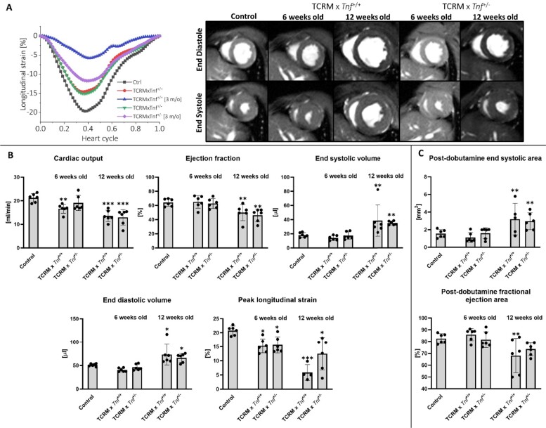
Next, we addressed functional changes in TCRM × Tnf+/+ and TCRM × Tnf+/− mice using echocardiographic imaging and MRI. Due to the sudden and unpredicted occurrence of the phenotype and the poor physical condition of ‘non-survivors’, cardiac function was not assessed in these mice. Echocardiography of 6-week-old TCRM × Tnf+/+ and TCRM × Tnf+/− revealed decreased stroke volume in TCRM mice, but no change in ejection fraction and fractional shortening was observed (see Supplementary material online, Figure S4). Measurements of heart function with MRI suggested progressive development of left ventricular dysfunction in the TCRM model, but despite the difference in myocarditis intensity, no significant difference between TCRM × Tnf+/+ and TCRM × Tnf+/− groups was detected in ejection fraction and end-systolic and end-diastolic volumes at 6 weeks of age (Figure 4B). The cardiac output was preserved for TCRM × Tnf+/− but was reduced for TCRM × Tnf+/+ at 6 weeks of age. Assessment of a left ventricle function in response to β1-adrenergic receptor agonist dobutamine confirmed uncompromised fractional area shortening and stress end-systolic area in 6-week-old TCRM mice (Figure 4C). The cardiac output, ejection fraction, and end-systolic and end-diastolic volumes were significantly worsened in both groups at 12 weeks of age. Correspondingly, the analysis of left ventricle deformability (as assessed via global deformation along longitudinal direction) revealed significantly reduced peak strain for both groups already at 6 weeks of age with a distinct worsening at the age of 12 weeks, especially in TCRM × Tnf+/+ animals (Figure 4A). The deterioration of cardiac global function at 12 weeks of age was accompanied by a marked increase in the post-dobutamine end-systolic area and fractional area shortening in TCRM × Tnf+/+ and TCRM × Tnf+/− groups underlying decreased contractility capability in response to increased workload (Figure 4C).
Figure 4.
Cardiac MRI (cMRI) of TCRM mice. Six-week-old TCRM wild-type (control), TCRM × Tnf+/+ and TCRM × Tnf+/−, and 12-week-old TCRM × Tnf+/+ and TCRM × Tnf+/− were taken into the analysis. (A) Representative changes in left ventricle longitudinal strain during a single heart cycle and representative frames of matched layers from the cMRI during diastole and systole in all groups. (B) Mean values of cardiac output, ejection fraction, end-systolic volume, end-diastolic volume, and peak longitudinal strain, n = 6. (C) Mean end-systolic area and fractional ejection area after dobutamine injection, n = 6. P values calculated with one-way ANOVA followed by multiple comparison using Fisher’s LSD test. ns P > 0.05, *P < 0.05, **P < 0.01, ***P < 0.001.
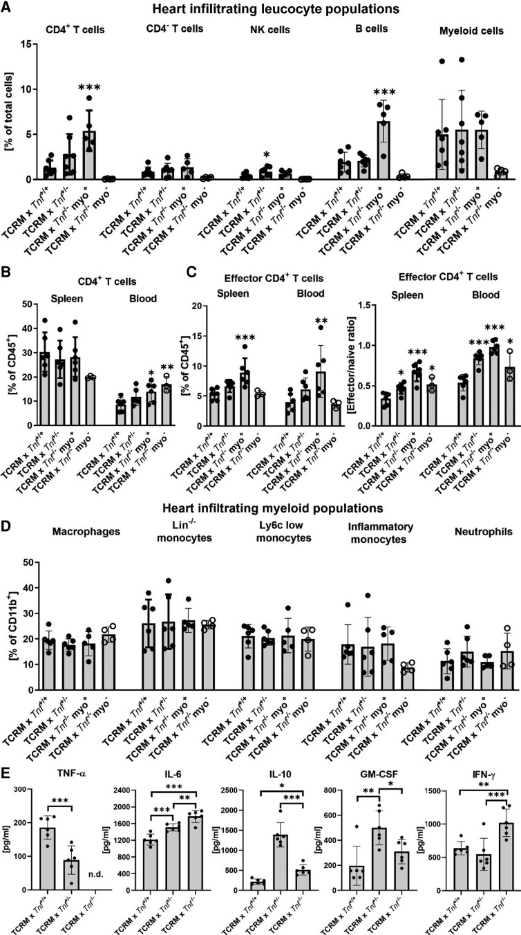
Flow cytometry analysis confirmed increased percentage of CD45+ leucocytes in myocarditis-positive (myo+) TCRM × Tnf−/− hearts (see Supplementary material online, Figure S1). Subset analysis of cardiac infiltrates showed an increased prevalence of CD4+ T and B cells in the hearts of TCRM × Tnf+/− and myo+ TCRM × Tnf−/− mice (Figure 5A). As expected, CD45+ leucocytes and CD4+ T lymphocytes were nearly absent in the myocarditis-negative (myo−) TCRM × Tnf−/− hearts (Figure 5A, Supplementary material online, Figure S1). Although TNF-α deficiency did not affect the prevalence of total CD4+ T cells in the spleen, the percentage of circulating CD4+ T cells was increased in myo+ TCRM × Tnf−/− mice. In line with these findings, the Teff/Tn ratio was significantly increased in TCRM × Tnf+/− and myo+ TCRM × Tnf−/− mice (Figure 5B, C). On the other hand, analysis of CD11b+ cells showed no major effect of TNF-α deficiency on myeloid cell subpopulations (Figure 5D). To address how the lack of TNF-α influenced the cytokine response of activated CD4+ T cells, we stimulated TCRM splenocytes with MyHC antigen and measured cytokines in supernatants after 72 h. TNF-α deficiency was associated with increased production of IL-6, IL-10, GM-CSF, and IFN-γ (Figure 5E). All of these findings indicate that TNF-α is negatively regulating the adaptive immune response.
Figure 5.
Leucocyte populations in the TCRM model. Cells isolated from hearts, spleen, and blood of 6-week-old TCRM × Tnf+/+, TCRM × Tnf+/−, and TCRM × Tnf−/− mice were analysed using flow cytometry. TCRM × Tnf−/− mice were divided into myocarditis-positive (TCRM × Tnf−/− myo+, >3% of CD45+ cells in the heart) and myocarditis-negative (TCRM × Tnf−/− myo−, <3% of CD45+ cells in the heart; see Supplementary material online, Figure S3). (A) Main heart-infiltrating leucocyte populations (n = 4–7). (B) Percentages of splenic CD4+ T cells. (C) Prevalence of CD4+ T effector cells and the ratio of effector to naïve CD4+ T cells in spleens and in blood (n = 3–7). (D) Analysis of heart-infiltrating populations of myeloid lineage (n = 4–7). Gating strategies used in the experiments are presented in Supplementary material online, Figure S3. (E) TNF-α, IL-6, IL-10, GM-CSF, and IFN-γ levels in supernatants of TCRM splenocytes activated with MyHC for 3 days, n = 6. P values calculated with one-way ANOVA followed by multiple comparison using Fisher’s LSD test. In A–D, statistical significance was calculated against TCRM × Tnf+/+ group. ns P > 0.05, *P < 0.05, **P < 0.01, ***P < 0.001.
3.3. TNF-α produced by cMVECs regulates cell adhesion
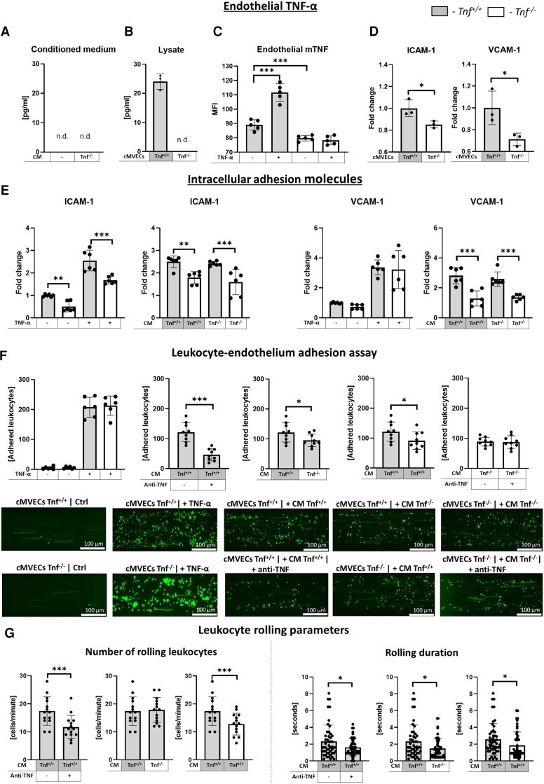
Our data obtained with the inducible EAM model suggested that TNF-α secreted by Teff played a role in cMVEC activation (Figure 2). In the next step, we addressed the relevance of TNF-α produced by cMVECs. No soluble TNF-α was detected in supernatants of cMVECs, but instead, we observed its presence in cell lysates and on the cellular membrane (Figure 6A–C). Interestingly, membrane TNF-α was up-regulated upon stimulation with exogenous TNF-α (Figure 6C). Analysis of adhesion molecules showed that naïve cMVECs in the hearts of Tnf−/− mice were characterized by reduced ICAM-1 and VCAM-1 level (Figure 6D). A reduced ICAM-1 level was further confirmed in cultured Tnf−/− cMVECs in unstimulated and TNF-α-stimulated conditions as well as following treatment with conditioned medium of MyHC-stimulated CD44hiCD62Llow Teff cells (Figure 6E). Next, we addressed the role of TNF-α signalling in adhesion of splenocytes to activated cMVECs under shear flow. As expected, this process was TNF-α dependent, and in the case of activation of cMVECs with the secretome of TCRM Teff, neutralizing anti-TNF-α antibody effectively reduced splenocyte adhesion (Figure 6F). Experiments with Tnf−/− cells confirmed that TNF-α produced not only by Teff but also by cMVECs contributed to this process (Figure 6F). A detailed cell motion analysis revealed in the case of Tnf−/− cMVECs also a reduced number of rolling splenocytes, suggesting impaired tethering and shorter rolling duration lowering the chance for firm adhesion (Figure 6G). All these data suggest that TNF-α produced by cMVECs might play an active role in the development of CD4+ T cell–mediated myocarditis.
Figure 6.
Contribution of soluble and membrane TNF-α in cell adhesion. (A) Concentration of TNF-α (measured by ELISA) in supernatants of resting cMVECs and cMVECs stimulated with medium conditioned of activated Tnf−/− Teff cells and (B) in Tnf+/+ and Tnf−/− cMVECs lysates (n = 3). (C) Surface expression of TNF-α (measured by flow cytometry) on resting and TNF-α-stimulated Tnf+/+ and Tnf−/− cMVECs (n = 5). (D) Surface expression of ICAM-1 and VCAM-1 on freshly isolated CD31+ endothelial cells from wild-type (Tnf+/+) and Tnf−/− hearts (n = 3) and (E) in cMVECs stimulated for 16 h with 5 ng/mL TNF-α or with supernatants of MyHC-activated TCRM × Tnf+/+ or TCRM × Tnf−/− Teff cells cultured in the presence of the respective APCs for 3 days. (F) Quantification of firmly adhered splenocytes to cMVECs in the adhesion assay for indicated conditions. Fluorescence microphotographs show representative CFSE-labelled splenocytes in microcapillaries coated with cMVECs. Frequency of rolling splenocytes on the surface of cMVECs (n = 14) and the distance of the rolling splenocytes (n = 50) are presented for the indicated conditions in G. P values calculated with one-way ANOVA followed by multiple comparison using Fisher’s LSD test or with unpaired Student’s t-test. ns P > 0.05, *P < 0.05, **P < 0.01, ***P < 0.001.
3.4. TNF-α triggers activation-induced cell death of heart-reactive T cells
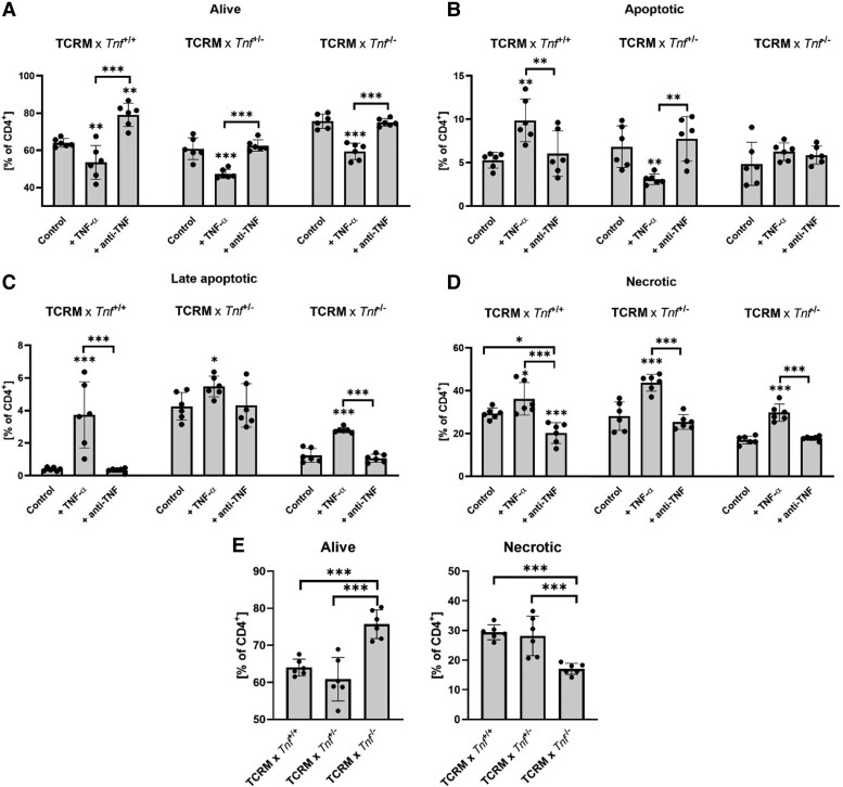
In two models of myocarditis TNF-α, deficient mice were characterized by an increased fraction of Teff cells in inflamed hearts and the periphery. TNF-α has been previously recognized to trigger activation-induced cell death; therefore, we hypothesized that in our disease models, TNF-α deficiency led to an uncontrolled expansion of autoreactive CD4+ T cells. Reactivation of MyHC-reactive Teff cells (but also Tn cells) from inducible EAM model and TCRM mice for 3 days showed no difference in proliferation and survival between Tnf+/+ and Tnf−/− cells (see Supplementary material online, Figure S5). Instead, TCRM Tnf−/− CD4+ T lymphocytes showed significantly less cell death when stimulated with MyHC antigen for 7 days (Figure 7A–E). In line with these data, the addition of TNF-α cytokine to antigen-activated TCRM CD4+ T cells induced apoptosis and cell death, whereas the addition of neutralizing anti-TNF-α antibody resulted in increased cell survival (Figure 7A–D). These data confirmed that TNF-α could effectively regulate the survival of heart-reactive Teff cells by an activation-induced cell death mechanism.
Figure 7.
TNF-α triggers activation-induced cell death of heart-reactive CD4+ T cells. TCRM splenocytes were pulsed with α-MyHC peptide in the absence (control) or presence of 5 ng/mL TNF-α (+TNF) or 10 µg/mL anti-TNF-α neutralizing antibody (+anti-TNF) for 7 days. (A–D) Quantification of live (A), apoptotic (B), late apoptotic (C), and necrotic (D) CD4+ T cells at Day 7 (n = 6). (E) Comparison of live and necrotic CD4+ T cells of control groups. P values calculated with one-way ANOVA followed by multiple comparison using Fisher’s LSD test. ns P > 0.05, *P < 0.05, **P < 0.01, ***P < 0.001.
4. Discussion
Despite decades of extensive research, the role of TNF-α in cardiovascular disease is still puzzling. In particular, outcomes of the anti-TNF-α therapy in various cardiac conditions provided an ambiguous answer. On the one hand, results of clinical trials showed worsening of cardiac function in some heart failure patients treated with a high dose of the TNF-α inhibitor infliximab.21 On the other hand, clinical studies on rheumatoid arthritis patients receiving TNF-α blockers reported no increased risk of heart failure and effective reduction in the incidence of major cardiovascular events suggesting mild cardioprotective effects.22 Unlike heart failure patients, most patients with systemic autoimmune diseases show normal cardiac function and no serious heart pathologies. However, analysis in a subgroup of rheumatic patients with post-ischaemic events showed no benefits of anti-TNF-α therapy and in the case of an elderly population exacerbation of heart failure and reduced survival.23,24 Thus, these clinical findings suggested a dual role of TNF-α signalling in cardiovascular disease. Our data from two EAM models strongly support this hypothesis. Reduced myocarditis prevalence of Tnf−/− mice indicated a supportive role of TNF-α in disease induction, whereas increased cardiac death of TCRM × Tnf−/+ and TCRM × Tnf−/− mice and exacerbated myocarditis in TCRM × Tnf−/+ mice suggested a cardioprotective effect in established disease. Previous works in various experimental myocarditis models also reported both cardioprotective and harmful effects following the blockade of TNF-α signalling. For example, preventive treatment with an anti-TNF-α antibody effectively reduced the severity of myocarditis caused by infection with a cardiotropic virus,25 protozoan parasite Trypanosoma cruzi,26 or by immunization with cardiac myosin.27 Similar results were observed in Tnf−/− and Tnfrsf1a−/− mice.17,28,29 However, in the case of T. cruzi infection, increased accumulation of CD4+ T cells in the heart and enhanced mortality at the late stage of the disease were observed in Tnfrsf1a−/− mice.29 In line with these data, in a hamster model of T. cruzi–induced myocarditis, a long-term treatment with anti-TNF-α antibody enhanced left ventricular dysfunction.30 All these data lead to the conclusion that TNF-α signalling during the early phase of myocarditis contributes to the onset of the disease, whereas during the course of the disease, in the already inflamed heart, it is counteracting disease exacerbation.
The pro-inflammatory effect of TNF-α signalling is well established, and our data confirmed its role in regulating the adhesion of inflammatory cells to cMVECs - a critical step during the onset of tissue inflammation. In line with our findings, reduced inflammatory responses were reported in Tnf−/− mice in other experimental cardiovascular disease conditions including atherosclerosis,31 myocardial infarction,32 hypertrophy,33 and viral myocarditis.28 These data underline the importance of TNF-α signalling in the induction of the immune response and explain the cardioprotective effects of anti-TNF-α therapy in patients with a still healthy, unaffected cardiovascular system. Soluble TNF-α has been well recognized to activate endothelial cells. We found that not only soluble TNF-α but also its membrane form expressed by cMVECs was crucial for cell adhesion. In line with previous reports, the lack of membrane TNF-α resulted in lower levels of adhesion molecules on the surface of endothelial cells.32 We were able to demonstrate that this phenotype correlated with the impaired ability of cMVECs to tether flowing cells and with reduced rolling phase preceding firm adhesion. Although expression of TNF-α in endothelial cells is very low, it has been acknowledged to regulate their physiology and promote atherosclerosis through interaction with TNFR2.34,35 Taken together, endogenous expression of TNF-α in cMVECs not only regulates their physiology but plays an important role in the process of leucocyte recruitment. These data may also explain major differences in the efficacy and side effects of the commonly used anti-TNF-α drugs etanercept and infliximab, as the latter not only acts through neutralization of soluble TNF-α but also binds to the membrane form of TNF-α.36 Transmembrane TNF-α can act both as a ligand for TNFR2 and as a receptor capable of reverse-signaling,37 but which exact molecular mechanism plays a dominant role in activated cMVECs remains unclear.
In contrast to the well-characterized pro-inflammatory aspects of TNF-α signalling, cellular mechanisms behind its cardioprotective effect are less understood. We demonstrated that the anti-inflammatory TNF-α activity was independent of endothelial cell activation but, instead, linked with its potential to induce cell death of pathogenic heart-reactive Teff cells. We think that due to the abnormally high number of these heart-reactive CD4+ T cells in the TCRM mice, the cardioprotective effect of TNF-α was well-pronounced and evident in this model. TNF-α is known to sensitize T lymphocytes for enhanced activation-induced cell death, and our data confirmed TNF-α-induced death of heart-reactive CD4+ T cells in the TCRM model. In this process, cells undergo apoptosis in a controlled manner upon activation of death receptors, which belong to the TNF receptor superfamily. Therefore, TNF-α next to FasL and TRAIL represents a classic example of death receptor agonists.38 Interestingly, only soluble but not transmembrane TNF-α sensitizes T lymphocytes for enhanced activation-induced cell death.39 TNF-α induces activation-induced cell death not only in CD4+ but also in CD8+ T cells.40 In viral myocarditis, the clearance of cardiotropic viruses relies on the effective elimination of infected cardiomyocytes by CD8+ T cells.41 By blocking TNF-α signalling, one can expect an increase in the number and activity of CD8+ T cells, resulting in enhanced viral clearance. Thus, the role of the TNF-α-dependent activation-induced cell death mechanism in myocarditis progression could also depend on the underlying viral or autoimmune cause of the disease. Furthermore, in the context of COVID-19, where myocarditis can occur as a complication of SARS-CoV-2 cardiotropic infection or cytokine storm, understanding the interplay between TNF-α signalling and CD4+ and CD8+ T cell response becomes particularly relevant.42
In addition to the activation-induced cell death mechanisms, TNF-α can limit T cell expansion by enhancing, in cooperation with IFN-γ, nitric oxide production.43 This regulatory role of TNF-α is, however, not limited to the heart. Data from experimental autoimmune encephalomyelitis and arthritis demonstrated that Tnf−/− mice in those models also developed enhanced T cell–dependent autoimmune responses.44,45 Furthermore, commensal bacteria were recently shown to boost heart-specific T cell response and cause lethal inflammatory cardiomyopathy,46 further underlining the destructive potential of unleashed heart-reactive T cells. Our study also revealed an intriguing relationship between TNF-α and IL-6. We observed that TNF-α exerts a negative regulatory effect on the production of IL-6. In the light of the inhibitory effect of IL-6 on the Fas/FasL system47 and an indispensable role of IL-6 in promoting the development of heart-specific autoimmunity,18,48 our findings add another layer to interplay between TNF-α and IL-6.
So far, little is known about the involvement of cardiac autoimmunity in the development of heart failure in humans. In the case of myocarditis and dilated cardiomyopathy, some patients develop high titres of heart-specific autoantibodies and the presence of anti-MyHC antibodies has been associated with worse left ventricular systolic function.49,50 Clinical results from the treatment of myocarditis occurring as an adverse event under immune checkpoint inhibitor therapy in cancer patients shed more light on the protective role of TNF-α in the inflamed heart. Immune checkpoint inhibitors targeting PD-1/PD-1L and CTLA-4 prevent activated T cell de-activation and anergy, thereby increasing the risk of autoimmune adverse effects. Due to these reasons, this therapy is generally not recommended for patients with pre-existing autoimmune disorders. It is therefore plausible that there is a high involvement of heart-specific autoimmunity in myocarditis incidence in patients receiving immune checkpoint inhibitors. Strikingly, a recent clinical study demonstrated that myocarditis patients receiving infliximab showed a strongly increased risk of cardiovascular death [odds ratio (OR) 12.0, P = 0.005].14 In light of our data, these discouraging findings are, however, not surprising and could be linked to the protective effect of eliminating activated heart-specific Teff cells by TNF-α. Of note, similar unfavourable outcomes of anti-TNF-α therapy were obtained in multiple sclerosis, which is also considered an autoimmune disease. A randomized, placebo-controlled multicentre study reported exacerbated disease severity in multiple sclerosis patients receiving TNFR1 fusion protein Lenercept.51
In conclusion, our data suggest that TNF-α signalling plays a dual role in myocarditis by promoting inflammation in the early phase through the regulation of endothelial activation and suppressing immune response during the acute and chronic stages by suppressing the expansion of autoreactive T cells. However, it should be acknowledged that the latter mechanism remains associative due to the nature of the models used and the dual role of TNF-α signalling. Moreover, the observed heterogeneity in the phenotype of both models may suggest the existence of an unrecognized factor that determines whether TNF-α signalling has a pro- or anti-inflammatory effect. Therefore, the involvement of TNF-α signalling in myocarditis may extend beyond the proposed mechanisms. Experiments employing lineage-specific and time-specific blockade of TNF-α signalling in myocarditis could provide a more thorough understanding of the complexities associated with TNF-α signalling and its impact on disease progression. Understanding these cellular and molecular mechanisms enables us to better design future therapies. Accordingly, targeting TNF-α should be therefore limited to patients without significant involvement of heart-specific autoimmunity or be performed in combination with T cell immune regulators, such as alemtuzumab. Furthermore, it seems that more attention should be paid to heart-reactive T cells and the immune component in myocarditis and other heart failure patients. Targeted therapy against intrinsic cardiac-specific autoimmune mechanisms might represent a promising option to attenuate pathogenic inflammatory processes in the heart.
Supplementary material
Supplementary material is available at Cardiovascular Research online.
Supplementary Material
Acknowledgements
We would like to kindly thank Burkard Ludewig (Kantonsspital St. Gallen) for providing us with the TCRM mice.
Contributor Information
Filip Rolski, Department of Clinical Immunology, Jagiellonian University Medical College, Wielicka 265, Cracow 30-663, Poland.
Karolina Tkacz, Department of Clinical Immunology, Jagiellonian University Medical College, Wielicka 265, Cracow 30-663, Poland.
Kazimierz Węglarczyk, Department of Clinical Immunology, Jagiellonian University Medical College, Wielicka 265, Cracow 30-663, Poland.
Grzegorz Kwiatkowski, Jagiellonian Centre for Experimental Therapeutics, Jagiellonian University, Cracow, Poland.
Paweł Pelczar, Center for Transgenic Models, University of Basel, Basel, Switzerland.
Agnieszka Jaźwa-Kusior, Department of Medical Biotechnology, Jagiellonian University, Cracow, Poland.
Anna Bar, Jagiellonian Centre for Experimental Therapeutics, Jagiellonian University, Cracow, Poland.
Gabriela M Kuster, Clinic of Cardiology, University Hospital Basel, Basel, Switzerland; Department of Biomedicine, University Hospital and University of Basel, Basel, Switzerland.
Stefan Chłopicki, Jagiellonian Centre for Experimental Therapeutics, Jagiellonian University, Cracow, Poland.
Maciej Siedlar, Department of Clinical Immunology, Jagiellonian University Medical College, Wielicka 265, Cracow 30-663, Poland.
Gabriela Kania, Department of Rheumatology, University Hospital Zurich, University of Zurich, Wagistrasse 14, 8952 Schlieren, Switzerland.
Przemysław Błyszczuk, Department of Clinical Immunology, Jagiellonian University Medical College, Wielicka 265, Cracow 30-663, Poland; Department of Rheumatology, University Hospital Zurich, University of Zurich, Wagistrasse 14, 8952 Schlieren, Switzerland.
Author contributions
F.R., K.T., and K.W. contributed to data collection, A.J.-K. performed echocardiography, P.B. conceived the idea of the study, P.P. generated TNF-α knockout mice, and F.R. conducted the statistical analysis. Gr.K. performed cMRI analysis. A.B. and G.M.K. contributed to mouse experiments. S.C., M.S., and Ga.K. interpreted the data and reviewed it. F.R. and P.B. drafted the original manuscript. All authors reviewed the manuscript draft and revised it critically with respect to the intellectual content. All authors approved the final version of the manuscript to be published.
Funding
This work has been supported by the National Science Centre (Poland), grant 2016/21/B/NZ5/01397.
Data availability
The data underlying this article are available in the article and in its online Supplementary material.
References
- 1. Groenewegen A, Rutten FH, Mosterd A, Hoes AW. Epidemiology of heart failure. Eur J Heart Fail 2020;22:1342–1356. [DOI] [PMC free article] [PubMed] [Google Scholar]
- 2. Chen W, Frangogiannis NG. The role of inflammatory and fibrogenic pathways in heart failure associated with aging. Heart Fail Rev 2010;15:415–422. [DOI] [PMC free article] [PubMed] [Google Scholar]
- 3. Bracamonte-Baran W, Čiháková D. Cardiac autoimmunity: myocarditis. Adv Exp Med Biol 2017;1003:187–221. [DOI] [PMC free article] [PubMed] [Google Scholar]
- 4. Boehmer TK, Kompaniyets L, Lavery AM, Hsu J, Ko JY, Yusuf H, Romano SD, Gundlapalli AV, Oster ME, Harris AM. Association between COVID-19 and myocarditis using hospital-based administrative data—United States, March 2020-January 2021. MMWR Morb Mortal Wkly Rep 2021;70:1228–1232. [DOI] [PMC free article] [PubMed] [Google Scholar]
- 5. Lampejo T, Durkin SM, Bhatt N, Guttmann O. Acute myocarditis: aetiology, diagnosis and management. Clin Med 2021;21:e505–e510. [DOI] [PMC free article] [PubMed] [Google Scholar]
- 6. Satoh M, Nakamura M, Satoh H, Saitoh H, Segawa I, Hiramori K. Expression of tumor necrosis factor-alpha-converting enzyme and tumor necrosis factor-alpha in human myocarditis. J Am Coll Cardiol 2000;36:1288–1294. [DOI] [PubMed] [Google Scholar]
- 7. Rolski F, Błyszczuk P. Complexity of TNF-α signaling in heart disease. J Clin Med 2020;9:3267. [DOI] [PMC free article] [PubMed] [Google Scholar]
- 8. Moss ML, Jin S-LC, Milla ME, Bickett DM, Burkhart W, Carter HL, Chen W-J, William C, Didsbury JR, Hassler D, Hoffman CR, Kost TA, Lambert MH, Leesnitzer MA, McCauley P, McGeehan G, Mitchell J, Moyer M, Pahel G, Rocque W, Overton LK, Schoenen F, Seaton T, Su J-L, Warner J, Willard D, Becherer JD. Erratum: cloning of a disintegrin metalloproteinase that processes precursor tumour-necrosis factor-α. Nature 2003;386:738–738. [DOI] [PubMed] [Google Scholar]
- 9. Ardestani S, Deskins DL, Young PP. Membrane TNF-alpha-activated programmed necrosis is mediated by ceramide-induced reactive oxygen species. J Mol Signal 2013;8:12. [DOI] [PMC free article] [PubMed] [Google Scholar]
- 10. Cabal-Hierro L, Lazo PS. Signal transduction by tumor necrosis factor receptors. Cell Signal 2012;24:1297–1305. [DOI] [PubMed] [Google Scholar]
- 11. Willrich MAV, Murray DL, Snyder MR. Tumor necrosis factor inhibitors: clinical utility in autoimmune diseases. Transl Res 2015;165:270–282. [DOI] [PubMed] [Google Scholar]
- 12. Mann DL, McMurray JJV, Packer M, Swedberg K, Borer JS, Colucci WS, Djian J, Drexler H, Feldman A, Kober L, Krum H, Liu P, Nieminen M, Tavazzi L, Van VD, Waldenstrom A, Warren M, Westheim A, Zannad F, Fleming T. Targeted anticytokine therapy in patients with chronic heart failure: results of the Randomized EtaNErcept Worldwide Evaluation (RENEWAL). Circulation 2004;109:1594–1602. [DOI] [PubMed] [Google Scholar]
- 13. Chung ES, Packer M, Lo KH, Fasanmade AA, Willerson JT. Randomized, double-blind, placebo-controlled, pilot trial of infliximab, a chimeric monoclonal antibody to tumor necrosis factor-α, in patients with moderate-to-severe heart failure: results of the anti-TNF therapy against congestive heart failure (ATTACH). Circulation 2003;107:3133–3140. [DOI] [PubMed] [Google Scholar]
- 14. Cautela J, Zeriouh S, Gaubert M, Bonello L, Laine M, Peyrol M, Paganelli F, Lalevee N, Barlesi F, Thuny F. Intensified immunosuppressive therapy in patients with immune checkpoint inhibitor-induced myocarditis. J Immunother Cancer 2020;8:e001887. [DOI] [PMC free article] [PubMed] [Google Scholar]
- 15. Rose NR, Herskowitz A, Neumann DA, Neu N. Autoimmune myocarditis: a paradigm of post-infection autoimmune disease. Immunol Today 1988;9:117–120. [DOI] [PubMed] [Google Scholar]
- 16. Błyszczuk P. Myocarditis in humans and in experimental animal models. Front Cardiovasc Med 2019;6:64. [DOI] [PMC free article] [PubMed] [Google Scholar]
- 17. Bachmaier K, Pummerer C, Kozieradzki I, Pfeffer K, Mak TW, Neu N, Penninger JM. Low-molecular-weight tumor necrosis factor receptor p55 controls induction of autoimmune heart disease. Circulation 1997;95:655–661. [DOI] [PubMed] [Google Scholar]
- 18. Nindl V, Maier R, Ratering D, De GR, Züst R, Thiel V, Scandella E, Di PF, Kopf M, Rudin M, Rülicke T, Ludewig B. Cooperation of Th1 and Th17 cells determines transition from autoimmune myocarditis to dilated cardiomyopathy. Eur J Immunol 2012;42:2311–2321. [DOI] [PubMed] [Google Scholar]
- 19. Rolski F, Czepiel M, Tkacz K, Fryt K, Siedlar M, Kania G, Błyszczuk P. T lymphocyte-derived exosomes transport MEK1/2 and ERK1/2 and induce NOX4-dependent oxidative stress in cardiac microvascular endothelial cells. Oxid Med Cell Longev 2022;2022:2457687. [DOI] [PMC free article] [PubMed] [Google Scholar]
- 20. Zarak-Crnkovic M, Kania G, Jaźwa-Kusior A, Czepiel M, Wijnen WJ, Czyż J, Müller-Edenborn B, Vdovenko D, Lindner D, Gil-Cruz C, Bachmann M, Westermann D, Ludewig B, Distler O, Lüscher TF, Klingel K, Eriksson U, Błyszczuk P. Heart non-specific effector CD4+ T cells protect from postinflammatory fibrosis and cardiac dysfunction in experimental autoimmune myocarditis. Basic Res Cardiol 2019;115:6. [DOI] [PMC free article] [PubMed] [Google Scholar]
- 21. Coletta AP, Clark AL, Banarjee P, Cleland JGF. Clinical trials update: RENEWAL (RENAISSANCE and RECOVER) and ATTACH. Eur J Heart Fail 2002;4:559–561. [DOI] [PubMed] [Google Scholar]
- 22. Ayyildiz YO, Vural MG, Efe TH, Ertem AG, Koseoglu C, Ayturk M, Yeter E, Keskin G, Akdemir R. Effect of long-term TNF-α inhibition with infliximab on left ventricular torsion in patients with rheumatoid arthritis. Hellenic J Cardiol 2015;56:406–413. [PubMed] [Google Scholar]
- 23. Setoguchi S, Schneeweiss S, Avorn J, Katz JN, Weinblatt ME, Levin R, Solomon DH. Tumor necrosis factor-α antagonist use and heart failure in elderly patients with rheumatoid arthritis. Am Heart J 2008;156:336–341. [DOI] [PMC free article] [PubMed] [Google Scholar]
- 24. Low ASL, Symmons DPM, Lunt M, Mercer LK, Gale CP, Watson KD, Dixon WG, Hyrich KL. Relationship between exposure to tumour necrosis factor inhibitor therapy and incidence and severity of myocardial infarction in patients with rheumatoid arthritis. Ann Rheum Dis 2017;76:654–660. [DOI] [PMC free article] [PubMed] [Google Scholar]
- 25. Yamada T, Matsumori A, Sasayama S. Therapeutic effect of anti-tumor necrosis factor-alpha antibody on the murine model of viral myocarditis induced by encephalomyocarditis virus. Circulation 1994;89:846–851. [DOI] [PubMed] [Google Scholar]
- 26. Pérez AR, Fontanella GH, Nocito AL, Revelli S, Bottasso OA. Short treatment with the tumour necrosis factor-alpha blocker infliximab diminishes chronic chagasic myocarditis in rats without evidence of Trypanosoma cruzi reactivation. Clin Exp Immunol 2009;157:291–299. [DOI] [PMC free article] [PubMed] [Google Scholar]
- 27. Smith SC, Allen PM. Neutralization of endogenous tumor necrosis factor ameliorates the severity of myosin-induced myocarditis. Circ Res 1992;70:856–863. [DOI] [PubMed] [Google Scholar]
- 28. Huber SA, Sartini D. Roles of tumor necrosis factor alpha (TNF-α) and the p55 TNF receptor in CD1d induction and Coxsackievirus B3-induced myocarditis. J Virol 2005;79:2659–2665. [DOI] [PMC free article] [PubMed] [Google Scholar]
- 29. Kroll-Palhares K, Silvério JC, da Silva AA, Michailowsky V, Marino AP, Silva NM, Carvalho CME, Pinto LMDO, Gazzinelli RT, Lannes-Vieira J. TNF/TNFR1 signaling up-regulates CCR5 expression by CD8+ T lymphocytes and promotes heart tissue damage during Trypanosoma cruzi infection: beneficial effects of TNF-alpha blockade. Mem Inst Oswaldo Cruz 2008;103:375–385. [DOI] [PubMed] [Google Scholar]
- 30. Bilate AMB, Salemi VM, Ramires FJ, de Brito T, Russo M, Fonseca SG, Faé KC, Martins DG, Silva AM, Mady C, Kalil J, Cunha-Neto E. TNF blockade aggravates experimental chronic Chagas disease cardiomyopathy. Microbes Infect 2007;9:1104–1113. [DOI] [PubMed] [Google Scholar]
- 31. Xiao N, Yin M, Zhang L, Qu X, Du H, Sun X, Mao L, Ren G, Zhang C, Geng Y, An L, Pan J. Tumor necrosis factor-alpha deficiency retards early fatty-streak lesion by influencing the expression of inflammatory factors in apoE-null mice. Mol Genet Metab 2009;96:239–244. [DOI] [PubMed] [Google Scholar]
- 32. Sato T, Suzuki H, Shibata M, Kusuyama T, Omori Y, Soda T, Shoji M, Iso Y, Koba S, Geshi E, Katagiri T, Shioda S, Sekikawa K. Tumor-necrosis-factor-alpha-gene-deficient mice have improved cardiac function through reduction of intercellular adhesion molecule-1 in myocardial infarction. Circ J 2006;70:1635–1642. [DOI] [PubMed] [Google Scholar]
- 33. Sun M, Chen M, Dawood F, Zurawska U, Li JY, Parker T, Kassiri Z, Kirshenbaum LA, Arnold M, Khokha R, Liu PP. Tumor necrosis factor-α mediates cardiac remodeling and ventricular dysfunction after pressure overload state. Circulation 2007;115:1398–1407. [DOI] [PubMed] [Google Scholar]
- 34. Green LA, Njoku V, Mund J, Case J, Yoder M, Murphy MP, Clauss M. Endogenous transmembrane TNF-alpha protects against premature senescence in endothelial colony forming cells. Circ Res 2016;118:1512–1524. [DOI] [PMC free article] [PubMed] [Google Scholar]
- 35. Nicolaou A, Zhao Z, Northoff BH, Sass K, Herbst A, Kohlmaier A, Chalaris A, Wolfrum C, Weber C, Steffens S, Rose-John S, Teupser D, Holdt LM. Adam17 deficiency promotes atherosclerosis by enhanced TNFR2 signaling in mice. Arterioscler Thromb Vasc Biol 2017;37:247–257. [DOI] [PubMed] [Google Scholar]
- 36. Zhang M, Wang J, Jia L, Huang J, He C, Hu F, Yuan L, Wang G, Yu M, Li Z. Transmembrane TNF-α promotes activation-induced cell death by forward and reverse signaling. Oncotarget 2017;8:63799–63812. [DOI] [PMC free article] [PubMed] [Google Scholar]
- 37. Qu Y, Zhao G, Li H. Forward and reverse signaling mediated by transmembrane tumor necrosis factor-alpha and TNF receptor 2: potential roles in an immunosuppressive tumor microenvironment. Front Immunol 2017;8:1675. [DOI] [PMC free article] [PubMed] [Google Scholar]
- 38. Rossin A, Miloro G, Hueber A-O. TRAIL and FasL functions in cancer and autoimmune diseases: towards an increasing complexity. Cancers (Basel) 2019;11:639. [DOI] [PMC free article] [PubMed] [Google Scholar]
- 39. Müller S, Rihs S, Schneider JMD, Paredes BE, Seibold I, Brunner T, Mueller C. Soluble TNF-alpha but not transmembrane TNF-alpha sensitizes T cells for enhanced activation-induced cell death. Eur J Immunol 2009;39:3171–3180. [DOI] [PubMed] [Google Scholar]
- 40. Otano I, Alvarez M, Minute L, Ochoa MC, Migueliz I, Molina C, Azpilikueta A, de Andrea CE, Etxeberria I, Sanmamed MF, Teijeira Á, Berraondo P, Melero I. Human CD8 T cells are susceptible to TNF-mediated activation-induced cell death. Theranostics 2020;10:4481–4489. [DOI] [PMC free article] [PubMed] [Google Scholar]
- 41. Lasrado N, Reddy J. An overview of the immune mechanisms of viral myocarditis. Rev Med Virol 2020;30:1–14. [DOI] [PubMed] [Google Scholar]
- 42. Fairweather D, Beetler DJ, Di Florio DN, Musigk N, Heidecker B, Cooper LT Jr. COVID-19, myocarditis and pericarditis. Circ Res 2023;132:1302–1319. [DOI] [PMC free article] [PubMed] [Google Scholar]
- 43. Kania G, Siegert S, Behnke S, Prados-Rosales R, Casadevall A, Lüscher TF, Luther SA, Kopf M, Eriksson U, Blyszczuk P. Innate signaling promotes formation of regulatory nitric oxide-producing dendritic cells limiting t-cell expansion in experimental autoimmune myocarditis. Circulation 2013;127:2285–2294. [DOI] [PubMed] [Google Scholar]
- 44. Kassiotis G, Pasparakis M, Kollias G, Probert L. TNF accelerates the onset but does not alter the incidence and severity of myelin basic protein-induced experimental autoimmune encephalomyelitis. Eur J Immunol 1999;29:774–780. [DOI] [PubMed] [Google Scholar]
- 45. Campbell IK, O’Donnell K, Lawlor KE, Wicks IP. Severe inflammatory arthritis and lymphadenopathy in the absence of TNF. J Clin Invest 2001;107:1519–1527. [DOI] [PMC free article] [PubMed] [Google Scholar]
- 46. Gil-Cruz C, Perez-Shibayama C, De Martin A, Ronchi F, van der Borght K, Niederer R, Onder L, Lütge M, Novkovic M, Nindl V, Ramos G, Arnoldini M, Slack EMC, Boivin-Jahns V, Jahns R, Wyss M, Mooser C, Lambrecht BN, Maeder MT, Rickli H, Flatz L, Eriksson U, Geuking MB, McCoy KD, Ludewig B. Microbiota-derived peptide mimics drive lethal inflammatory cardiomyopathy. Science 2019;366:881–886. [DOI] [PubMed] [Google Scholar]
- 47. Ayroldi E, Zollo O, Cannarile L, D’Adamio F, Grohmann U, Delfino DV, Riccardi C. Interleukin-6 (IL-6) prevents activation-induced cell death: IL-2- independent inhibition of Fas/fasL expression and cell death. Blood 1998;92:4212–4219. [PubMed] [Google Scholar]
- 48. Poffenberger MC, Horwitz MS. IL-6 during viral-induced chronic autoimmune myocarditis: is it protective or pathogenic? Ann Y Acad Sci 2009;1173:318–325. [DOI] [PubMed] [Google Scholar]
- 49. Caforio ALP, Mahon NJ, Tona F, McKenna WJ. Circulating cardiac autoantibodies in dilated cardiomyopathy and myocarditis: pathogenetic and clinical significance. Eur J Heart Fail 2002;4:411–417. [DOI] [PubMed] [Google Scholar]
- 50. Lauer B, Schannwell M, Kühl U, Strauer BE, Schultheiss HP. Antimyosin autoantibodies are associated with deterioration of systolic and diastolic left ventricular function in patients with chronic myocarditis. J Am Coll Cardiol 2000;35:11–18. [DOI] [PubMed] [Google Scholar]
- 51. Lenercept T, Sclerosis M, Group S. TNF neutralization in MS: results of a randomized, placebo-controlled multicenter study. Neurology 2012;53:457–457. [PubMed] [Google Scholar]
Translational perspective.
Clinical trials implementing the use of TNF-α inhibitors in myocarditis and heart failure patients reported health worsening and cardiovascular-related death. Our data indicated that in addition to its well-known pro-inflammatory effect, TNF-α negatively regulated the expansion of heart-specific T cells and thereby protected from myocarditis-associated cardiac death. Thus, the use of anti-TNF-α agents could be re-considered for heart failure patients without active cardiac autoimmunity. Furthermore, targeted therapy against intrinsic cardiac-specific autoimmune mechanisms might represent a promising option to attenuate pathogenic inflammatory processes in the heart.
Associated Data
This section collects any data citations, data availability statements, or supplementary materials included in this article.
Supplementary Materials
Data Availability Statement
The data underlying this article are available in the article and in its online Supplementary material.
References
- 1. Groenewegen A, Rutten FH, Mosterd A, Hoes AW. Epidemiology of heart failure. Eur J Heart Fail 2020;22:1342–1356. [DOI] [PMC free article] [PubMed] [Google Scholar]
- 2. Chen W, Frangogiannis NG. The role of inflammatory and fibrogenic pathways in heart failure associated with aging. Heart Fail Rev 2010;15:415–422. [DOI] [PMC free article] [PubMed] [Google Scholar]
- 3. Bracamonte-Baran W, Čiháková D. Cardiac autoimmunity: myocarditis. Adv Exp Med Biol 2017;1003:187–221. [DOI] [PMC free article] [PubMed] [Google Scholar]
- 4. Boehmer TK, Kompaniyets L, Lavery AM, Hsu J, Ko JY, Yusuf H, Romano SD, Gundlapalli AV, Oster ME, Harris AM. Association between COVID-19 and myocarditis using hospital-based administrative data—United States, March 2020-January 2021. MMWR Morb Mortal Wkly Rep 2021;70:1228–1232. [DOI] [PMC free article] [PubMed] [Google Scholar]
- 5. Lampejo T, Durkin SM, Bhatt N, Guttmann O. Acute myocarditis: aetiology, diagnosis and management. Clin Med 2021;21:e505–e510. [DOI] [PMC free article] [PubMed] [Google Scholar]
- 6. Satoh M, Nakamura M, Satoh H, Saitoh H, Segawa I, Hiramori K. Expression of tumor necrosis factor-alpha-converting enzyme and tumor necrosis factor-alpha in human myocarditis. J Am Coll Cardiol 2000;36:1288–1294. [DOI] [PubMed] [Google Scholar]
- 7. Rolski F, Błyszczuk P. Complexity of TNF-α signaling in heart disease. J Clin Med 2020;9:3267. [DOI] [PMC free article] [PubMed] [Google Scholar]
- 8. Moss ML, Jin S-LC, Milla ME, Bickett DM, Burkhart W, Carter HL, Chen W-J, William C, Didsbury JR, Hassler D, Hoffman CR, Kost TA, Lambert MH, Leesnitzer MA, McCauley P, McGeehan G, Mitchell J, Moyer M, Pahel G, Rocque W, Overton LK, Schoenen F, Seaton T, Su J-L, Warner J, Willard D, Becherer JD. Erratum: cloning of a disintegrin metalloproteinase that processes precursor tumour-necrosis factor-α. Nature 2003;386:738–738. [DOI] [PubMed] [Google Scholar]
- 9. Ardestani S, Deskins DL, Young PP. Membrane TNF-alpha-activated programmed necrosis is mediated by ceramide-induced reactive oxygen species. J Mol Signal 2013;8:12. [DOI] [PMC free article] [PubMed] [Google Scholar]
- 10. Cabal-Hierro L, Lazo PS. Signal transduction by tumor necrosis factor receptors. Cell Signal 2012;24:1297–1305. [DOI] [PubMed] [Google Scholar]
- 11. Willrich MAV, Murray DL, Snyder MR. Tumor necrosis factor inhibitors: clinical utility in autoimmune diseases. Transl Res 2015;165:270–282. [DOI] [PubMed] [Google Scholar]
- 12. Mann DL, McMurray JJV, Packer M, Swedberg K, Borer JS, Colucci WS, Djian J, Drexler H, Feldman A, Kober L, Krum H, Liu P, Nieminen M, Tavazzi L, Van VD, Waldenstrom A, Warren M, Westheim A, Zannad F, Fleming T. Targeted anticytokine therapy in patients with chronic heart failure: results of the Randomized EtaNErcept Worldwide Evaluation (RENEWAL). Circulation 2004;109:1594–1602. [DOI] [PubMed] [Google Scholar]
- 13. Chung ES, Packer M, Lo KH, Fasanmade AA, Willerson JT. Randomized, double-blind, placebo-controlled, pilot trial of infliximab, a chimeric monoclonal antibody to tumor necrosis factor-α, in patients with moderate-to-severe heart failure: results of the anti-TNF therapy against congestive heart failure (ATTACH). Circulation 2003;107:3133–3140. [DOI] [PubMed] [Google Scholar]
- 14. Cautela J, Zeriouh S, Gaubert M, Bonello L, Laine M, Peyrol M, Paganelli F, Lalevee N, Barlesi F, Thuny F. Intensified immunosuppressive therapy in patients with immune checkpoint inhibitor-induced myocarditis. J Immunother Cancer 2020;8:e001887. [DOI] [PMC free article] [PubMed] [Google Scholar]
- 15. Rose NR, Herskowitz A, Neumann DA, Neu N. Autoimmune myocarditis: a paradigm of post-infection autoimmune disease. Immunol Today 1988;9:117–120. [DOI] [PubMed] [Google Scholar]
- 16. Błyszczuk P. Myocarditis in humans and in experimental animal models. Front Cardiovasc Med 2019;6:64. [DOI] [PMC free article] [PubMed] [Google Scholar]
- 17. Bachmaier K, Pummerer C, Kozieradzki I, Pfeffer K, Mak TW, Neu N, Penninger JM. Low-molecular-weight tumor necrosis factor receptor p55 controls induction of autoimmune heart disease. Circulation 1997;95:655–661. [DOI] [PubMed] [Google Scholar]
- 18. Nindl V, Maier R, Ratering D, De GR, Züst R, Thiel V, Scandella E, Di PF, Kopf M, Rudin M, Rülicke T, Ludewig B. Cooperation of Th1 and Th17 cells determines transition from autoimmune myocarditis to dilated cardiomyopathy. Eur J Immunol 2012;42:2311–2321. [DOI] [PubMed] [Google Scholar]
- 19. Rolski F, Czepiel M, Tkacz K, Fryt K, Siedlar M, Kania G, Błyszczuk P. T lymphocyte-derived exosomes transport MEK1/2 and ERK1/2 and induce NOX4-dependent oxidative stress in cardiac microvascular endothelial cells. Oxid Med Cell Longev 2022;2022:2457687. [DOI] [PMC free article] [PubMed] [Google Scholar]
- 20. Zarak-Crnkovic M, Kania G, Jaźwa-Kusior A, Czepiel M, Wijnen WJ, Czyż J, Müller-Edenborn B, Vdovenko D, Lindner D, Gil-Cruz C, Bachmann M, Westermann D, Ludewig B, Distler O, Lüscher TF, Klingel K, Eriksson U, Błyszczuk P. Heart non-specific effector CD4+ T cells protect from postinflammatory fibrosis and cardiac dysfunction in experimental autoimmune myocarditis. Basic Res Cardiol 2019;115:6. [DOI] [PMC free article] [PubMed] [Google Scholar]
- 21. Coletta AP, Clark AL, Banarjee P, Cleland JGF. Clinical trials update: RENEWAL (RENAISSANCE and RECOVER) and ATTACH. Eur J Heart Fail 2002;4:559–561. [DOI] [PubMed] [Google Scholar]
- 22. Ayyildiz YO, Vural MG, Efe TH, Ertem AG, Koseoglu C, Ayturk M, Yeter E, Keskin G, Akdemir R. Effect of long-term TNF-α inhibition with infliximab on left ventricular torsion in patients with rheumatoid arthritis. Hellenic J Cardiol 2015;56:406–413. [PubMed] [Google Scholar]
- 23. Setoguchi S, Schneeweiss S, Avorn J, Katz JN, Weinblatt ME, Levin R, Solomon DH. Tumor necrosis factor-α antagonist use and heart failure in elderly patients with rheumatoid arthritis. Am Heart J 2008;156:336–341. [DOI] [PMC free article] [PubMed] [Google Scholar]
- 24. Low ASL, Symmons DPM, Lunt M, Mercer LK, Gale CP, Watson KD, Dixon WG, Hyrich KL. Relationship between exposure to tumour necrosis factor inhibitor therapy and incidence and severity of myocardial infarction in patients with rheumatoid arthritis. Ann Rheum Dis 2017;76:654–660. [DOI] [PMC free article] [PubMed] [Google Scholar]
- 25. Yamada T, Matsumori A, Sasayama S. Therapeutic effect of anti-tumor necrosis factor-alpha antibody on the murine model of viral myocarditis induced by encephalomyocarditis virus. Circulation 1994;89:846–851. [DOI] [PubMed] [Google Scholar]
- 26. Pérez AR, Fontanella GH, Nocito AL, Revelli S, Bottasso OA. Short treatment with the tumour necrosis factor-alpha blocker infliximab diminishes chronic chagasic myocarditis in rats without evidence of Trypanosoma cruzi reactivation. Clin Exp Immunol 2009;157:291–299. [DOI] [PMC free article] [PubMed] [Google Scholar]
- 27. Smith SC, Allen PM. Neutralization of endogenous tumor necrosis factor ameliorates the severity of myosin-induced myocarditis. Circ Res 1992;70:856–863. [DOI] [PubMed] [Google Scholar]
- 28. Huber SA, Sartini D. Roles of tumor necrosis factor alpha (TNF-α) and the p55 TNF receptor in CD1d induction and Coxsackievirus B3-induced myocarditis. J Virol 2005;79:2659–2665. [DOI] [PMC free article] [PubMed] [Google Scholar]
- 29. Kroll-Palhares K, Silvério JC, da Silva AA, Michailowsky V, Marino AP, Silva NM, Carvalho CME, Pinto LMDO, Gazzinelli RT, Lannes-Vieira J. TNF/TNFR1 signaling up-regulates CCR5 expression by CD8+ T lymphocytes and promotes heart tissue damage during Trypanosoma cruzi infection: beneficial effects of TNF-alpha blockade. Mem Inst Oswaldo Cruz 2008;103:375–385. [DOI] [PubMed] [Google Scholar]
- 30. Bilate AMB, Salemi VM, Ramires FJ, de Brito T, Russo M, Fonseca SG, Faé KC, Martins DG, Silva AM, Mady C, Kalil J, Cunha-Neto E. TNF blockade aggravates experimental chronic Chagas disease cardiomyopathy. Microbes Infect 2007;9:1104–1113. [DOI] [PubMed] [Google Scholar]
- 31. Xiao N, Yin M, Zhang L, Qu X, Du H, Sun X, Mao L, Ren G, Zhang C, Geng Y, An L, Pan J. Tumor necrosis factor-alpha deficiency retards early fatty-streak lesion by influencing the expression of inflammatory factors in apoE-null mice. Mol Genet Metab 2009;96:239–244. [DOI] [PubMed] [Google Scholar]
- 32. Sato T, Suzuki H, Shibata M, Kusuyama T, Omori Y, Soda T, Shoji M, Iso Y, Koba S, Geshi E, Katagiri T, Shioda S, Sekikawa K. Tumor-necrosis-factor-alpha-gene-deficient mice have improved cardiac function through reduction of intercellular adhesion molecule-1 in myocardial infarction. Circ J 2006;70:1635–1642. [DOI] [PubMed] [Google Scholar]
- 33. Sun M, Chen M, Dawood F, Zurawska U, Li JY, Parker T, Kassiri Z, Kirshenbaum LA, Arnold M, Khokha R, Liu PP. Tumor necrosis factor-α mediates cardiac remodeling and ventricular dysfunction after pressure overload state. Circulation 2007;115:1398–1407. [DOI] [PubMed] [Google Scholar]
- 34. Green LA, Njoku V, Mund J, Case J, Yoder M, Murphy MP, Clauss M. Endogenous transmembrane TNF-alpha protects against premature senescence in endothelial colony forming cells. Circ Res 2016;118:1512–1524. [DOI] [PMC free article] [PubMed] [Google Scholar]
- 35. Nicolaou A, Zhao Z, Northoff BH, Sass K, Herbst A, Kohlmaier A, Chalaris A, Wolfrum C, Weber C, Steffens S, Rose-John S, Teupser D, Holdt LM. Adam17 deficiency promotes atherosclerosis by enhanced TNFR2 signaling in mice. Arterioscler Thromb Vasc Biol 2017;37:247–257. [DOI] [PubMed] [Google Scholar]
- 36. Zhang M, Wang J, Jia L, Huang J, He C, Hu F, Yuan L, Wang G, Yu M, Li Z. Transmembrane TNF-α promotes activation-induced cell death by forward and reverse signaling. Oncotarget 2017;8:63799–63812. [DOI] [PMC free article] [PubMed] [Google Scholar]
- 37. Qu Y, Zhao G, Li H. Forward and reverse signaling mediated by transmembrane tumor necrosis factor-alpha and TNF receptor 2: potential roles in an immunosuppressive tumor microenvironment. Front Immunol 2017;8:1675. [DOI] [PMC free article] [PubMed] [Google Scholar]
- 38. Rossin A, Miloro G, Hueber A-O. TRAIL and FasL functions in cancer and autoimmune diseases: towards an increasing complexity. Cancers (Basel) 2019;11:639. [DOI] [PMC free article] [PubMed] [Google Scholar]
- 39. Müller S, Rihs S, Schneider JMD, Paredes BE, Seibold I, Brunner T, Mueller C. Soluble TNF-alpha but not transmembrane TNF-alpha sensitizes T cells for enhanced activation-induced cell death. Eur J Immunol 2009;39:3171–3180. [DOI] [PubMed] [Google Scholar]
- 40. Otano I, Alvarez M, Minute L, Ochoa MC, Migueliz I, Molina C, Azpilikueta A, de Andrea CE, Etxeberria I, Sanmamed MF, Teijeira Á, Berraondo P, Melero I. Human CD8 T cells are susceptible to TNF-mediated activation-induced cell death. Theranostics 2020;10:4481–4489. [DOI] [PMC free article] [PubMed] [Google Scholar]
- 41. Lasrado N, Reddy J. An overview of the immune mechanisms of viral myocarditis. Rev Med Virol 2020;30:1–14. [DOI] [PubMed] [Google Scholar]
- 42. Fairweather D, Beetler DJ, Di Florio DN, Musigk N, Heidecker B, Cooper LT Jr. COVID-19, myocarditis and pericarditis. Circ Res 2023;132:1302–1319. [DOI] [PMC free article] [PubMed] [Google Scholar]
- 43. Kania G, Siegert S, Behnke S, Prados-Rosales R, Casadevall A, Lüscher TF, Luther SA, Kopf M, Eriksson U, Blyszczuk P. Innate signaling promotes formation of regulatory nitric oxide-producing dendritic cells limiting t-cell expansion in experimental autoimmune myocarditis. Circulation 2013;127:2285–2294. [DOI] [PubMed] [Google Scholar]
- 44. Kassiotis G, Pasparakis M, Kollias G, Probert L. TNF accelerates the onset but does not alter the incidence and severity of myelin basic protein-induced experimental autoimmune encephalomyelitis. Eur J Immunol 1999;29:774–780. [DOI] [PubMed] [Google Scholar]
- 45. Campbell IK, O’Donnell K, Lawlor KE, Wicks IP. Severe inflammatory arthritis and lymphadenopathy in the absence of TNF. J Clin Invest 2001;107:1519–1527. [DOI] [PMC free article] [PubMed] [Google Scholar]
- 46. Gil-Cruz C, Perez-Shibayama C, De Martin A, Ronchi F, van der Borght K, Niederer R, Onder L, Lütge M, Novkovic M, Nindl V, Ramos G, Arnoldini M, Slack EMC, Boivin-Jahns V, Jahns R, Wyss M, Mooser C, Lambrecht BN, Maeder MT, Rickli H, Flatz L, Eriksson U, Geuking MB, McCoy KD, Ludewig B. Microbiota-derived peptide mimics drive lethal inflammatory cardiomyopathy. Science 2019;366:881–886. [DOI] [PubMed] [Google Scholar]
- 47. Ayroldi E, Zollo O, Cannarile L, D’Adamio F, Grohmann U, Delfino DV, Riccardi C. Interleukin-6 (IL-6) prevents activation-induced cell death: IL-2- independent inhibition of Fas/fasL expression and cell death. Blood 1998;92:4212–4219. [PubMed] [Google Scholar]
- 48. Poffenberger MC, Horwitz MS. IL-6 during viral-induced chronic autoimmune myocarditis: is it protective or pathogenic? Ann Y Acad Sci 2009;1173:318–325. [DOI] [PubMed] [Google Scholar]
- 49. Caforio ALP, Mahon NJ, Tona F, McKenna WJ. Circulating cardiac autoantibodies in dilated cardiomyopathy and myocarditis: pathogenetic and clinical significance. Eur J Heart Fail 2002;4:411–417. [DOI] [PubMed] [Google Scholar]
- 50. Lauer B, Schannwell M, Kühl U, Strauer BE, Schultheiss HP. Antimyosin autoantibodies are associated with deterioration of systolic and diastolic left ventricular function in patients with chronic myocarditis. J Am Coll Cardiol 2000;35:11–18. [DOI] [PubMed] [Google Scholar]
- 51. Lenercept T, Sclerosis M, Group S. TNF neutralization in MS: results of a randomized, placebo-controlled multicenter study. Neurology 2012;53:457–457. [PubMed] [Google Scholar]


