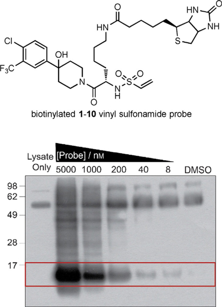
Figure 2.
The structure of the biotinylated 1–10d probe is shown above. This probe was added to HEK293T lysate (100 μg) containing purified GACKIX N627C (25 nm). Any biotin-containing proteins isolated on NeutrAvidin agarose beads were visualized by means of western blotting through chemiluminescence with streptavidin–HRP. The bands within the red box represent the expected mass for GACKIX N627C (12 kDa).

