Abstract
The Y-incision technique introduced by Dr. Bo Yang in 2021 is a very innovative technique that can enlarge the aortic annulus by two or more sizes without violating the left atrium or mitral valve. However, we encountered a case in which the left coronary artery ostium was located close to the left-non commissure. Therefore, we considered it would be dangerous to expand the incision to the left coronary annulus. We therefore devised a new technique that enlarges only the noncoronary annulus in an “L” fashion instead of a “Y” fashion. In performing this surgery, preoperative three-dimensional images were useful for understanding the anatomy when planning the aortic annular enlargement procedure. The L-incision technique can be a useful alternative method of aortic annulus enlargement.
Keywords: aortic annular enlargement, L-incision technique, modified Y-incision technique
Introduction
The Nicks and Manouguian procedures have been used as aortic annulus enlargement techniques. The Nicks procedure generally enables aortic annulus enlargement by only one valve size.1) Although the Manouguian procedure enables more enlargement, this procedure is complicated and carries the risk of mitral regurgitation in the long term.2) The Y-incision technique to enlarge the aortic annulus proposed by Dr. Bo Yang in 2021 solved these limitations.3) This technique widely opens the aortic root; however, it requires an extended subannular incision toward the nadir of the left coronary cusp. We modified the incision in an “L” fashion instead of a “Y” fashion, because the left coronary artery (LCA) ostium was close to the left-non commissure, as demonstrated by preoperative three-dimensional computed tomography (3D-CT) images.
Case Report
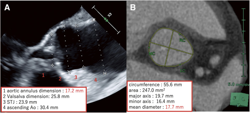
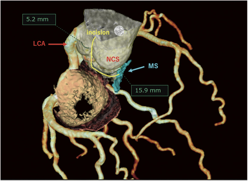
A 53-year-old female with a height of 139 cm and a body surface area (BSA) of 1.19 m2 complained of dyspnea on exertion. Echocardiography revealed severe aortic stenosis (AS) and a very narrow aortic annulus. CT and transesophageal echo examination showed that the annulus diameter was only 17 mm (Fig. 1), and aortic annular enlargement was necessary to implant a minimal-sized mechanical valve. Initially, we planned to apply the Y-incision technique. The 3D-CT showed that the distance between the ostium of the LCA and the left-non commissure was only 5.2 mm (Fig. 2); thus, a sufficient gain toward the left coronary cusp with the Y-incision technique was not achievable. Additionally, we determined that this approach carried the risk of coronary artery injury and would have little benefit. Thus, we planned to apply the annular enlargement with an “L”-fashioned incision of only the noncoronary area instead of a “Y” fashion. The distance between the bottom of the left-non commissure and the membranous septum (MS) was 15.9 mm using 3D-CT (Fig. 2).
Fig. 1. (A) The transesophageal echo examination shows the aortic root. The aortic annulus dimension is 17.2 mm. (B) CT shows that the mean diameter of aortic annulus is 17.7 mm. CT: computed tomography.
Fig. 2. Three-dimensional CT image. The blue area represents the MS under the NCS. The distance from the bottom of the left non-crossing to the MS is 15.9 mm, and the distance of 5.2 mm from the left-non commissure to the LCA. The yellow line is the “L”-fashioned incision. CT: computed tomography; MS: membranous septum; NCS: noncoronary sinus; LCA: left coronary artery.
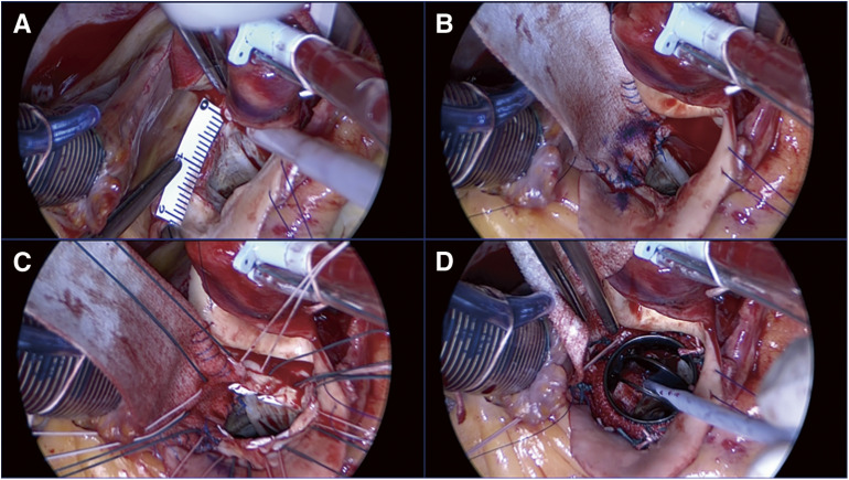
A partial transverse aortotomy was performed above the sinotubular junction (Video). After excision of the aortic valve, the annular calcification was completely debrided. The LCA ostium was very close to the left-non commissure and the Valsalva wall was thinner than expected. First, we made a vertical incision in the noncoronary Valsalva 2 mm from the left-non commissure, which extended across the annulus. We then changed the direction and advanced the incision along the annulus toward the MS. We incised 15 mm in an “L” fashion beneath the noncoronary annulus (Figs. 2 and 3A) considering the preoperative 3D-CT. The patch width to be met was set at 17 mm with a 2 mm seam allowance. A rectangular-shaped Hemashield Dacron patch (Boston Scientific Corp, Natick, MA, USA) 17 mm in width was sewn to the mitral annulus with running 5-0 Prolene suture (Ethicon, Somerville, NJ, USA), and the patch was sewn up to the incisional edges of the noncoronary sinus Valsalva (Fig. 3B). By applying this patch, the area of a right-angled isosceles triangle with one side of 15 mm is added to the existing valve opening area, so the valve circumference can theoretically be enlarged by 15mm at the maximum. In reality, it would be impossible to enlarge the image to 15mm due to distortion and other factors, but we thought it was possible to enlarge the image by 15 mm at least. A 16-mm-sizer of Medtronic Open Pivot AP360 valve (Medtronic, Minneapolis, MN, USA) was fitted to the enlarged aortic annulus. Twelve pledgeted mattress sutures were placed along the aortic annulus. Nine mattress sutures were placed from the ventricular side to the aortic side, and three mattress sutures were placed from the outside of the patch (Fig. 3C). A 16-mm Medtronic Open Pivot AP360 was placed in the supra-annular position (Fig. 3D). We verified that the patch did not interfere with the movement of valve leaflets. The aortotomy was closed with the patch trimmed into a teardrop shape.
Fig. 3. (A) The incision was extended 15 mm in an “L” fashion from left-non commissure’s bottom to the MS. (B) A rectangular patch was sewn. (C) 2-0 pledgeted sutures were placed along the aortic annulus, 9 by non-everting mattress and 3 by everting mattress from the outside to the inside the patch. (D) The mechanical valve was seated supra-annularly. The pivot guard faced the center of the patch and the center of the mechanical valve leaflets faced the left and right coronary arteries.
Discussion and Conclusions
The Nicks procedure can enlarge the aortic annulus by only one size up at most.1,3) The Y-incision technique, which uses rectangular patches, allows for a very convenient enlargement of two or more sizes. The Manouguian procedure requires an incision into the mitral valve or left atrium, which the Y-incision technique does not.2–4) The concern with the Y-incision technique is the risk of torsion of the LCA by the rectangular patch when the incision toward the left coronary annulus is too close to the left coronary ostium. Dr. Bo yang also mentioned the possibility of LCA distortion by a rectangular patch, and that the transverse sinus behind the rectangular patch allows the patch and left coronary sinus to extend posteriorly without significantly distorting the LCA.3) However, in the present case, the LCA was only 5.2 mm away from the left-non commissure. If the Y-incision technique had been performed by adding an incision up to the nadir of the left coronary sinus, as he suggested, we believed the LCA could be twisted. The L-incision technique, which does not require an incision under the left coronary annulus, can avoid this problem. However, since aortic annulus enlargement is performed only on the noncoronary annulus side, it is necessary to preoperatively estimate whether sufficient aortic annulus enlargement can be achieved using this method. In the present study, preoperative 3D-CT was analyzed, and the distance between the bottom of the non-left commissure and the MS was measured to be 15.9 mm. Considering the patient’s BSA (1.19 cm2), we preoperatively determined that prosthesis–patient mismatch could be avoided if a 16-mm Medtronic Open Pivot AP360 with an outer diameter of 23.8 mm could be implanted with a 15-mm aortic valve circumference enlargement. Preoperative measurements allowed us to safely and smoothly perform the procedure. There is also a risk of conduction disorders if the subannular incision extends into the MS. The patient was discharged from the hospital without any conduction disorders.
For patients with AS in whom the aortic annulus is so narrow that minimal prosthetic valve insertion by the Nicks procedure is expected to be difficult, the L incision, a modification of the Y incision, can be a simple and useful alternative method. From the 3D-CT analysis, it is possible to predict in advance the extent of enlargement that can be achieved before the surgery, thus predicting whether the target prosthetic valve size can be inserted. This is very useful in terms of safety for the surgeon.
Declarations
Ethics approval and consent to participate
Informed consent was obtained from the patient for this case report.
Consent for publication
Informed consent was obtained from the patient for this case report.
Data availability
The datasets used and/or analyzed during the current study are available from the corresponding author on reasonable request.
Authors’ contributions
GI involved in the initial assessment of the patient, analyzed data, and wrote the main manuscript. YT performed the surgery. YT, GI, AM, TK, and TS made contributions to the conceptual ideas. KN constructed a three-dimensional image, which contributed to the conception. MN and TK performed the postoperative treatment. All authors also read and approved the final manuscript.
Funding
Not applicable.
Disclosure Statement
The authors declare that they have no conflict of interest.
References
- 1).Nicks R, Cartmill T, Bernstein L. Hypoplasia of the aortic root. The problem of aortic valve replacement. Thorax 1970; 25: 339–46. [DOI] [PMC free article] [PubMed] [Google Scholar]
- 2).Imanaka K, Takamoto S, Furuse A. Mitral regurgitation late after Manouguian’s anulus enlargement and aortic valve replacement. J Thorac Cardiovasc Surg 1998; 115: 727–9. [DOI] [PubMed] [Google Scholar]
- 3).Yang B. A novel simple technique to enlarge the aortic annulus by two valve sizes. JTCVS Tech 2021; 5: 13–6. [DOI] [PMC free article] [PubMed] [Google Scholar]
- 4).Yang B, Naeem A. A Y incision and rectangular patch to enlarge the aortic annulus by three valve sizes. Ann Thorac Surg 2021; 112: e139–41. [DOI] [PMC free article] [PubMed] [Google Scholar]
Associated Data
This section collects any data citations, data availability statements, or supplementary materials included in this article.
Data Availability Statement
The datasets used and/or analyzed during the current study are available from the corresponding author on reasonable request.


