


Abstract
Human cardiac fibroblasts (HCFs) have mRNA transcripts that encode different mechanosensitive ion channels and channel regulatory proteins whose functions are not known yet. The primary goal of this work was to define the mechanosensitive ion channelome of HCFs. The most common type of cationic channel is the transient receptor potential (TRP) family, which is followed by the TWIK-related K+ channel (TREK), transmembrane protein 63 (TMEM63), and PIEZO channel (PIEZO) families. In the sodium-dependent NON-voltage-gated channel (SCNN) subfamily, only SCNN1D was shown to be highly expressed. Particular members of the acid-sensing ion channel (ASIC) (ASIC1 and ASIC3) subfamilies were also significantly expressed. The transcripts per kilobase million (TPMs) for Piezo 2 were almost 100 times less abundant than those for Piezo 1. The tandem of P domains in a weak inward rectifying K+ channel (TWIK)-2 channel, TWIK-related acid-sensitive K+ channel (TASK)-5, TASK-1, and the TWIK-related K1 (TREK-1) channel were the four most prevalent types in the K2P subfamily. The highest expression in the TRPP subfamily was found for PKD2 and PKD1, while in the TRPM subfamily, it was found for TRPM4, TRPM7, and TRPM3. TRPV2, TRPV4, TRPV3, and TRPV6 (all members of the TRPV subfamily) were also substantially expressed. A strong expression of the TRPC1, TRPC4, TRPC6, and TRPC2 channels and all members of the TRPML subfamily (MCOLN1, MCOLN2, and MCOLN3) was also shown. In terms of the transmembrane protein 16 (TMEM16) family, the HCFs demonstrated significant expression of the TMEM16H, TMEM16F, TMEM16J, TMEM16A, and TMEM16G channels. TMC3 is the most expressed channel in HCFs of all known members of the transmembrane channel-like protein (TMC) family. This analysis of the mechanosensitive ionic channel transcriptome in HCFs: (1) agrees with previously documented findings that all currently identified mechanosensitive channels play a significant and well recognized physiological function in elucidating the mechanosensitive characteristics of HCFs; (2) supports earlier preliminary reports that point to the most common expression of the TRP mechanosensitive family in HCFs; and (3) points to other new mechanosensitive channels (TRPC1, TRPC2, TWIK-2, TMEM16A, ASIC1, and ASIC3).
Keywords: Human cardiac fibroblasts, mechanosensitive cation channels, stretch-activated channels, mRNA, channelome, transcriptomic profile
Impact statement
Understanding the various types of channels is crucial for understanding the ionic mechanisms that trigger intracellular cascades, which are important for the regulation of myocardial function. Most of the recently published works do not address all possible genes and proteins in the channelome because they implement the reverse transcription polymerase chain reaction (RT-PCR) technique with predefined DNA primers for specific channel genes. The obtained data concerning the mechanosensitive ionic channel transcriptome in HCFs in this study support earlier preliminary reports that point to the most common expression of the transient receptor potential (TRP) mechanosensitive family and point to other new mechanosensitive channels (TRPC1, TRPC2, TWIK-2, TMEM16A, ASIC1, and ASIC3). We expect that newly classified participants will contribute to the understanding of the mechanoelectrical relationship between cardiac fibroblasts (CFs) and cardiomyocytes in special conditions like changed osmolarity, pH, or even the redox status of the extracellular environment.
Introduction
Mechanosensitive cation channels in cardiac fibroblasts
The ionic channels of cardiac myocytes have been pretty well investigated, but there is no complete information about the channelome of human cardiac fibroblasts (HCFs). Understanding the different classes of channels 1 is important for understanding the ionic mechanisms that trigger intracellular cascades,2–4 which are important for the regulation of myocardial function. 5 The channelomic properties of HCFs appear to be important for the excitability of the whole heart because cardiac fibroblasts (CFs) and cardiomyocytes can form gap junctions between each other.5–7 These gap junctions between cardiomyocytes and CFs (together with gap junctions between myocytes and endothelial cells and myocytes or macrophages) 8 work as modulators of cardiac electrophysiology 9 and substrates of mechanoelectrical feedback. 10 This relationship is especially important in different pathological conditions, such as atrial fibrillation. 11
One of the first attempts to estimate the main currents and their underlying channels in CFs was made by Li et al. 12 They reported that human ventricular fibroblasts contain the following currents: a large conductance Ca2+-activated K+ current (BKCa) 13 expressed in 88% of fibroblasts and represented by KCa.1.1 channel; two types of voltage-gated Na+ currents (INa) with different characteristics, represented by NaV1.2, NaV1.3, NaV1.6, and NaV1.7 channels for INa.TTX and NaV1.5 channels for INa.TTXR expressed in 61% of CFs; a delayed rectifier K+ current (IKDR), represented by Kv1.5, and Kv1.6 channels registered in 15% of CFs; a transient outward K+ current (Ito) represented by Kv4.2, and Kv4.3 channels registered in 14% of CFs, an inward rectifier K+ current (IKir) represented by Kir2.1 and Kir2.3 channels registered in 24% of CFs, and chloride current (ICl) represented by Clnc3 channels registered in 7% of CFs. 12
Another kind of channel identified in CFs is transient receptor potential (TRP) channels.14–24 These channels respond weekly to changes in voltage and can usually be turned on by different ligands, the accumulation of metabolites inside the cell, oxidative stress, thermal stimuli, or mechanical stimulation. 14 The presence of TRPC2, TRPC3, and TRPC5 (canonical subtype) channel transcripts was shown in rat ventricle fibroblasts. 12 TRPC1, TRPC3, TRPC6, and TRPC7 expression was also verified in rat ventricular fibroblasts. 13 HCFs contain TRPC1, TRPC4, and TRPC6, 14 while human atrial fibroblasts have been reported to have TRPC1 and TRPC6. 15 Furthermore, quantitative real-time polymerase chain reaction (qPCR) revealed that all TRP transcripts are present in murine ventricular fibroblasts. 15 Both TRPC3 and TRPC6 mediate fibrogenesis in the heart. 17 TRPC7 channels probably participate in many cell metabolic processes and can be inactivated by phospholipase C. 16
Three isoforms of TRPM channels (melastatin subtype) were found in CFs: TRPM2, TRPM4, and TRPM7.20–22 Simard et al. found that Ca2+-activated TRPM4 channels are very important for the growth of atrial fibroblasts in humans. 21 Overexpression of TRPM4 in human ventricle fibroblasts is probably one of the reasons for cardiac fibrogenesis in some diseases. 21 The presence of TRPM7 in CFs has been shown to have a key role in the response to angiotensin II. 22 In addition, during atrial fibrillation, these channels contribute to transforming growth factor β1 (TGF-β1)-induced fibrogenesis since they are the major Ca2+-permeable channel in human atrial fibroblasts. 22
TRPV2, TRPV3, and TRPV4 channels (vanilloid subtypes) are expressed in CFs.23–25 TRPV2 is an intracellular channel. Its role in CF is unclear, but it is known that it participates in the process of differentiation in dermal fibroblasts. 23 TRPV3 is involved in TGF-β1/cyclin-dependent kinase 2/cyclin E (TGF-β1/CDK2/cyclin E)-induced fibroblast proliferation during cardiac fibrosis under conditions of pressure overload. 24 The regulating role of TRPV4 in the process of fibroblast differentiation into myofibroblasts has been shown in rats. 25 It has the ability to react to both TGF-β1 signals and mechanical stimuli. 25
TRPA1 (subfamily A, member 1) is the only class of TRPA receptors. It exhibits sensitivity to TGF-β1 signals and is involved in the control of fibroblast differentiation. This regulatory function is mediated by the calcineurin-dependent stimulation of the nuclear factor of activated T cells (NFAT3). 26 Some of the TRP channels described above show mechanical sensitivity, but one of the most important classes of mechanically gated channels is the Piezo family. 27 Several studies showed expression of the Piezo1 channel in adult murine, rat, and human CFs at levels similar to those of endothelial cells.28–30
It is worth noting that the authors implemented the reverse transcription polymerase chain reaction (RT-PCR) technique with predefined DNA primers for specific channel genes. Therefore, they were mostly unable to find all possible genes and proteins in the channelome. Table 1 shows the ion channel-encoding genes and the corresponding cationic channel proteins.
Table 1.
Ion channel-encoding genes and the corresponding cationic channel proteins.
| Family | Subfamily | Gene | Channel | Family | Subfamily | Gene | Channel | Family | Subfamily | Gene | Channel |
|---|---|---|---|---|---|---|---|---|---|---|---|
| ENaC | ASIC | ASIC1 | ASIC1 | TRP | TRPA | TRPA1 | TRPA1 | TMEM16 | TMEM16 | ANO1 | Anoctamin-1 |
| ASIC2 | ASIC2 | TRPC | TRPC1 | TRPC1 | ANO2 | Anoctamin-2 | |||||
| ASIC3 | ASIC3 | TRPC2 | TRPC2 | ANO3 | Anoctamin-3 | ||||||
| ASIC4 | ASIC4 | TRPC3 | TRPC3 | ANO4 | Anoctamin-4 | ||||||
| ASIC5 | ASIC5 | TRPC4 | TRPC4 | ANO5 | Anoctamin-5 | ||||||
| SCNN | SCNN1A | SCNN1A | TRPC5 | TRPC5 | ANO6 | Anoctamin-6 | |||||
| SCNN1B | SCNN1B | TRPC6 | TRPC6 | ANO7 | Anoctamin-7 | ||||||
| SCNN1G | SCNN1G | TRPC7 | TRPC7 | ANO9 | Anoctamin-9 | ||||||
| SCNN1D | SCNN1D | TRPV | TRPV1 | TRPV1 | ANO8 | Anoctamin-8 | |||||
| TRPV2 | TRPV2 | ANO10 | Anoctamin-10 | ||||||||
| PIEZO | PIEZO | PIEZO1 | PIEZO1 | TRPV3 | TRPV3 | ||||||
| PIEZO2 | PIEZO2 | TRPV4 | TRPV4 | TMC | TMC | TMC1 | TMC1 | ||||
| TRPV5 | TRPV5 | TMC2 | TMC2 | ||||||||
| K2P | K2P | KCNK1 | TWIK-1 | TRPV6 | TRPV6 | TMC3 | TMC3 | ||||
| KCNK2 | TREK-1 | TRPM | TRPM1 | TRPM1 | TMC4 | TMC4 | |||||
| KCNK3 | TASK-1 | TRPM2 | TRPM2 | TMC5 | TMC5 | ||||||
| KCNK4 | TRAAK | TRPM3 | TRPM3 | TMC6 | TMC6 | ||||||
| KCNK5 | TASK-2 | TRPM4 | TRPM4 | TMC7 | TMC7 | ||||||
| KCNK6 | TWIK-2 | TRPM5 | TRPM5 | TMC8 | TMC8 | ||||||
| KCNK7 | KCNK7 | TRPM6 | TRPM6 | ||||||||
| KCNK9 | TASK-3 | TRPM7 | TRPM7 | ||||||||
| KCNK10 | TREK-2 | TRPM8 | TRPM8 | ||||||||
| KCNK12 | THIK-2 | TRPML | MCOLN1 | TRPML1 | |||||||
| KCNK13 | THIK-1 | MCOLN2 | TRPML1 | ||||||||
| KCNK15 | TASK-5 | MCOLN3 | TRPML1 | ||||||||
| KCNK16 | TALK-1 | TRPP/PKD | PKD1 | PC1 | |||||||
| KCNK17 | TALK-2 | PKDREJ | PKDREJ | ||||||||
| KCNK18 | TRESK | PKD1L1 | PKD1L1 | ||||||||
| TMEM63 | TMEM63 | TMEM63A | CSC1-like protein 1 | PKD1L2 | PKD1L2 | ||||||
| TMEM63B | CSC1-like protein 2 | PKD1L3 | PKD1L3 | ||||||||
| TMEM63C | Calcium permeable stress-gated cation channel 1 |
PKD2 | PKD2 | ||||||||
| PKD2L1 | PKD2L1 | ||||||||||
| PKD2L2 | PKD2L2 |
ENaC: epithelial sodium channels; ASIC: acid-sensing ion channels; SCNN: sodium-dependent NON-voltage-gated channels; TWIK: tandem of P domains in a weak inward rectifying K+ channel; K2P: two-pore domain potassium channels; TREK: TWIK-related K+ channel; TRAAK: TWIK-related arachidonic acid-activated K+ channel; TASK: TWIK-related acid-sensitive K+ channel; TALK: TWIK-related alkaline-sensitive K+ channel; THIK: TWIK-related halothane-sensitive K+ channel; TRESK: TWIK-related spinal cord-expressed K+ channel; TMEM63: transmembrane protein 63; TRPC: transient receptor potential—canonical; TRPV: vanilloid; TRPM: melastatin; TRPP/PKD: polycystin; TRPML: mucolipin; TRPA: ankyrin; TRPN: “nompC”; TMEM16: transmembrane protein 16; TMC: transmembrane channel-like protein; PIEZO: PIEZO channel; KCNK7: potassium channel subfamily K member 7.
Proteomics is a powerful method that has recently been utilized to identify the huge number of membrane proteins in CFs. 31 Despite the huge number of revealed proteins, ion channels and regulatory channel proteins represent only a small percentage. 32 This finding may be due to the difficulties in utilizing typical proteomic techniques to visualize proteins that have numerous transmembrane domains, such as ion channels. 33 HCF contains mRNA despite being anucleate; hence, screening the transcriptome is an alternate method to identify HCF proteins. Gene expression microarray experiments over the last decade have revealed an ever-growing number of transcripts inside HCFs. 34 Next-generation sequencing methods have recently revealed a huge number of genes expressed in HCFs. 34
Based on all of the above, the primary objective of this work was to define the mechanosensitive ion channelome of HCFs using transcriptomic analysis. Taking into account that HCFs contain mRNA transcripts that encode various mechanosensitive ion channels and channel regulatory proteins that have no recognized function yet, this basic study will help us investigate the mechanosensitive channelome of HCFs in depth. This research can also serve as a reference map for future research into this huge protein family in the HCF cell line.
Materials and methods
Cell culture
A primary culture of HCFs (adult ventricles, Catalog # 306-05A) was purchased from Sigma-Aldrich (St. Louis, MI, USA). The cells were cultured as monolayers in full Dulbecco’s Modified Eagle Medium (DMEM), which consisted of 10% fetal bovine serum (Invitrogen, Hong Kong) and antibiotics (100 U/mL penicillin G and 100 μg/mL streptomycin). The cells were incubated at a temperature of 37°C in a humidified atmosphere containing 95% air and 5% CO2. In order to reduce variations in the expression of genes and functional ion channels, the cells used in this study were from early passages (between 2 and 4).
RNA isolation, sequencing, and analysis
TRIzol (Invitrogen, Thermo Fisher Scientific, Inc., Dreieich, Germany) was used to isolate RNA for sequencing. It was extracted using chloroform (Sigma-Aldrich, Schnelldorf, Germany), in accordance with the manufacturer’s recommendations. The assessment of RNA concentration and purity was conducted using the Nanodrop (Thermo Fisher Scientific, Inc.).
The RNA that was extracted underwent purification using the RNeasy mini kit, manufactured by Qiagen (Hilden, Germany), in accordance with the procedure provided by the manufacturer. The assessment of RNA quantity and quality was conducted using the Nanodrop (Thermo Fisher Scientific, Inc.) and the Qi-RNA kit (Thermo Fisher Scientific, Inc.). The samples were prepared using the NEB Ultra II RNA kit (New England Biolabs, Ipswich, MA, USA), following the provided instructions. The NEBNext Poly (A) magnetic isolation module of mRNA (New England Biolabs) and unique dual-indexing were included throughout the preparation process. The concentration, size distribution, and quality of the resulting libraries were evaluated on a Qubit 4 fluorometer (Thermo Fisher Scientific, Inc.) using a high-sensitivity dsDNA kit (Invitrogen, Carlsbad, CA, USA), as well as a 4200 TapeStation with a high-sensitivity D5000 kit (Agilent, Santa Clara, CA, USA). On the basis of these assessments, the libraries were first normalized in accordance with their molarity, pooled, and then quantified using a library quantification kit developed specifically for Illumina platforms (Roche, Basel, Switzerland) on a StepOnePlus qPCR machine (Thermo Fisher Scientific, Inc.). The pooled libraries were loaded onto an S2 FlowCell at a concentration of 350 pm, along with 1% PhiX. Paired-end sequencing (2 × 150 bp) was performed using a NovaSeq 6000 next-generation sequencer (Illumina, San Diego, CA, USA). The RNA-Seq analysis was performed in triplicate.
The quality of raw FASTQ-sequenced reads was first evaluated using FastQC v0.11.5 (http://www.bioinformatics.babraham.ac.uk/projects/fastqc/). 35 We observed that our sequencing data were of high quality, with a high Q30 value and a good mapping rate, indicating a successful sequencing run and reliable base calling. Base quality (Phred scores) along the length of the reads in each sample are presented in the Supplementary Materials. Subsequently, the reads underwent a series of processing steps for quality enhancement and alignment. Initially, Trimmomatic v0.36 was employed to trim reads for quality and remove adapter sequences. 36 Surviving trimmed read pairs then underwent further processing using Fastp to eliminate poly-G tails and address artifacts specific to Novaseq/Nextseq platforms. 37 After implementing the quality trimming protocols, the reads underwent a further assessment of their quality via FastQC. 37 Following the completion of quality control and trimming procedures, the reads were aligned to the human reference genome GRCh38.p4 using HISAT2 with default settings. 38 The resulting alignments in SAM format were subsequently converted to BAM format and organized by coordinates using SAM tools v1.3.1. 39 These sorted alignment files were then processed through HTSeq-count v0.6.1p1 40 with specific options (-s no -t exon -I gene_id) to generate raw counts for downstream analysis. Subsequently, normalization of the raw counts was performed using the TPM (Transcripts per Kilobase Million) method to account for gene length and total read count differences across samples. This step allows for a more accurate comparison of gene expression levels across different genes and samples.
Unless specifically indicated otherwise, all experiments were conducted with a minimum of three replicates, and the presented data are expressed as the mean value ± standard deviation (SD).
Results and discussion
Consideration of ion channel stoichiometry in mRNA sequencing data analysis
When examining the mRNA sequencing data, it is important to consider the stoichiometry of the ion channel subunits in the formation of functional ion channels. Some cationic channels can form functional homo- or heteromeric channel tetramers, as described by Hofmann et al. 41 This means that the channel can be composed of identical subunits (homomeric) or different subunits (heteromeric), both in groups of four, to create a functioning channel.
In heteromeric channels, different genes encode for different subunits. Therefore, the number of functional channels that can be formed in a cell is often limited by the gene with the minimum number of transcripts. This is because even if one gene is highly expressed, the formation of functional channels would still be limited if another gene encoding for a different subunit in the same channel was expressed at lower levels.
Therefore, in our study, we made the assumption that the total number of functional channels is limited by the gene with the minimum number of transcripts. This approach allowed us to estimate the potential abundance of functional channels in the cell and get a better understanding of the transcriptional landscape of ion channels in the experimental context.
The families and subfamilies of mRNAs that encode the alpha subunits responsible for the formation of cationic channel pores are presented in Table 1. The TRP channels are the most abundant family of cationic channels, followed by the TWIK-related K+ channel (TREK), transmembrane protein 63 (TMEM63), PIEZO channel (PIEZO), and epithelial sodium channels (ENaC), and to a lesser extent, the transmembrane protein 16 (TMEM16) and transmembrane channel-like protein (TMC) channels (Figure 1). As expected, significant disparities were seen in the relative levels of gene expression across several cationic channels in HCFs, providing a preliminary assessment of the respective significance of each channel.
Figure 1.
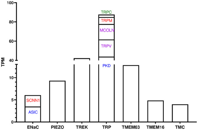
Profile analysis of channel gene expression in human cardiac fibroblasts. Families and subfamilies of mRNAs encoding pore-forming alpha subunits of cationic channels in human cardiac fibroblasts.
ASIC: acid-sensing ion channels; ENaC: epithelial sodium channels; SCNN: sodium-dependent NON-voltage-gated channels; PIEZO: PIEZO channel; TMC: transmembrane channel-like protein; TMEM16: transmembrane protein 16; TMEM63: transmembrane protein 63; TREK: TWIK-related K+ channel; TRPC: transient receptor potential—canonical; TRPM: melastatin; TRPML: mucolipin; TRPP/PKD: polycystin; TRPV: vanilloid; TWIK: tandem of P domains in a weak inward rectifying K+ channel; the values of the y-axis is the sum of the average TPM between three replicates.
Two subfamilies of ENaC have been identified: acid-sensing ion channels (ASIC) and sodium-dependent NON-voltage-gated channels (SCNN) (Figure 2(A)). ENaC is assembled as a heterotrimer composed of the following homologous subunits: a, b, and g or d, b, and g. 42 The subunits of ENaC have structural homology to a group of proteins found in Caenorhabditis elegans, including mec-4, mec-10, and deg-1. 43 These proteins have been hypothesized to function as ion channels involved in mechanosensation,43–45 which led to the assumption that ENaC is responsive to membrane stretching. 45 ENaC mechanosensitivity has also been investigated in epithelial cells. 46 Only SCNN1D and SCNN1A were shown to be highly expressed in this study, while the other two members of the SCNN subfamily, SCNN1G and SCNN1B, were represented only with a few TPMs (Figure 2(A)).
Figure 2.

Relative abundance of genes encoding ion channels in human cardiac fibroblasts. Bars represent the mean value ± SEM (n = 10).
The second subfamily ASIC is represented by genes with several splice variants, resulting in at least eight subunits: ASIC1a and -b; ASIC2a and -b; ASIC3a, -b, and -c (only in humans); other species have only one ASIC3 and ASIC4. 47 Chronologically, the majority of ASICs had various names due to the history of cloning. 47 Currently, there is enough scientific data indicating the expression of ASIC in primary sensory neurons and mechanoreceptor afferents. 47 Defects in mechanotransduction were seen in several tissues, including the arterial baroreceptors, stomach, colon, cochlea, and skin, as a result of disruption in the expression of different ASICs. 46 Interestingly, the present particular members of the ASIC subfamily (ASIC3, ASIC1, and ASIC4) were significantly expressed in HCFs (Figure 2(A)), while the remaining ASIC2 was weakly expressed (Figure 2(A)).
Lewis and Grandl 48 discovered that the nonselective cation channel Piezo is inherently mechanosensitive and functions as a primary force sensor in numerous cells and tissues. Piezo1 ion pore is selective for divalent (Ba2+, Ca2+, Mg2+, and Mn2+) and monovalent (K+, Na+, Cs+, and Li+) cations. 49 Piezo1 is expressed in both cardiomyocytes and fibroblasts inside the cardiac tissue, and its presence is required for normal ventricular outflow and aortic valve function, 50 the cyclic stretch response, 51 and the facilitation of homeostatic cardiac mechanochemical transduction. 52 Both Piezo 1 and Piezo 2 were detected in this study; however, the TPMs for Piezo 2 were almost 100 times less abundant than those for Piezo 1 (Figure 2(B)). In tissue culture, Piezo1 reacts to a variety of mechanical stimuli, including lateral membrane tension,53–55 compression,56,57 osmotic swelling, 58 and rhythmic mechanical stimulations. 59
K2P channels, also known as leak or background conductance channels, are a diverse group of voltage-independent K+ channels that regulate the resting Em in smooth muscle cells (SMCs). 60 Based on their sequence and functional properties, K2P channels may be divided into six separate subfamilies. The weak rectifying group (which includes TWIK-1, TWIK-2, and K2P7.1), the mechanogated group (which includes TREK-1, TREK-2, and TWIK-related arachidonic acid-activated K+ channel [TRAAK]), the acid-sensing group (which includes TWIK-related acid-sensitive K+ channel [TASK]-1, TASK-3, and TASK-5), the alkaline-sensitive group (which includes TWIK-related alkaline-sensitive K+ channel [TALK]-1, TALK-2, and TASK-2), the halothane-sensitive group (which includes TWIK-related halothane-sensitive K+ channel (THIK)-1 and THIK-2), and the TWIK-related spinal cord-expressed K+ channel (TRESK) channel, which is predominantly expressed in the spinal cord. Mechanosensitive ion channels have been identified among the 15 distinct kinds of K2P channels, specifically the arachidonic acid-activated K+ channel (TRAAK) and TWIK-related K1 and K2 (TREK-1 and TREK-2, respectively).60,61 Abraham et al. 61 reported that only TREK-1 has been identified as being expressed and functional in HCFs. Despite this, our results showed that in the cultured HCFs, the most expressed type was the TWIK-2 channel, followed by TASK-5, TASK-1, KCNK7, TREK-1, and TWIK-1 (Figure 2(C)). Within the mammalian contractile organs and tissues, TREK-1 plays a crucial role in inducing cellular hyperpolarization, followed by a reduction in cellular contraction in response to mechanical forces. 62 The inherent mechanosensitivity of TREK-1 has been confirmed by electrophysiological studies of reconstituted TREK-1 channels in liposomes. 63 Obtained recordings have shown that TREK-1 may be triggered by intrinsic tension of the membrane without the participation of additional cytoplasmic players.63,64 The obtained results based on the expression levels of the K2P channels represented by a low number of TPMs were in the following order: THIK-1, TASK-2, THIK-2, and TASK-3 (Figure 2(C)).
Furthermore, it was thought that TMEM63A (also called CSC1-like protein 1) was an ion channel that was activated by high osmolarity. 65 The same authors have shown that TMEM63s function as ion channels that respond to mechanical stimuli, inducing stretch-activated currents upon expression in non-specialized cells. 65 TMEM63A has two homologous counterparts, TMEM63B and TMEM63C, which exhibit a high degree of similarity. 66 The highly expressed members of the TMEM63 family were found to be TMEM63A, whose physiological role is incompletely understood but mechanical sensitivity is evidenced, 67 and TMEM63B (CSC1-like protein 2), a mammalian homolog of OSCA characterized as a mechanosensitive channel (Figure 2(F)). The Ca2+-permeable channel is responsible for facilitating the entry of Ca2+ ions into the cells in response to changes in the extracellular Ca2+ concentration and hyperosmolarity and can alter HCF mechanosensitivity. 65 In addition, TMEM63C (Ca2+ permeable stress-gated cation channel 1), as another member of the TMEM63 family, was found to be weakly expressed and, as a result, does not participate in the mechanosensitive properties of HCFs (Figure 2(F)).
The family of TRP channels consists of cationic channels (seven subfamilies) that exhibit a range of biophysical properties and play various physiological roles. These subfamilies include TRPA (ankyrin), TRPC (canonical), TRPM (melastatin), TRPML (mucolipin), TRPN (“nompC,” families lacking potential mechanoreceptors), TRPP/PPD (polycystin), and TRPV (vanilloid). TRP channels are involved in the modulation of intracellular Ca2+ levels via two mechanisms: direct facilitation of Ca2+ influx and indirect modification of the Em. 45 The TRPP channels PKD2 and PKD1 exhibited the highest levels of expression in this study, while in the TRPM subfamily, the TRPM4, TRPM7, TRPM3, TRPM8, and TRPM2 channels were also highly expressed, whereas TRPM6, TRPM5, and TRPM1 were represented with only a few TPMs (Figure 2(G)). Among the members of the subfamily TRPV, TRPV2, TRPV4, TRPV1, TRPV3, and TRPV6 were also highly expressed (Figure 2(G)). TRPV2 is involved in stretch-dependent responses in heart cells. 68 The expression of TRPV2 is concentrated in intercalated disks that allow synchronous contraction of the cardiomyocytes. 68 TRPV1 expression has been observed in many cell types derived from murine hearts, including afferent fibers and myocytes, 69 SMC, 70 endothelial cells, 70 and fibroblasts. 71 In mammals, TRPV1 functions as a mechanosensor in response to various mechanical stressors. 70 TRPV1 functions as an osmoreceptor in reaction to hypotonic conditions, 72 a thermosensor, 73 and an intravascular mechanosensor for variations in blood pressure, 74 and in the urinary bladder and the digestive tract.75–78 Functionally expressed TRPV1 channels in murine CFs were confirmed by Wang et al. 71 The TRPV4 channel is Ca2+ and Mg2+ permeable, and its activation is triggered by a variety of stimuli, including heat, osmotic swelling, mechanical stress, and the binding of the metabolite 5’,6’-epoxyeicosatrienoic acid and phorbol ester compounds.79–82 Measurements of the single-channel conductance of mammalian TRPV4 induced by applied puppet pressure indicated stretch insensitivity in the condition of their overexpression in HEK293 cells. 79 TRPV4 found in murine chondrocytes does not exhibit any changes in response to membrane stretching; however, the application of mechanical stress at sites where cells come into contact with the extracellular matrix seems to induce TRPV4-dependent electrical currents. 82 TRPV3 was also found to be expressed in CFs. 83 Carvacrol-induced activation of TRPV3 in a rat model of hypertrophy induced by pressure overload was found to exacerbate cardiac function and promote an increase in fibrosis. 83 This study confirms a strong expression of the TRPC1, TRPC4, TRPC6, and TRPC2 channels and all members of the TRPML subfamily (MCOLN1, MCOLN2, and MCOLN3) (Figure 2(G)). TRPC channels appear to play a significant role in cardiac dysfunction, and nearly all of them are upregulated in HCFs,84–86 except for TRPC3, TRPC7, and TRPC5. The study conducted by Yamaguchi et al. revealed the significance of TRPC3/6/7 channels in the cellular response to mechanical stressors such as flow, stretch, and osmotic pressure. 86 Among all TRPC channels expressed in cardiac tissue, only TRPC1 and TRPC6 exhibit mechanosensitivity.86,87 Despite published data by Huang et al. 88 that TRPC1 expression is not detected in HCFs; in this, study, we detected significant expression of TRPC1 (Figure 2(G)). Further studies have shown that TRPC6 exhibits mechanosensitivity upon activation, and it is postulated that the channel’s involvement in the response to mechanical stimuli requires PLA2/-hydroxylase metabolite pathway (20-HETE pathway) activation. 89 Further investigation is necessary to determine if TRPC6 functions as a main mechanosensor or whether its upregulation is a result of Ca2+ influx via other channels, such as Orai. Despite the fact that published data by Löf et al. 90 has not confirmed TRPC2 expression in humans; in this study, we confirmed TRPC2 expression in HCFs. It seems that TRPC channels do not play the role of major mechanosensors, but they still have an important role in the response to mechanical stressors. TRPML1 (TRP cation channel, mucolipin subfamily, member 1), as the best-expressed TRPML channel in this study, is believed to channel iron ions through the endosome/lysosome membrane into the cell. 91 It is especially important for lysosomal function and has a significant role in processes such as vesicular trafficking, exocytosis, and autophagy.92,93 TRPML2 is associated with the small GTPase (Arf6)-regulated trafficking pathway and is involved in intracellular transport through membranes and membrane proteins. 94 Computational analyses of the secondary structure predict the presence of six transmembrane domains, an ion transport motif, and a TRP motif.
The following TRP channels were also present with lower TPMs: TRPM6, TRPM5, TRPM1, TRPC3, TRPC7, and TRPC5 (Figure 2(G)). In the TMEM16 family, HCFs showed that the TMEM16H, TMEM16F, TMEM16J, TMEM16A, and TMEM16G were the most expressed channels (Figure 2(E)). TMEM16H stands as the only vertebrate homolog for which no investigation ha2s been made regarding ion conduction or lipid scrambling. 95 TMEM16G has significant expression levels in the prostate and plays an important role in the establishment of intercellular connections, 96 while TMEM16J is associated with the development of colorectal and pancreatic cancer. 97 When Ca2+ enters the cell through the mechanosensitive channel TMEM16F, also known as ANO-6, it activates the phospholipid scramblase. This causes the exchange of cytoplasmic anionic phospholipids for neutral phospholipids, which relaxes dynamin and opens the compartments of the reserve plasma membrane. 98 The findings show that shear stress or fluid flow upregulates Cl−-currents in human and mouse biliary cells and that the operative channel is TMEM16A-(ANO-1), a Ca2+-activated Cl−-channel (CaCC). 99 Identification of this unique, highly expressed channel may shed fresh light on the mechanosensitivity of HCFs. Despite extensive research on CaCC flux in different cell types, such as the SMC in the human airways, there is still ongoing debate on the precise molecular channel structure responsible for the Cl− conductance. The identification of a novel protein family, known as the TMEM16 family, has now emerged as a possible contender for the authentic CaCC. 99 This prompted us to examine whether the functional expression of members of this family may be seen in cultured HCFs. Based on the obtained results, all additional members of the TMEM16 family in HCFs were quantified in the following order: ANO4, ANO9, ANO3, ANO2, and ANO5 (Figure 2(E)).
The TMC family is part of a seven-branch superfamily. TMC3 was found to be the member of the TMC family that was most common in HCFs. It was found to be a protein with mechanosensitive ion channel activity (Figure 2(D)). The TMC6 gene in humans encodes a protein known as TMC protein 6 (TMC6), which is the second most highly expressed member in HCFs (Figure 2(D)). 100 In vivo, TMC6 and its homolog TMC8 mutually interact and combine with zinc transporter 1 (SLC30A1) to form a complex, and both are typically situated inside the endoplasmic reticulum, but they may also be found in the Golgi apparatus and nuclear membrane. 101 The use of electrophysiological techniques in the examination of HEK293T cells has shown that TMC4 functions as a voltage-dependent Cl− channel, facilitating the permeation of organic anions such as gluconate. 102 TMC7 was found to be involved in the proliferation of cancerogenic cells and oral squamous cell carcinoma metastasis. 103 Based on the obtained TPMs, this was the last seriously expressed channel from the TMC family found in HCFs. TMC1, TMC5, and TMC2 were characterized by weaker expression (Figure 2(D)).
Conclusions
In short, this analysis of the mechanosensitive ionic channel transcriptome in HCFs (1) agrees with previously published data about the expression of mechanosensitive channels that play an important role in describing the mechanosensitive properties of HCFs; (2) supports earlier preliminary reports that the TRP mechanosensitive family is the most represented in HCFs; and (3) points to other new mechanosensing channels (TRPC1, TRPC2, TWIK-2, TMEM16A, ASIC1, and ASIC3). We anticipate that the newly categorized mechanosensitive channels will help in understanding the mechanoelectrical relationship between fibroblasts and cardiomyocytes in certain situations, like when the osmolarity, pH, or redox status of the outside environment changes. Anyway, more research needs to be done to figure out the real meaning of the newly categorized channels with mechanosensitive properties.
In addition to its many strengths, this study also has a few weaknesses. One of the basic limitations is that the expression of mRNA does not consistently result in protein synthesis, and in some cases, even when protein is synthesized, it may not effectively translocate to the cellular membrane or form a conformation that enables its functionality as a channel. The basic problem lies in the fact that cell culture passage has the potential to modify channel expression.
Supplemental Material
Supplemental material, sj-pdf-1-ebm-10.1177_15353702231218488 for Transcriptomic profile of the mechanosensitive ion channelome in human cardiac fibroblasts by Vadim Mitrokhin, Andrei Bilichenko, Viktor Kazanski, Roman Schobik, Stanislav Shileiko, Veronika Revkova, Vladimir Kalsin, Olga Kamkina, Andre Kamkin and Mitko Mladenov in Experimental Biology and Medicine
Footnotes
Authors’ Contributions: VM and MM were responsible for the conception and design. VM, OK, AB, SS, VK, VR, and OK were responsible for developing and carrying out the experiments. AK, VM, and MM prepared and interpreted the table and figure plots. Both MM and VM wrote the first version of this paper. Each co-author has seen and approved the final text and is fully accountable for all parts of the work shown here.
The author(s) declared no potential conflicts of interest with respect to the research, authorship, and/or publication of this article.
Funding: The author(s) received no financial support for the research, authorship, and/or publication of this article.
ORCID iD: Mitko Mladenov  https://orcid.org/0000-0003-3475-2131
Data Availability Statement: The data were submitted to the NCBI Sequence Read Archive (SRA) (Data Citation 1). This SRA submission contains raw data from all samples. Sequence Read Archive (SRA): https://www.ncbi.nlm.nih.gov/sra/PRJNA991152 (2023).
Supplemental Material: Supplemental material for this article is available online.
References
- 1. Kamkin A, Kiseleva I, Isenberg G. Activation and inactivation of a non-selective cation conductance by local mechanical deformation of acutely isolated cardiac fibroblasts. Cardiovasc Res 2003;57:793–803 [DOI] [PubMed] [Google Scholar]
- 2. Mitrokhin V, Gorbacheva L, Mladenov M, Kamkin A. IL-2-induced NF-κB phosphorylation upregulates cation nonselective conductance in human cardiac fibroblasts. Int Immunopharmacol 2018;64:170–4 [DOI] [PubMed] [Google Scholar]
- 3. Mitrokhin V, Kazanski V, Kalsin V, Mladenov M, Kamkin A. Interleukin-6 induced activation of a non-selective outward cation conductance in human cardiac fibroblasts. Cytokine 2017;97:117–22 [DOI] [PubMed] [Google Scholar]
- 4. Kamkin GA, Kamkina VO, Shim LA, Bilichenko A, Mitrokhin MV, Kazansky EV, Filatova ST, Abramochkin VD, Mladenov IM. The role of activation of two different sGC binding sites by NO-dependent and NO-independent mechanisms in the regulation of SACs in rat ventricular cardiomyocytes. Physiol Rep 2022;10:e15246 [DOI] [PMC free article] [PubMed] [Google Scholar]
- 5. Kohl P, Kamkin GA, Kiseleva SI, Noble D. Mechanosensitive fibroblasts in the sino-atrial node region of rat heart: interaction with cardiomyocytes and possible role. Exp Physiol 1994;79:943–56 [DOI] [PubMed] [Google Scholar]
- 6. Camelliti P, Green RC, le Grice I, Kohl P. Fibroblast network in rabbit sinoatrial node: structural and functional identification of homogeneous and heterogeneous cell coupling. Circ Res 2004;94:828–35 [DOI] [PubMed] [Google Scholar]
- 7. Camelliti P, Green RC, Kohl P. Structural and functional coupling of cardiac myocytes and fibroblasts. Adv Cardiol 2006;42:132–49 [DOI] [PubMed] [Google Scholar]
- 8. Mahoney MV, Mezzano V, Mirams RG, Maass K, Li Z, Cerrone M, Morley EG. Connexin43 contributes to electrotonic conduction across scar tissue in the intact heart. Sci Rep 2016;6:26744. [DOI] [PMC free article] [PubMed] [Google Scholar]
- 9. Johnson DR, Camelliti P. Role of non-myocyte gap junctions and connexin hemichannels in cardiovascular health and disease: novel therapeutic targets. Int J Mol Sci 2018;19:866. [DOI] [PMC free article] [PubMed] [Google Scholar]
- 10. Kamkin A, Kiseleva I, Isenberg G, Wagner DK, Günther J, Theres H, Scholz H. Cardiac fibroblasts and the mechano-electric feedback mechanism in healthy and diseased hearts. Prog Biophys Mol Biol 2003;82:111–20 [DOI] [PubMed] [Google Scholar]
- 11. Yue L, Xie J, Nattel S. Molecular determinants of cardiac fibroblast electrical function and therapeutic implications for atrial fibrillation. Cardiovasc Res 2011;89:744–53 [DOI] [PMC free article] [PubMed] [Google Scholar]
- 12. Li GR, Sun HY, Chen JB, Zhou Y, Tse HF, Lau CP. Characterization of multiple ion channels in cultured human cardiac fibroblasts. PLoS ONE 2009;4:e7307 [DOI] [PMC free article] [PubMed] [Google Scholar]
- 13. Mitrokhin MV, Kalsin V, Kamkina O, Babkina I, Zotov A, Troitskiy AV, Mladenov IM, Kamkin GA. Participation of PKG and PKA-related pathways in the IFN-γ induced modulation of the BKCa channel activity in human cardiac fibroblasts. J Pharmacol Sci 2019;141:25–31 [DOI] [PubMed] [Google Scholar]
- 14. Rose AR, Hatano N, Ohya S, Imaizumi Y, Giles RW. C-type natriuretic peptide activates a non-selective cation current in acutely isolated rat cardiac fibroblasts via natriuretic peptide C receptor-mediated signaling. J Physiol 2007;580:255–74 [DOI] [PMC free article] [PubMed] [Google Scholar]
- 15. Nishida M, Onohara N, Sato Y, Suda R, Ogushi M, Tanabe S, Inoue R, Mori Y, Kurose H. Gα12/13-mediated up-regulation of TRPC6 negatively regulates endothelin-1-induced cardiac myofibroblast formation and collagen synthesis through the nuclear factor of activated T cells activation. J Biol Chem 2007;282:23117–28 [DOI] [PubMed] [Google Scholar]
- 16. Chen JB, Tao R, Sun HY, Tse HF, Lau CP, Li GR. Multiple Ca2+ signaling pathways regulate intracellular Ca2+ activity in human cardiac fibroblasts. J Cell Physiol 2010;223:68–75 [DOI] [PubMed] [Google Scholar]
- 17. Du J, Xie J, Zhang Z, Tsujikawa H, Fusco D, Silverman D, Yue L. TRPM7-mediated Ca2+ signals confer fibrogenesis in human atrial fibrillation. Circ Res 2010;106:992–1003 [DOI] [PMC free article] [PubMed] [Google Scholar]
- 18. Harada M, Luo X, Qi YX, Tadevosyan A, Maguy A, Ordog B, Ledoux J, Kato T, Naud P, Voigt N, Shi Y, Kamiya K, Murohara T, Kodama I, Tardif JC, Schotten U, van Wagoner RD, Dobrev D, Nattel S. Transient receptor potential canonical-3 channel-dependent fibroblast regulation in atrial fibrillation. Circulation 2012;126:2051–64 [DOI] [PMC free article] [PubMed] [Google Scholar]
- 19. Kiyonaka S, Kato K, Nishida M, Mio K, Numaga T, Sawaguchi Y, Yoshida T, Wakamori M, Mori E, Numata T, Ishii M, Takemoto H, Ojida A, Watanabe K, Uemura A, Kurose H, Morii T, Kobayashi T, Sato Y, Sato C, Hamachi I, Mori Y. Selective and direct inhibition of TRPC3 channels underlies biological activities of a pyrazole compound. Proc Natl Acad Sci U S A 2009;106:5400–5 [DOI] [PMC free article] [PubMed] [Google Scholar]
- 20. Takahashi K, Sakamoto K, Kimura J. Hypoxic stress induces transient receptor potential melastatin 2 (TRPM2) channel expression in adult rat cardiac fibroblasts. J Pharmacol Sci 2012;118:186–97 [DOI] [PubMed] [Google Scholar]
- 21. Simard C, Magaud C, Adjlane R, Dupas Q, Sallé L, Manrique A, Bois P, Faivre JF, Guinamard R. TRPM4 non-selective cation channel in human atrial fibroblast growth. Pflügers Arch 2020;472:1719–32 [DOI] [PubMed] [Google Scholar]
- 22. Li S, Li M, Yi X, Guo F, Zhou Y, Chen S, Wu X. TRPM7 channels mediate the functional changes in cardiac fibroblasts induced by angiotensin II. Int J Mol Med 2017;39:1291–8 [DOI] [PubMed] [Google Scholar]
- 23. Ishii T, Uchida K, Hata S, Hatta M, Kita T, Miyake Y, Okamura K, Tamaoki S, Ishikawa H, Yamazaki J. TRPV2 channel inhibitors attenuate fibroblast differentiation and contraction mediated by keratinocyte-derived TGF-β1 in an in vitro wound healing model of rats. J Dermatol Sci. 2018;90:332–42 [DOI] [PubMed] [Google Scholar]
- 24. Liu Y, Qi H, Mingyao E, Shi P, Zhang Q, Li S, Wang Y, Cao Y, Chen Y, Ba L, Gao J, Huang W, Sun H. Transient receptor potential vanilloid-3 (TRPV3) activation plays a central role in cardiac fibrosis induced by pressure overload in rats via TGF-β1 pathway. Naunyn Schmiedebergs Arch Pharmacol 2018;391:131–43 [DOI] [PubMed] [Google Scholar]
- 25. Adapala KR, Thoppil JR, Luther JD, Paruchuri S, Meszaros GJ, Chilian MW, Thodeti KC. TRPV4 channels mediate cardiac fibroblast differentiation by integrating mechanical and soluble signals. J Mol Cell Cardiol 2013;54:45–52 [DOI] [PMC free article] [PubMed] [Google Scholar]
- 26. Wang Z, Xu Y, Wang M, Ye J, Liu J, Jiang H, Wan J. TRPA1 inhibition ameliorates pressure overload-induced cardiac hypertrophy and fibrosis in mice. EBioMedicine 2018;36:54–62 [DOI] [PMC free article] [PubMed] [Google Scholar]
- 27. Blythe MN, Muraki K, Ludlow JM, Stylianidis V, Gilbert TJH, Evans LE, Cuthbertson K, Foster R, Swift J, Li J, Drinkhill JM, van Nieuwenhoven AF, Porter EK, Beech JD, Turner AN. Mechanically activated PIEZO1 channels of cardiac fibroblasts stimulate p38 mitogen-activated protein kinase activity and interleukin-6 secretion. J Biol Chem 2019;294:17395–408 [DOI] [PMC free article] [PubMed] [Google Scholar]
- 28. Ploeg CM, Munts C, Prinzen WF, Turner NA, van Bilsen M, van Nieuwenhoven AF. PIEZO1 mechanosensitive ion channel mediates stretch-induced Nppb expression in adult rat cardiac fibroblasts. Cells 2021;10:1745. [DOI] [PMC free article] [PubMed] [Google Scholar]
- 29. Emig R, Knodt W, Krussig JM, Zgierski-Johnston MC, Gorka O, Groß O, Peyronnet R. PIEZO1 channels contribute to the regulation of human atrial fibroblast mechanical properties and matrix stiffness sensing. Cells 2021;10:663. [DOI] [PMC free article] [PubMed] [Google Scholar]
- 30. Sebastião JM, Pereira R, Serra M, Gomes-Alves P, Alves MP. Unveiling human cardiac fibroblast membrane proteome. Proteomics 2018;18:e1700446 [DOI] [PubMed] [Google Scholar]
- 31. Moita RM, Silva MM, Diniz C, Serra M, Hoet MR, Barbas A, Simão D. Transcriptome and proteome profiling of activated cardiac fibroblasts support target prioritization in cardiac fibrosis. Front Cardiovasc Med 2022;9:1015473. [DOI] [PMC free article] [PubMed] [Google Scholar]
- 32. Qi R, Lin E, Song J, Wang Y, Lin L. Proteomic insights into cardiac fibrosis: from pathophysiological mechanisms to therapeutic opportunities. Molecules 2022;27:8784. [DOI] [PMC free article] [PubMed] [Google Scholar]
- 33. Kalyanasundaram A, Ling D, Gardner LM, Artiga JE, Hansen JB, Webb A, Freitas AM, Pietrzak M, Whitson AB, Mokadam AN, Janssen LMP, Mohler JP, Fedorov VV. Fibroblast-specific proteo-transcriptomes reveal distinct fibrotic signatures of human sinoatrial nodes in nonfailing and failing hearts. Circulation 2021;144:126–43 [DOI] [PMC free article] [PubMed] [Google Scholar]
- 34. Hesse J, Owenier C, Lautwein T, Zalfen R, Weber FJ, Ding Z, Alter C, Lang A, Grandoch M, Gerdes N, Fischer WJ, Klau WG, Dieterich C, Köhrer K, Schrader J. Single-cell transcriptomics defines heterogeneity of epicardial cells and fibroblasts within the infarcted murine heart. eLife 2021;10:e65921 [DOI] [PMC free article] [PubMed] [Google Scholar]
- 35. Andrews S. FASTQC. A quality control tool for high throughput sequence data. 2010, http://www.bioinformatics.babraham.ac.uk/projects/fastqc/
- 36. Bolger MA, Lohse M, Usadel B. Trimmomatic: a flexible trimmer for Illumina sequence data. Bioinformatics 2014;30:2114–20 [DOI] [PMC free article] [PubMed] [Google Scholar]
- 37. Chen S, Zhou Y, Chen Y, Gu J. Fastp: an ultra-fast all-in-one FASTQ preprocessor. Bioinformatics 2018;34:884–90 [DOI] [PMC free article] [PubMed] [Google Scholar]
- 38. Kim D, Langmead B, Salzberg SL. HISAT: a fast spliced aligner with low memory requirements. Nat Methods 2015;12:357–60 [DOI] [PMC free article] [PubMed] [Google Scholar]
- 39. Li H, Handsaker B, Wysoker A, Fennell T, Ruan J, Homer N, Marth G, Abecasis G, Durbin R, 1000. Genome Project Data Processing Subgroup. The sequence alignment/map format and SAMtools. Bioinformatics 2009;25:2078–9 [DOI] [PMC free article] [PubMed] [Google Scholar]
- 40. Anders S, Pyl TP, Huber W. HTSeq-a Python framework to work with high-throughput sequencing data. Bioinformatics 2015;31:166–9 [DOI] [PMC free article] [PubMed] [Google Scholar]
- 41. Hofmann T, Schaefer M, Schultz G, Gudermann T. Subunit composition of mammalian transient receptor potential channels in living cells. Proc Natl Acad Sci U S A 2002;99:7461–6 [DOI] [PMC free article] [PubMed] [Google Scholar]
- 42. Baldin JP, Barth D, Fronius M. Epithelial Na+ channel (ENaC) formed by one or two subunits forms functional channels that respond to shear force. Front Physiol 2020;11:141. [DOI] [PMC free article] [PubMed] [Google Scholar]
- 43. Canessa MC, Schild L, Buell G, Thorens B, Gautschi I, Horisberger DJ, Rossier CB. Amiloride-sensitive epithelial Na+ channel is made of three homologous subunits. Nature 1994;367:463–7 [DOI] [PubMed] [Google Scholar]
- 44. Chalfie M, Hart AC, Rankin CH, Goodman MB. Assaying mechanosensation. Worm Book, 2014, http://www.wormbook.org/ [DOI] [PMC free article] [PubMed]
- 45. Perez-Vizcaino F, Cogolludo A, Mondejar-Parreño G. Transcriptomic profile of cationic channels in human pulmonary arterial hypertension. Sci Rep 2021;11:15829. [DOI] [PMC free article] [PubMed] [Google Scholar]
- 46. Kellenberger S, Schild L. Epithelial sodium channel/degenerin family of ion channels: a variety of functions for a shared structure. Physiol Rev 2002;82:735–67 [DOI] [PubMed] [Google Scholar]
- 47. Chih-Cheng C, Chia-Wen W. Neurosensory mechanotransduction through acid-sensing ion channels. J Cell Mol Med 2013;17:337–49 [DOI] [PMC free article] [PubMed] [Google Scholar]
- 48. Lewis HA, Grandl J. PIEZO1 ion channels inherently function as independent mechanotransducers. eLife 2021;10:e70988 [DOI] [PMC free article] [PubMed] [Google Scholar]
- 49. Jiang F, Yin K, Wu K, Zhang M, Wang S, Cheng H, Zhou Z, Xiao B. The mechanosensitive PIEZO1 channel mediates heart mechano-chemo transduction. Nat Commun 2021;12:869. [DOI] [PMC free article] [PubMed] [Google Scholar]
- 50. Garoffolo G, Pesce M. Mechanotransduction in the cardiovascular system: from developmental origins to homeostasis and pathology. Cells 2019;8:1607. [DOI] [PMC free article] [PubMed] [Google Scholar]
- 51. Friedrich O, Wagner S, Battle RA, Schurmann S, Martinac B. Mechano-regulation of the beating heart at the cellular level–mechanosensitive channels in a normal and diseased heart. Prog Biophys Mol Biol 2012;110:226–38 [DOI] [PubMed] [Google Scholar]
- 52. Lewis HA, Grandl J. Mechanical sensitivity of PIEZO1 ion channels can be tuned by cellular membrane tension. eLife 2015;4:e12088 [DOI] [PMC free article] [PubMed] [Google Scholar]
- 53. Coste B, Xiao B, Santos SJ, Syeda R, Grandl J, Spencer SK, Kim ES, Schmidt M, Mathur J, Dubin EA. Piezo proteins are pore-forming subunits of mechanically activated channels. Nature 2012;483:176–81 [DOI] [PMC free article] [PubMed] [Google Scholar]
- 54. Cox DC, Bae C, Ziegler L, Hartley S, Nikolova-Krstevski V, Rohde RP, Ng CA, Sachs F, Gottlieb AP, Martinac B. Removal of the mechanoprotective influence of the cytoskeleton reveals PIEZO1 is gated by bilayer tension. Nat Commun 2016;7:10366. [DOI] [PMC free article] [PubMed] [Google Scholar]
- 55. Wang L, You X, Lotinun S, Zhang L, Wu N, Zou W. Mechanical sensing protein PIEZO1 regulates bone homeostasis via osteoblast-osteoclast crosstalk. Nat Commun 2020;11:282. [DOI] [PMC free article] [PubMed] [Google Scholar]
- 56. Lee W, Leddy AH, Chen Y, Lee HS, Zelenski AN, McNulty LA, Wu J, Beicker NK, Coles J, Zauscher S, Grandl J, Sachs F, Guilak F, Liedtke BW. Synergy between PIEZO1 and PIEZO2 channels confers high-strain mechanosensitivity to articular cartilage. Proc Natl Acad Sci U S A 2014;111:5114–22 [DOI] [PMC free article] [PubMed] [Google Scholar]
- 57. Deivasikamani V, Dhayalan S, Abudushalamu Y, Mughal R, Visnagri A, Cuthbertson K, Scragg LJ, Munsey ST, Viswambharan H, Muraki K, Foster R, Asipu Sivaprasadarao A, Kearney TM, David J, Beech JD, Sukumar P. PIEZO1 channel activation mimics high glucose as a stimulator of insulin release. Sci Rep 2019;9:16876. [DOI] [PMC free article] [PubMed] [Google Scholar]
- 58. Lewis HA, Cui FA, McDonald FM, Grandl J. Transduction of repetitive mechanical stimuli by PIEZO1 and PIEZO2 ion channels. Cell Rep 2017;19:2572–85 [DOI] [PMC free article] [PubMed] [Google Scholar]
- 59. Yang Q, Zhou Y, Wang J, Fu W, Li X. Study on the mechanism of excessive apoptosis of nucleus pulposus cells induced by shRNA-PIEZO1 under abnormal mechanical stretch stress. J Cell Biochem 2019;120:3989–97 [DOI] [PubMed] [Google Scholar]
- 60. Brohawn GS. How ion channels sense mechanical force: insights from mechanosensitive K2P channels TRAAK, TREK1, and TREK2. Ann N Y Acad Sci 2015;1352:20–32 [DOI] [PubMed] [Google Scholar]
- 61. Abraham MD, Lee ET, Watson JL, Mao L, Chandok G, Wang GH, Frangakis S, Pitt SG, Shah HS, Wolf MJ, Rockman AH. The two-pore domain potassium channel TREK-1 mediates cardiac fibrosis and diastolic dysfunction. J Clin Investig 2018;128:4843–55 [DOI] [PMC free article] [PubMed] [Google Scholar]
- 62. Wiedmann F, Rinne S, Donner B, Decher N, Katus AH, Schmidt C. Mechanosensitive TREK-1 two-pore-domain potassium (K2P) channels in the cardiovascular system. Prog Biophys Mol Biol 2021;159:126–35 [DOI] [PubMed] [Google Scholar]
- 63. Brohawn GS, Su Z, MacKinnon R. Mechanosensitivity is mediated directly by the lipid membrane in TRAAK and TREK1 K+ channels. Proc Natl Acad Sci U S A 2014;111:3614–9 [DOI] [PMC free article] [PubMed] [Google Scholar]
- 64. Berrier C, Pozza A, de Lavalette AdL, Chardonnet S, Mesneau A, Jaxel C, le Maire M, Ghazi A. The purified mechanosensitive channel TREK-1 is directly sensitive to membrane tension. J Biol Chem 2013;288:27307–14 [DOI] [PMC free article] [PubMed] [Google Scholar]
- 65. Murthy ES, Dubin EA, Whitwam T, Jojoa-Cruz S, Cahalan MS, Mousavi ARS, Ward BA, Patapoutian A. OSCA/TMEM63 are an evolutionarily conserved family of mechanically activated ion channels. eLife 2018;7:e41844 [DOI] [PMC free article] [PubMed] [Google Scholar]
- 66. Zhao X, Yan X, Liu Y, Zhang P, Ni X. Coexpression of mouse TMEM63A, TMEM63B and TMEM63C confers hyperosmolarity activated ion currents in HEK293 cells. Cell Biochem Funct 2016;34:238–41 [DOI] [PubMed] [Google Scholar]
- 67. Yan H, Helman G, Murthy ES, Ji H, Crawford J, Kubisiak T, Bent JS, Xiao J, Taft JR, Coombs A, Wu Y, Pop A, Li D, de Vries SL, Jiang Y, Salomons SG, van der Knaap SM, Patapoutian A, Simons C, Burmeister M, Wang J, Wolf IN. Heterozygous variants in the mechanosensitive ion channel TMEM63A result in transient hypomyelination during infancy. Am J Hum Genet 2019;105:996–1004 [DOI] [PMC free article] [PubMed] [Google Scholar]
- 68. Iwata Y, Matsumura T. Blockade of TRPV2 is a novel therapy for cardiomyopathy in muscular dystrophy. Int J Mol Sci. 2019;20:3844. [DOI] [PMC free article] [PubMed] [Google Scholar]
- 69. Gao F, Liang Y, Wang X, Lu Z, Li L, Zhu S, Liu D, Yan Z, Zhu Z. TRPV1 activation attenuates high-salt diet-induced cardiac hypertrophy and fibrosis through PPAR-δ upregulation. PPAR Res 2014;2014:491963. [DOI] [PMC free article] [PubMed] [Google Scholar]
- 70. Zhang C, Ye L, Zhang Q, Wu F, Wang L. The role of TRPV1 channels in atherosclerosis. Channels 2020;14:141–50 [DOI] [PMC free article] [PubMed] [Google Scholar]
- 71. Wang Q, Ma S, Li D, Zhang Y, Tang B, Qiu C, Yang Y, Yang D. Dietary capsaicin ameliorates pressure overload–induced cardiac hypertrophy and fibrosis through the transient receptor potential vanilloid type 1. Am J Hypertens 2014;27:1521–9 [DOI] [PubMed] [Google Scholar]
- 72. Liedtke W, Choe Y, Martí-Renom AM, Bell MA, Denis SC, Hudspeth JA, Friedman MJ, Heller S. Vanilloid receptor–related osmotically activated channel (VR-OAC), a candidate vertebrate osmoreceptor. Cell 2000;103:525–35 [DOI] [PMC free article] [PubMed] [Google Scholar]
- 73. Rosenbaum T, Simon AS. TRPV1 receptors and signal transduction. In: Liedtke WB, Heller S. (eds) TRP ion channel functions in sensory transduction and cellular signaling cascades. Boca Raton, FL: CRC Press/Taylor & Francis, 2007, pp.69–84 [Google Scholar]
- 74. Baylie LR, Brayden EJ. TRPV channels and vascular function. Acta Physiol 2011;203:99–116 [DOI] [PMC free article] [PubMed] [Google Scholar]
- 75. Chan HLC, Facer P, Davis BJ, Smith DG, Egerton J, Bountra C, Williams SN, Anand P. Sensory fibres expressing capsaicin receptor TRPV1 in patients with rectal hypersensitivity and fecal urgency. Lancet 2003;361:385–91 [DOI] [PubMed] [Google Scholar]
- 76. Lazzeri M, Vannucchi GM, Zardo C, Spinelli M, Beneforti P, Turini D, Faussone-Pellegrini MS. Immunohistochemical evidence of vanilloid receptor 1 in the normal human urinary bladder. Eur Urol 2004;46:792–8 [DOI] [PubMed] [Google Scholar]
- 77. Liedtke W. TRPV channels’ function in osmo- and mechanotransduction. In: Liedtke WB, Heller S. (eds) TRP ion channel function in sensory transduction and cellular signaling cascades. Boca Raton, FL: CRC Press, 2007, pp. 2–24 [PubMed] [Google Scholar]
- 78. Apostolidis A, Brady MC, Yiangou Y, Davis J, Fowler CJ, Anand P. Capsaicin receptor TRPV1 in the urothelium of neurogenic human bladders and effect of intravesical resiniferatoxin. Urology 2005;65:400–5 [DOI] [PubMed] [Google Scholar]
- 79. Nikolaev AY, Cox DC, Ridone P, Rohde RP, Cordero-Morales FJ, Vásquez V, Laver RD, Martinac B. Mammalian TRP ion channels are insensitive to membrane stretch. J Cell Sci 2019;132:jcs238360 [DOI] [PMC free article] [PubMed] [Google Scholar]
- 80. Swain MS, Romac M-JJ, Shahid AR, Pandol JS, Liedtke W, Vigna RS, Liddle AR. TRPV4 channel opening mediates pressure-induced pancreatitis initiated by PIEZO1 activation. J Clin Investig 2020;130:2527–41 [DOI] [PMC free article] [PubMed] [Google Scholar]
- 81. Vriens J, Watanabe H, Janssens A, Droogmans G, Voets T, Nilius B. Cell swelling, heat, and chemical agonists use distinct pathways for the activation of the cation channel TRPV4. Proc Natl Acad Sci U S A 2004;101:396–401 [DOI] [PMC free article] [PubMed] [Google Scholar]
- 82. Servin-Vences RM, Moroni M, Lewin RG, Poole K. Direct measurement of TRPV4 and PIEZO1 activity reveals multiple mechanotransduction pathways in chondrocytes. eLife 2017;6:e21074 [DOI] [PMC free article] [PubMed] [Google Scholar]
- 83. Liu Y, Qi H, E M, Shi P, Zhang Q, Li S, Wang Y, Cao Y, Chen Y, Ba L, Gao J, Huang W, Sun H. Transient receptor potential vanilloid-3 (TRPV3) activation plays a central role in cardiac fibrosis induced by pressure overload in rats via TGF-beta1 pathway. Naunyn Schmiedebergs Arch Pharmacol 2018;391:131–43 [DOI] [PubMed] [Google Scholar]
- 84. Morine JK, Paruchuri V, Qiao X, Aronovitz M, Huggins SG, DeNofrio D, Kiernan SM, Karas HR, Kapur KN. Endoglin selectively modulates transient receptor potential channel expression in left and right heart failure. Cardiovasc Pathol 2016;25:478–82 [DOI] [PMC free article] [PubMed] [Google Scholar]
- 85. Bush WE, Hood BD, Papst JP, Chapo AJ, Minobe W, Bristow RM, Olson NE, McKinsey AT. Canonical transient receptor potential channels promote cardiomyocyte hypertrophy through the activation of calcineurin signaling. J Biol Chem 2006;281:33487–96 [DOI] [PubMed] [Google Scholar]
- 86. Yamaguchi Y, Iribe G, Nishida M, Naruse K. Role of TRPC3 and TRPC6 channels in the myocardial response to stretch: linking physiology and pathophysiology. Prog Biophys Mol Biol 2017;130:264–72 [DOI] [PubMed] [Google Scholar]
- 87. Spassova AM, Hewavitharana T, Xu W, Soboloff J, Gill LD. A common mechanism underlies stretch activation and receptor activation of TRPC6 channels. Proc Natl Acad Sci U S A 2006;103:16586–91 [DOI] [PMC free article] [PubMed] [Google Scholar]
- 88. Huang H, Wang W, Liu P, Jiang Y, Zhao Y, Wei H, Niu W. TRPC1 expression and distribution in rat hearts. Eur J Histochem 2009;53:e26 [DOI] [PMC free article] [PubMed] [Google Scholar]
- 89. Inoue R, Jensen JL, Jian Z, Shi J, Hai L, Lurie IA, Henriksen HF, Salomonsson M, Morita H, Kawarabayashi Y, Mori M, Mori Y, Ito I. Synergistic activation of vascular TRPC6 channel by receptor and mechanical stimulation via phospholipase C/diacylglycerol and phospholipase a2/omega-hydroxylase/20-HETE pathways. Circ Res 2009;104:1399–409 [DOI] [PubMed] [Google Scholar]
- 90. Löf C, Viitanen T, Sukumaran P, Törnquist K. TRPC2: of mice but not men. Adv Exp Med Biol 2011;704:125–34 [DOI] [PubMed] [Google Scholar]
- 91. Dong XP, Cheng X, Mills E, Delling M, Wang F, Kurz T, Xu H. The type IV mucolipidosis-associated protein TRPML1 is an endo-lysosomal iron release channel. Nature 2008;455:992–6 [DOI] [PMC free article] [PubMed] [Google Scholar]
- 92. Wang W, Zhang X, Gao Q, Xu H. TRPML1: an ion channel in the lysosome. Handb Exp Pharmacol 2014;222:631–45 [DOI] [PubMed] [Google Scholar]
- 93. Di Paola S, Scotto-Rosato A, Medina LD. TRPML1: the Ca(2+) retaker of the lysosome. Cell Calcium 2018;69:112–21 [DOI] [PubMed] [Google Scholar]
- 94. Karacsonyi C, Miguel SA, Puertollano R. Mucolipin-2 localizes to the Arf6-associated pathway and regulates the recycling of GPI-APs. Traffic 2007;8:1404–14 [DOI] [PubMed] [Google Scholar]
- 95. Gyobu S, Ishihara K, Suzuki J, Segawa K, Nagata S. Characterization of the scrambling domain of the TMEM16 family. Proc Natl Acad Sci U S A 2017;114:6274–9 [DOI] [PMC free article] [PubMed] [Google Scholar]
- 96. Das S, Hahn Y, Walker AD, Nagata S, Willingham CM, Peehl MD, Bera TK, Lee B, Pastan I. Topology of NGEP, a prostate-specific cell:cell junction protein widely expressed in many cancers of different grade level. Cancer Res 2008;68:6306–12 [DOI] [PMC free article] [PubMed] [Google Scholar]
- 97. Jun I, Park SH, Piao H, Han WJ, An JM, Yun GB, Zhang X, Cha HY, Shin KY, Yook IJ, Jung J, Gee YH, Park SJ, Yoon SD, Jeung HC, Lee GM. ANO9/TMEM16J promotes tumourigenesis via EGFR and is a novel therapeutic target for pancreatic cancer. Br J Cancer 2017;117:1798–809 [DOI] [PMC free article] [PubMed] [Google Scholar]
- 98. Bricogne C, Fine M, Pereira MP, Sung J, Tijani M, Wang Y, Henriques R, Collins KM, Hilgemann WD. TMEM16F activation by Ca2+ triggers plasma membrane expansion and directs PD-1 trafficking. Sci Rep 2019;9:619. [DOI] [PMC free article] [PubMed] [Google Scholar]
- 99. Gallos G, Remy EK, Danielsson J, Funayama H, Fu WX, Chang SHY, Yim P, Xu D, Sr EWC. Functional expression of the TMEM16 family of calcium-activated chloride channels in airway smooth muscle. Am J Physiol Lung Cell Mol Physiol 2013;305:625–34 [DOI] [PMC free article] [PubMed] [Google Scholar]
- 100. Ramoz N, Rueda AL, Bouadjar B, Montoya SL, Orth G, Favre M. Mutations in two adjacent novel genes are associated with epidermodysplasia verruciformis. Nat Genet 2002;32:579–81 [DOI] [PubMed] [Google Scholar]
- 101. Lazarczyk M, Pons C, Mendoza JA, Cassonnet P, Jacob Y, Favre M. Regulation of cellular zinc balance as a potential mechanism of EVER-mediated protection against pathogenesis by cutaneous oncogenic human papillomaviruses. J Exp Med 2008;205:35–42 [DOI] [PMC free article] [PubMed] [Google Scholar]
- 102. Kasahara Y, Narukawa M, Ishimaru Y, Kanda S, Umatani C, Takayama Y, Tominaga M, Oka Y, Kondo K, Kondo T, Takeuchi A, Misaka T, Abe K, Asakura T. TMC4 is a novel chloride channel involved in high-concentration salt taste sensation. J Physiol Sci 2021;71:23. [DOI] [PMC free article] [PubMed] [Google Scholar]
- 103. Jia B, Zheng X, Qiu X, Jiang X, Liu J, Huang Z, Xiang S, Chen G, Zhao J. Long non-coding RNA MIR4713HG aggravates malignant behaviors in oral tongue squamous cell carcinoma via binding with microRNA let-7c-5p. Int J Mol Med 2021;47:84. [DOI] [PMC free article] [PubMed] [Google Scholar]
Associated Data
This section collects any data citations, data availability statements, or supplementary materials included in this article.
Supplementary Materials
Supplemental material, sj-pdf-1-ebm-10.1177_15353702231218488 for Transcriptomic profile of the mechanosensitive ion channelome in human cardiac fibroblasts by Vadim Mitrokhin, Andrei Bilichenko, Viktor Kazanski, Roman Schobik, Stanislav Shileiko, Veronika Revkova, Vladimir Kalsin, Olga Kamkina, Andre Kamkin and Mitko Mladenov in Experimental Biology and Medicine