Abstract
Background/Aim
Interim positron emission tomography/computed tomography (PET/CT) scan is a valuable tool for assessing the early metabolic response to chemotherapy in diffuse large B-cell lymphoma (DLBCL). Although radiotherapy is an effective treatment for lymphoma, especially for local tumor control, the role of consolidative radiotherapy in diffuse large B-cell lymphoma (DLBCL) remains controversial. This study analyzed the clinical outcomes of patients with DLBCL treated with rituximab, cyclophosphamide, doxorubicin, vincristine, and prednisone (R-CHOP), stratified by interim PET response and the administration of radiotherapy.
Patients and Methods
We conducted a retrospective review of 107 patients with DLBCL treated with R-CHOP chemotherapy between January 2012 and December 2016. Overall survival (OS), recurrence-free survival (RFS), and freedom from disease progression (FFDP) were calculated using the Kaplan–Meier method and compared using the log-rank test.
Results
Forty-six patients were included in this analysis, with a median follow-up time of 65.9 months (range=4.7-125.3 months). The metabolic CR (mCR) group exhibited superior OS, RFS, and FFDP compared with the metabolic PR (mPR) group (p=0.003, p=0.001, and p=0.008, respectively). The 1-, 2-, and 5-year FFDP were 92.97%, 89.3%, and 85.6%, respectively, in the mCR group and 78.6%, 61.9%, and 44.2%, respectively, in the mPR group. In subgroup analysis, the FFDP of the mPR group without radiotherapy was significantly lower than that of the other groups (mCR with/without radiotherapy and mPR with radiotherapy, p=0.001).
Conclusion
Consolidative radiation therapy using interim PET can benefit patients who do not achieve mCR. Further well-controlled prospective randomized trials are required.
Keywords: Diffuse large B cell lymphoma, interim PET/CT, consolidative radiation therapy, consolidation, radiotherapy, early response
Diffuse large B-cell lymphoma (DLBCL), the most common subtype of non-Hodkin’s lymphoma, is characterized by its aggressive clinical course (1,2). The standard chemotherapy regimen for DLBCL is rituximab, cyclophosphamide, doxorubicin, vincristine, and prednisone (R-CHOP) (3,4). Although approximately two-thirds of patients achieve remission with first-line R-CHOP therapy, up to 30% face a poor prognosis if the initial treatment fails, even with salvage therapy (5-7).
For patients unresponsive to R-CHOP, radiotherapy emerges as a potential treatment option. In the rituximab era, studies evaluating the role of radiotherapy have produced conflicting results (8-10), attributed to reduced survival benefits from radiotherapy due to radiation-related toxicity and the heterogeneous nature of DLBCL. Given recent developments in radiotherapy techniques, current treatment strategies employing smaller target volume contours and radiation dose reduction therapy are anticipated to further diminish radiation-related toxicities (11-13). Therefore, we posit that the incorporation of optimized radiotherapy in selective patients with DLBCL will result in improved clinical outcomes.
Meanwhile, F-18 fluorodeoxyglucose (FDG) positron emission tomography (PET)/computed tomography (CT) imaging can assist in the detection, staging, and remission assessment of patients with DLBCL (14,15). In recent years, numerous studies have demonstrated that patients negative for 18FDG-PET (interim PET; iPET) during treatment exhibit superior clinical outcomes compared with patients who are iPET-positive (15,16). We hypothesized that the efficacy of radiotherapy would vary depending on iPET results (iPET-negative or iPET-positive). However, limited information is available on how the incorporation of radiotherapy based on iPET results influences the oncological outcomes of DLBCL. This study aimed to stratify patients according to whether or not they received radiotherapy and iPET response and to compare oncological outcomes.
Patients and Methods
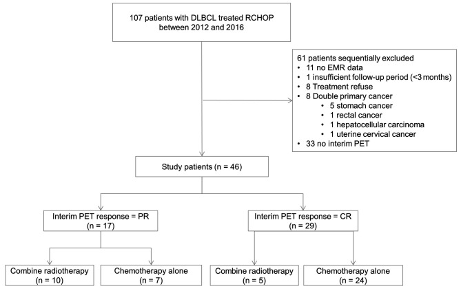
Study population. We performed a retrospective review of 107 patients with DLBCL treated with R-CHOP at our hospital between January 2012 and December 2016. The inclusion criteria were as follows: (a) histologically proven DLBCL; (b) age ≥18 years; (c) receipt of first-line R-CHOP chemotherapy; (d) underwent baseline PET-CT; and (e) underwent iPET scan after two or three cycles of R-CHOP chemotherapy. Exclusion criteria included: (a) double primary cancer; (b) negative baseline PET-CT; (c) incomplete R-CHOP chemotherapy with <6 cycles; and (d) insufficient follow-up duration (<3 months). Ultimately, 46 patients were included in the analysis (Figure 1).
Figure 1. Flow diagram. DLBCL: Diffuse large B-cell lymphoma; RCHOP: rituximab, cyclophosphamide, doxorubicin, vincristine, and prednisone; EMR: electronic medical record; PET: positron emission tomography; PR: partial remission; CR: complete remission.
This study received approval from the Kosin University Gospel Hospital Ethics Committee and Review Board, and the requirement for informed consent was waived owing to the retrospective nature of the study.
Treatment. Chemotherapy. All patients received six cycles of R-CHOP, consisting of rituximab (375 mg/m2 on day 1, then every 3 weeks), cyclophosphamide (750 mg/m2 intravenously on day 1), doxorubicin (50 mg/m2 intravenously on day 1), vincristine (1.4 mg/m2, ≤2.0 mg intravenously on day 1), and prednisolone (100 mg daily, orally on days 1 to 5, every 3 weeks).
Radiation therapy. Each patient was positioned in supine position and immobilized using a vacuum cushion. CT with intravenous contrast enhancement (GE LightSpeed RT; GE Healthcare, Waukesha, WI, USA) was employed. The CT slice thickness was set at 2.5-5 mm. Delineation of the gross target volume was aided using CT, magnetic resonance imaging MRI, and PET. In some patients, a clinical target volume covering the adjacent nodal area in the same axial section as the gross tumor volume (GTV) was established. The planning target volume included a 5-7 mm margin from the GTV or clinical target volume. Radiation therapy underwent verification with kV imaging guidance using an onboard imager (Varian Medical Systems, Palo Alto, CA, USA) once or twice weekly. Setup corrections were based on anatomical landmarks, including bones and organs. Radiation therapy was administered using 6-10 MV X-rays from a linear accelerator (Clinac IX; Varian Medical Systems).
PET-CT imaging. FDG PET-CT scans were performed thrice using the same imaging protocol and PET-CT: at baseline, after two or three cycles (iPET), and upon completion of six cycles of RCHOP chemotherapy. All patients fasted for ≤6 h before the FDG PET/CT scan, and serum glucose levels were measured before F18-FDG injection. Prior to the PET scan, non-contrast-enhanced CT (3 mm slice thickness) was performed for anatomical co-registration. FDG (370-444MBq) was injected intravenously, and scanning commenced 50-70 min later using a Siemens Biograph mCT-64 PET/CT scanner (Siemens Healthcare, Knoxville, TN, USA). PET images were acquired in three-dimensional mode with an acquisition time of 3 min for each table position and 1.5 min for each bed position. Iterative reconstruction of PET images was performed with ordered subset expectation maximization, and attenuation-correction was performed using CT-derived transmittance maps. In our study, a complete metabolic remission (mCR) was defined as FDG uptake in DLBCL lesions indistinguishable from that in adjacent normal tissue.
Statistical analysis. To compare clinical factors between patients with and without metabolic remission on iPET, the t-test was employed for continuous variables, whereas the chi-square test or Fisher’s exact test was used for categorical variables. Overall survival (OS) was estimated from the date of diagnosis to the date of death or the last follow-up, and recurrence-free survival (RFS) and freedom from disease progression (FFDP) were estimated from the date of diagnosis to the date of tumor recurrence or the last follow-up. OS, RFS, and FFDP were calculated using the Kaplan–Meier (KM) method and compared using log-rank tests. Univariate and multivariate analyses were conducted using a Cox proportional hazards model. Backward-elimination Cox regression was applied to select principal risk factors in the multivariate model. For all statistical tests, p-values <0.05 were considered statistically significant. All statistical analyses were carried out using R software (version 3.6.1; R Foundation for Statistical Computing, Vienna, Austria).
Results
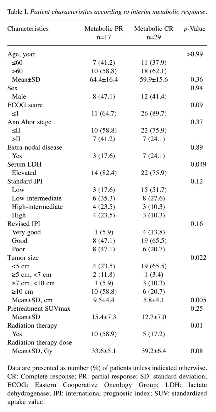
Patients characteristics. Of 46 patients, 29 (63.0%) achieved mCR on iPET. The baseline characteristics of the patients are summarized in Table I. The mean tumor size was 9.5 cm in the metabolic partial remission (mPR) group and 5.8 cm in the mCR group, with this difference being statistically significant (p=0.005). Serum lactate dehydrogenase (LDH) levels were higher in the mPR group (82.4% vs. 75.9%, p=0.049). A higher percentage of patients in the mPR group received consolidative radiotherapy compared with the mCR group (58.9% vs. 17.2%, p=0.01). No statistically significant differences were observed between groups in terms of age, sex, ECOG scores, Ann Abor stage, extra-nodal disease status, IPI score, pretreatment SUVmax, and dose of radiation therapy.
Table I. Patient characteristics according to interim metabolic response.
Data are presented as number (%) of patients unless indicated otherwise. CR: Complete response; PR: partial response; SD: standard deviation; ECOG: Eastern Cooperative Oncology Group; LDH: lactate dehydrogenase; IPI: international prognostic index; SUV: standardized uptake value.
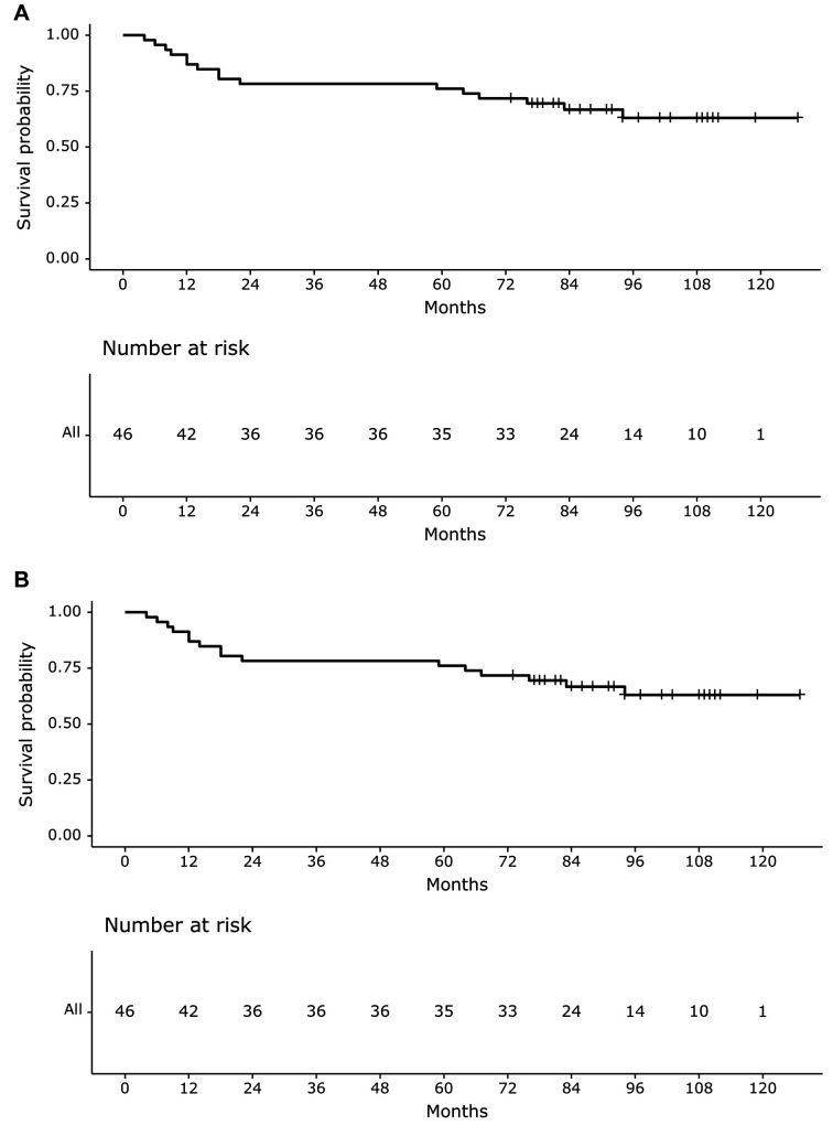
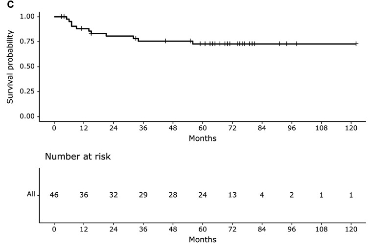
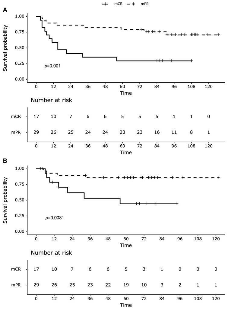
Oncologic outcome. As of the October 2023 analysis, 30 (65.2%) patients were alive (mCR group, 23; mPR group, seven) and 16 patients had died, with a median follow-up time of 65.9 months (range=4.7-125.3 months). The median time to OS, RFS, and FFDP was not reached. The mean OS was 94.3 months (Figure 2A). The RFS rates at 1, 2, and 5-years were 78.3%, 69.6%, and 60.9%, respectively (Figure 2B). Eleven patients experienced relapse after treatment, and the 1-, 2-, and 5-year FFDP rates were 88.1%, 80.7%, and 72.8%, respectively (Figure 2C). The mCR group exhibited superior OS, RFS, and FFDP compared with the mPR group (p=0.003, p=0.001, and p=0.008, respectively). The 1-, 2-, and 5-year OS rates according to the KM curve were 100%, 89.7%, and 86.2%, respectively, in the mCR group, and 64.7 %, 58.8 %, and 58.8 %, respectively, in the mCR group. The 1-, 2-, and 5-year RFS rates were 89.7%, 86.2%, and 79.3%, respectively, in the mCR group and 58.8 %, 41.2 %, and 29.4 %, respectively, in the mPR group (Figure 3A). The 1-, 2-, and 5-year FFDF were 92.97%, 89.3%, and 85.6% in the mCR group, and 78.6%, 61.9%, and 44.2% in the mPR group, respectively (Figure 3B).
Figure 2. Clinical outcome for all patients, by months of diagnosis. (A) Overall survival (OS), (B) recurrence-free survival (RFS), and (C) freedom from disease progression (FFDP).
Figure 3. Kaplan–Meier survival analysis for (A) recurrence-free survival (RFS) and (B) Freedom from disease progression (FFDP) stratified by interim positron emission tomography (PET) response [metabolic complete remission (mCR) vs. metabolic partial remission (mPR)].
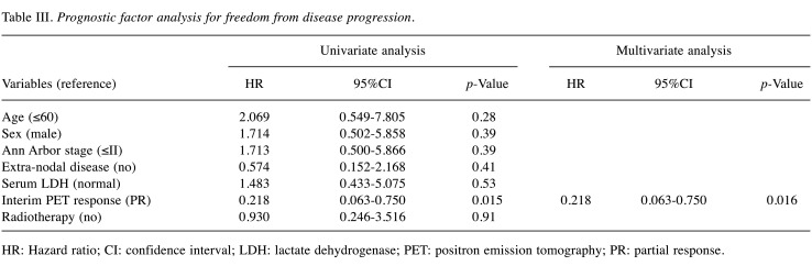
In both the univariate and multivariate analyses, only mPR was associated with poorer OS (Table II) and FFDF (Table III). The multivariate analysis of RFS indicated that mPR (HR=0.164, 95%CI=0.062-0.434; p<0.001) and older age (HR=3.372, 95%CI=0.053-10.799) were associated with a decreased RFS. Similarly, in the multivariate analysis of RFS, mPR (HR=0.164, 95%CI=0.062-0.434; p<0.001) and age >60 (HR=3.372, 95%CI=0.053-10.799) were associated with poorer RFS (Table IV).
Table II. Prognostic factor analysis for overall survival.
HR: Hazard ratio; CI: confidence interval; LDH: lactate dehydrogenase; PET: positron emission tomography; PR: partial response.
Table III. Prognostic factor analysis for freedom from disease progression.
HR: Hazard ratio; CI: confidence interval; LDH: lactate dehydrogenase; PET: positron emission tomography; PR: partial response.
Table IV. Prognostic factor analysis for recurrence-free survival.
HR: Hazard ratio; CI: confidence interval; LDH: lactate dehydrogenase; PET: positron emission tomography; PR: partial response.
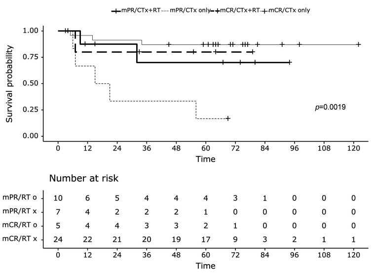
Subgroup analysis. To explore the impact of consolidative radiation therapy based on early treatment response to iPET, the data was stratified into four groups (Figure 1): Group A (mPR with consolidative radiation therapy), Group B (mPR without consolidative radiation therapy), Group C (mCR with consolidative radiation therapy), Group D (mCR without consolidative radiation therapy). Analysis of these four groups indicate that Group B exhibited significantly poorer FFDP than the other groups (Figure 4, p=0.001).
Figure 4. Kaplan–Meier survival analysis for freedom from disease progression (FFDP) stratified by interim positron emission tomography (PET) response [metabolic complete remission (mCR) vs. metabolic partial remission (mPR)] and treatment [chemotherapy (CTx) + radiation therapy (RT) vs. chemotherapy only].
mCR group. The mPR group, with or without consolidative radiation therapy, and the mCR group, with or without consolidative radiation therapy, were compared. In the mCR group, no significant differences were noted in OS, RFS, or FFDP between patients who received radiotherapy and those who did not.
mPR group. Similar to the mCR group, the mPR group showed no significant differences in OS or RFS. However, in the subgroup analysis of FFDP, patients who received consolidative radiation therapy exhibited a trend that did not reach the threshold of statistical significance (p=0.052).
Discussion
Despite the improvement in oncological outcomes with the addition of rituximab to first-line chemotherapy, a subset of patients with DLBCL still require consolidation treatment. The role of consolidation radiation therapy in both pre-rituximab and rituximab eras remains a topic of debate. The concerns about the long-term toxicity of radiotherapy, including an elevated risk of secondary malignancies, have led to caution in its use (10,17,18). A multi-institutional study involving 334 patients with stage I-II non-bulky (<7 cm) DLBCL found no significant difference in OS between the R-CHOP and R-CHOP plus radiotherapy groups (p=0.28) (19). A meta-analysis that analyzed 11 trials of consolidation radiotherapy following chemotherapy (N=4,584) published from June 1966 to December 2018, indicated that there was no survival benefit when consolidation radiotherapy was given to unselected DLBCL patients following chemotherapy (20).
Contrary to the hesitation, some reports highlight the efficacy of consolidation radiation therapy in patients with DLBCL. Haque et al. (17) conducted a population-based propensity score matching study utilizing US Surveillance, Epidemiology, and End Results (SEER) data of patients with early stage DLBCL. They reported that patients who received radiation therapy demonstrated improved OS both before and after the rituximab era. Similarly, Phan et al. (9) reported in their study that consolidation radiation therapy after R-CHOP significantly improved OS and PFS in stage 1 or 2 DLBCL. The apparent contradiction in these results can be attributed to the high heterogeneity of DLBCL, including factors, such as immunophenotype, gene expression, and prognosis (21,22). Building on the findings from the RICOVER-60 trial (23), where additional radiation therapy emerged as an independent prognostic factor for event-free survival (p=0.005), we posit that consolidation radiation holds promise for yielding favorable outcomes in a subset of patients with selective DLBCL.
Simultaneously, recent studies have underscored the independent prognostic value of the response on iPET in patients with DLBCL (24,25). Furthermore, PET scan has proved valuable in early identification of “treatment non-responsive” patients with a poor prognosis (26).
According to the National Comprehensive Cancer Network (NCCN) guideline (version 6. 2023), it is recommended to consider radiotherapy after chemotherapy in patients with stage I or II bulky disease (7.5 cm or more), especially in those with iPET PR. In advanced stages, consolidation radiotherapy is recommended for early bulky disease or isolated skeletal sites.
In our study, consolidation radiotherapy had no effect on oncologic outcome in all patients. However, subgroup analysis revealed that consolidative radiation therapy improved FFDP in patients who did not to attain metabolic remission on iPET. This result supports the NCCN guideline and the effectiveness of consolidation radiotherapy in selected patients. We contend that the decision to proceed with radiotherapy based on iPET scan findings has the potential to enhance clinical outcomes, particularly in individuals who do not achieve complete mCR on iPET CT.
Study limitations. This study has several limitations, primarily stemming from its retrospective design, introducing an inherent selection bias within the patient cohort. A major constraint was the small sample size obtained from a single institution. Consequently, we acknowledge that this analysis may have been insufficient to detect potentially subtle differences between subgroups because of the limited number of patients.
Conclusion
In summary, our data suggest potential benefits of consolidation radiation therapy for patients with DLBCL displaying mPR on iPET scans. We aspire to see our results serve as a foundation for future well-controlled, large-scale, and prospective randomized trials.
Conflicts of Interest
The Authors have no conflicts of interest to declare in relation to this study.
Authors’ Contributions
J Yu, DJ Kim designed the study. SU Jung, JH Choi, and S Jun contributed data and data analysis. J Yu wrote the original draft of the manuscript. DJ Kim, HS Lee reviewed and edited manuscript. All Authors approved the final version of the manuscript and agreed to be accountable for all aspects of the work.
Funding
This study was supported by a grant from the Kosin University College of Medicine.
References
- 1.Morton LM, Wang SS, Devesa SS, Hartge P, Weisenburger DD, Linet MS. Lymphoma incidence patterns by WHO subtype in the United States, 1992-2001. Blood. 2006;107(1):265–276. doi: 10.1182/blood-2005-06-2508. [DOI] [PMC free article] [PubMed] [Google Scholar]
- 2.Teras LR, DeSantis CE, Cerhan JR, Morton LM, Jemal A, Flowers CR. 2016 US lymphoid malignancy statistics by World Health Organization subtypes. CA Cancer J Clin. 2016;66(6):443–459. doi: 10.3322/caac.21357. [DOI] [PubMed] [Google Scholar]
- 3.Pfreundschuh M, Kuhnt E, Trümper L, Österborg A, Trneny M, Shepherd L, Gill DS, Walewski J, Pettengell R, Jaeger U, Zinzani P, Shpilberg O, Kvaloy S, de Nully Brown P, Stahel R, Milpied N, López-Guillermo A, Poeschel V, Grass S, Loeffler M, Murawski N. CHOP-like chemotherapy with or without rituximab in young patients with good-prognosis diffuse large-B-cell lymphoma: 6-year results of an open-label randomised study of the MabThera International Trial (MInT) Group. Lancet Oncol. 2011;12(11):1013–1022. doi: 10.1016/s1470-2045(11)70235-2. [DOI] [PubMed] [Google Scholar]
- 4.Delarue R, Tilly H, Mounier N, Petrella T, Salles G, Thieblemont C, Bologna S, Ghesquières H, Hacini M, Fruchart C, Ysebaert L, Fermé C, Casasnovas O, Van Hoof A, Thyss A, Delmer A, Fitoussi O, Molina TJ, Haioun C, Bosly A. Dose-dense rituximab-CHOP compared with standard rituximab-CHOP in elderly patients with diffuse large B-cell lymphoma (the LNH03-6B study): a randomised phase 3 trial. Lancet Oncol. 2013;14(6):525–533. doi: 10.1016/s1470-2045(13)70122-0. [DOI] [PubMed] [Google Scholar]
- 5.Gisselbrecht C, Glass B, Mounier N, Singh Gill D, Linch DC, Trneny M, Bosly A, Ketterer N, Shpilberg O, Hagberg H, Ma D, Brière J, Moskowitz CH, Schmitz N. Salvage regimens with autologous transplantation for relapsed large B-cell lymphoma in the rituximab era. J Clin Oncol. 2010;28(27):4184–4190. doi: 10.1200/JCO.2010.28.1618. [DOI] [PMC free article] [PubMed] [Google Scholar]
- 6.Ng AK, Yahalom J, Goda JS, Constine LS, Pinnix CC, Kelsey CR, Hoppe B, Oguchi M, Suh CO, Wirth A, Qi S, Davies A, Moskowitz CH, Laskar S, Li Y, Mauch PM, Specht L, Illidge T. Role of radiation therapy in patients with relapsed/refractory diffuse large B-cell lymphoma: Guidelines from the International Lymphoma Radiation Oncology group. Int J Radiat Oncol Biol Phys. 2018;100(3):652–669. doi: 10.1016/j.ijrobp.2017.12.005. [DOI] [PubMed] [Google Scholar]
- 7.Crump M, Neelapu SS, Farooq U, Van Den Neste E, Kuruvilla J, Westin J, Link BK, Hay A, Cerhan JR, Zhu L, Boussetta S, Feng L, Maurer MJ, Navale L, Wiezorek J, Go WY, Gisselbrecht C. Outcomes in refractory diffuse large B-cell lymphoma: results from the international SCHOLAR-1 study. Blood. 2017;130(16):1800–1808. doi: 10.1182/blood-2017-03-769620. [DOI] [PMC free article] [PubMed] [Google Scholar]
- 8.Miller TP, Dahlberg S, Cassady JR, Adelstein DJ, Spier CM, Grogan TM, LeBlanc M, Carlin S, Chase E, Fisher RI. Chemotherapy alone compared with chemotherapy plus radiotherapy for localized intermediate- and high-grade non-Hodgkin’s lymphoma. N Engl J Med. 1998;339(1):21–26. doi: 10.1056/nejm199807023390104. [DOI] [PubMed] [Google Scholar]
- 9.Phan J, Mazloom A, Medeiros LJ, Zreik TG, Wogan C, Shihadeh F, Rodriguez MA, Fayad L, Fowler N, Reed V, Horace P, Dabaja BS. Benefit of consolidative radiation therapy in patients with diffuse large B-cell lymphoma treated with R-CHOP chemotherapy. J Clin Oncol. 2010;28(27):4170–4176. doi: 10.1200/jco.2009.27.3441. [DOI] [PubMed] [Google Scholar]
- 10.Vargo JA, Gill BS, Balasubramani GK, Beriwal S. Treatment selection and survival outcomes in early-stage diffuse large B-cell lymphoma: Do we still need consolidative radiotherapy. J Clin Oncol. 2015;33(32):3710–3717. doi: 10.1200/jco.2015.61.7654. [DOI] [PubMed] [Google Scholar]
- 11.Illidge T, Specht L, Yahalom J, Aleman B, Berthelsen AK, Constine L, Dabaja B, Dharmarajan K, Ng A, Ricardi U, Wirth A. Modern radiation therapy for nodal non-Hodgkin lymphoma – target definition and dose guidelines from the International Lymphoma Radiation Oncology group. Int J Radiat Oncol Biol Phys. 2014;89(1):49–58. doi: 10.1016/j.ijrobp.2014.01.006. [DOI] [PubMed] [Google Scholar]
- 12.Eismann J, Elsayad K, Rolf D, Sarif I, Wardelmann E, BerssenbrÜgge H, Lenz G, Eich HT. Intensity-modulated radiotherapy in patients with aggressive extranodal non-Hodgkin lymphoma of the head and neck. Anticancer Res. 2021;41(10):5131–5135. doi: 10.21873/anticanres.15330. [DOI] [PubMed] [Google Scholar]
- 13.Chattopadhyay S, Zheng G, Sud A, Sundquist K, Sundquist J, Försti A, Houlston R, Hemminki A, Hemminki K. Second primary cancers in non-Hodgkin lymphoma: Family history and survival. Int J Cancer. 2020;146(4):970–976. doi: 10.1002/ijc.32391. [DOI] [PubMed] [Google Scholar]
- 14.Barrington SF, Mikhaeel NG, Kostakoglu L, Meignan M, Hutchings M, Müeller SP, Schwartz LH, Zucca E, Fisher RI, Trotman J, Hoekstra OS, Hicks RJ, O’Doherty MJ, Hustinx R, Biggi A, Cheson BD. Role of imaging in the staging and response assessment of lymphoma: consensus of the International Conference on Malignant Lymphomas Imaging Working Group. J Clin Oncol. 2014;32(27):3048–3058. doi: 10.1200/JCO.2013.53.5229. [DOI] [PMC free article] [PubMed] [Google Scholar]
- 15.Zaman MU, Fatima N, Zaman A, Zaman U, Zaman S, Tahseen R. Progression free survival and predictor of recurrence in DLBCL patients with negative interim 18FDG PET/CT using standardized imaging and reporting protocols. Asian Pac J Cancer Prev. 2020;21(8):2343–2348. doi: 10.31557/APJCP.2020.21.8.2343. [DOI] [PMC free article] [PubMed] [Google Scholar]
- 16.Gupta N, Singh N. To evaluate prognostic significance of metabolic-derived tumour volume at staging 18-flurodeoxyglucose PET-CT scan and to compare it with standardized uptake value-based response evaluation on interim 18-flurodeoxyglucose PET-CT scan in patients of non-Hodgkin’s lymphoma (diffuse large B-cell lymphoma) Nucl Med Commun. 2020;41(4):395–404. doi: 10.1097/mnm.0000000000001159. [DOI] [PubMed] [Google Scholar]
- 17.Haque W, Dabaja B, Tann A, Khan M, Szeja S, Butler EB, Teh BS. Changes in treatment patterns and impact of radiotherapy for early stage diffuse large B cell lymphoma after Rituximab: A population-based analysis. Radiother Oncol. 2016;120(1):150–155. doi: 10.1016/j.radonc.2016.05.027. [DOI] [PubMed] [Google Scholar]
- 18.Bruno Ventre M, Ferreri AJ, Gospodarowicz M, Govi S, Messina C, Porter D, Radford J, Heo DS, Park Y, Martinelli G, Taylor E, Lucraft H, Hong A, Scarfò L, Zucca E, Christie D, International Extranodal Lymphoma Study Group Clinical features, management, and prognosis of an international series of 161 patients with limited-stage diffuse large B-cell lymphoma of the bone (the IELSG-14 study) Oncologist. 2014;19(3):291–298. doi: 10.1634/theoncologist.2013-0249. [DOI] [PMC free article] [PubMed] [Google Scholar]
- 19.Lamy T, Damaj G, Soubeyran P, Gyan E, Cartron G, Bouabdallah K, Gressin R, Cornillon J, Banos A, Le Du K, Benchalal M, Moles MP, Le Gouill S, Fleury J, Godmer P, Maisonneuve H, Deconinck E, Houot R, Laribi K, Marolleau JP, Tournilhac O, Branger B, Devillers A, Vuillez JP, Fest T, Colombat P, Costes V, Szablewski V, Béné MC, Delwail V, LYSA Group R-CHOP 14 with or without radiotherapy in nonbulky limited-stage diffuse large B-cell lymphoma. Blood. 2018;131(2):174–181. doi: 10.1182/blood-2017-07-793984. [DOI] [PMC free article] [PubMed] [Google Scholar]
- 20.Berger MD, Trelle S, Büchi AE, Jegerlehner S, Ionescu C, Lamy de la Chapelle T, Novak U. Impact on survival through consolidation radiotherapy for diffuse large B-cell lymphoma: a comprehensive meta-analysis. Haematologica. 2021;106(7):1923–1931. doi: 10.3324/haematol.2020.249680. [DOI] [PMC free article] [PubMed] [Google Scholar]
- 21.Alizadeh AA, Eisen MB, Davis RE, Ma C, Lossos IS, Rosenwald A, Boldrick JC, Sabet H, Tran T, Yu X, Powell JI, Yang L, Marti GE, Moore T, Hudson J Jr, Lu L, Lewis DB, Tibshirani R, Sherlock G, Chan WC, Greiner TC, Weisenburger DD, Armitage JO, Warnke R, Levy R, Wilson W, Grever MR, Byrd JC, Botstein D, Brown PO, Staudt LM. Distinct types of diffuse large B-cell lymphoma identified by gene expression profiling. Nature. 2000;403(6769):503–511. doi: 10.1038/35000501. [DOI] [PubMed] [Google Scholar]
- 22.Crombie JL, Armand P. Diffuse large B-cell lymphoma and high-grade B-cell lymphoma. Hematol Oncol Clin North Am. 2019;33(4):575–585. doi: 10.1016/j.hoc.2019.03.001. [DOI] [PubMed] [Google Scholar]
- 23.Held G, Murawski N, Ziepert M, Fleckenstein J, Pöschel V, Zwick C, Bittenbring J, Hänel M, Wilhelm S, Schubert J, Schmitz N, Löffler M, Rübe C, Pfreundschuh M. Role of radiotherapy to bulky disease in elderly patients with aggressive B-cell lymphoma. J Clin Oncol. 2014;32(11):1112–1118. doi: 10.1200/jco.2013.51.4505. [DOI] [PubMed] [Google Scholar]
- 24.Haioun C, Itti E, Rahmouni A, Brice P, Rain JD, Belhadj K, Gaulard P, Garderet L, Lepage E, Reyes F, Meignan M. [18F]fluoro-2-deoxy-D-glucose positron emission tomography (FDG-PET) in aggressive lymphoma: an early prognostic tool for predicting patient outcome. Blood. 2005;106(4):1376–1381. doi: 10.1182/blood-2005-01-0272. [DOI] [PubMed] [Google Scholar]
- 25.Baek DW, Cho HJ, Kim JH, Sohn SK, Song GY, Ahn SY, Jung SH, Ahn JS, Lee JJ, Kim HJ, Jeong SY, Hong CM, Min JJ, Moon JH, Yang DH. Quantitative assessment of interim PET/CT could have more prognostic relevance than visual assessment for predicting clinical outcome of extranodal diffuse large B cell lymphoma. In Vivo. 2020;34(4):2127–2134. doi: 10.21873/invivo.12018. [DOI] [PMC free article] [PubMed] [Google Scholar]
- 26.Nyilas R, Farkas B, Bicsko RR, Magyari F, Pinczes LI, Illes A, Gergely L. Interim PET/CT in diffuse large B-cell lymphoma may facilitate identification of good-prognosis patients among IPI-stratified patients. Int J Hematol. 2019;110(3):331–339. doi: 10.1007/s12185-019-02690-2. [DOI] [PubMed] [Google Scholar]


